Implications
Animal welfare includes health as well as immune competence, which is the ability of an animal to fight off infections. Stress may negatively affect immune responses. Testing immune competence by infecting piglets is often neither feasible nor desired in terms of animal welfare and disease control. We propose an experimental tetanus vaccination instead, using our optimized immunization protocol for piglets. While general immunoglobulin levels in piglets are highly age dependent because of their developing immune systems, the immune response to tetanus vaccination is independent of age, which makes it an ideal test of immune competence in piglets.
Introduction
Immune competence is defined as the ability of an organism to mount an appropriate immune response to antigenic stimulation. It is of interest as a parameter for research into systemic responses to challenges such as stress. In nature, immune competence is revealed during infections. However, experimental infection of pigs is both unfeasible for disease control reasons and undesirable for ethical reasons. Moreover, many experimental animal facilities are not equipped to handle infectious diseases.
Tests for immune competence should address both humoral and cellular adaptive immune responses as well as innate immune responses, which is not an easy task to complete with a single infection. Experimental vaccination addresses many components of the immune system with adjuvants in the vaccine that activate innate immunity (Ahlberg et al., Reference Ahlberg, Lovgren, Wallgren and Fossum2012; Di Pasquale et al., Reference Di Pasquale, Preiss, Tavares DaSilva and Garcon2015). In such immunization approaches, antigen-specific adaptive immune responses with antibody production and T cell proliferation are initiated as potential readouts. Both cellular T helper cell (Th) 1 and humoral Th2 responses are induced by tetanus toxoid (TT) immunization (elGhazali et al., Reference elGhazali, Paulie, Andersson, Hansson, Holmquist, Sun, Olsson, Ekre and Troye-Blomberg1993; Livingston et al., Reference Livingston, Jiang and Stephensen2013). While humoral responses are reflected by antibody concentrations in plasma, antigen-specific T lymphocyte responses may be evoked in vitro using isolated peripheral blood mononuclear cells from vaccinated animals. Moreover, TT vaccines represent a non-ubiquitous antigen to which piglets can be considered naïve (Ponsuksili et al., Reference Ponsuksili, Murani and Wimmers2008).
In our study, we used a commercial TT vaccine to optimize an immunization protocol for young piglets to trigger a strong immune response by both branches of the immune system. We varied the age at first immunization, the intervals between the first and booster immunizations and the times of blood sampling to analyze the in vivo response of specific antibody production following vaccination and the in vitro proliferation response. The immunizations were timed around litterwise weaning that we found not to compromise vaccination efficiency.
Material and methods
Animals and housing
All procedures involving animal handling and treatment were approved by the Committee for Animal Use and Care of the Agricultural Department of Mecklenburg-West Pomerania, Germany (AZ LALLF M-V/TSD/7221.3-2-002/14).
A total of 97 piglets from 10 German Landrace litters were used, with 58 non-castrated males and 39 females. The piglets were born and raised in the experimental pig unit of the Leibniz Institute for Farm Animal Biology in Dummerstorf, Germany. During the suckling period, sows and their piglets were housed in a loose farrowing pen (6 m2) with a plastic floor covered with sawdust and a water-heated lying area for the piglets. All piglets were kept at constant room temperature (28±1°C) under controlled lighting (12/12 h light/dark cycle, lights on at 0600 h) with unrestricted access to food and water. At the age of 28 days, the piglets were weaned litterwise to avoid social stress induced by mixing. Each litter was kept in a pen of 4.5 m2, of which 3.5 m2 was slatted.
Experimental setup
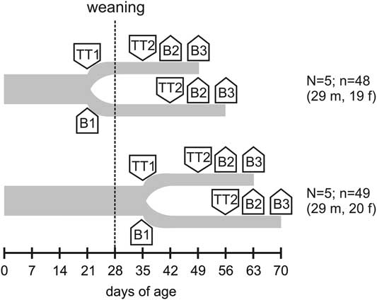
The piglets were randomly assigned to four experimental treatments: (1) primary immunization at 21 days of age, booster immunization 14 days later; (2) primary immunization at 21 days of age, booster immunization 21 days later (3) primary immunization at 35 days of age, booster immunization 14 days later or (4) primary immunization at 35 days of age, booster immunization 21 days later. Group allocation was performed with respect to gender to obtain approximately equal distribution of males and females. Piglets were immunized by intramuscular injection of Equilis® Te vaccine containing 40 limiting flocculation units per ml (Lf/ml) of TT and a saponin/cholesterol/phosphatidylcholine mixture (immunostimulating complex; ISCOM) as an adjuvant (MSD Tiergesundheit/Intervet GmbH, Unter-schleissheim, Germany) into the musculature of the neck directly behind the right ear. Blood samples were collected from the piglets by anterior vena cava puncture while they were in a supine position, immediately before the first immunization as well as 1 and 2 weeks after the booster immunization. For an overview, see Figure 1. The blood samples (5 ml) were immediately transferred to precooled heparinized propylene tubes and placed on ice. Part of each sample was subsequently centrifuged at 2000×g for 15 min at 4°C for plasma extraction. The plasma was stored at −20°C until further analysis by ELISA.
Experimental setup. First blood sampling (B1) and primary immunizations (TT1) were performed at 21 or 35 days of age. Booster immunizations (TT2) were performed 14 or 21 days later. Blood samples 2 and 3 were taken 1 (B2) and 2 weeks (B3) after TT2, respectively. N=number of litters; n=number of piglets; m=males; f=females.

Tetanus toxoid antibody detection
Plasma antibody titers (all isotypes) against TT were determined using an anti-tetanus antibody ELISA kit (RE57441; IBL International, Hamburg, Germany) according to the manufacturer’s directions. This kit uses TT-coated wells. Both TT-specific IgG and IgM in the samples bind to this antigen and to TT-peroxidase conjugate, which makes the kit species-independent.
Isolation of peripheral blood mononuclear cells and proliferation/viability assay
Peripheral blood mononuclear cells were isolated from heparinized blood by density gradient centrifugation, and the cell density was adjusted to 2×10⁶ cells/ml complete RPMI 1640 medium, as previously described (Tuchscherer et al., Reference Tuchscherer, Kanitz, Otten and Tuchscherer2002). To test the proliferative response to TT, the cell suspensions were added in triplicate to flat-bottom 96-well plates containing TT (Statens Serum Institut, Copenhagen, Denmark) to a final cell density of 1×10⁶ cells/ml and a final concentration of 0, 1.25 or 5 Lf/ml TT. Likewise, the proliferative response to phytohemagglutinin (PHA; 6.25 µg/ml) was tested. The plates were incubated for 72 h in a 5% CO2 humidified incubator. Cell proliferation/viability was evaluated using a 3-[4,5-dimethylthiazol-2-yl]-2,5 diphenyl tetrazolium bromide assay (Roche Diagnostics, Mannheim, Germany) as described before (Tuchscherer et al., Reference Tuchscherer, Kanitz, Otten and Tuchscherer2002). The optical density (OD) was measured with a microplate reader (Dynatech, Denkendorf, Germany) using a test wavelength of 550 nm and a reference wavelength of 690 nm. The results were expressed as the TT-specific proliferation index (PI) (OD in the presence of TT divided by OD in the absence of TT).
Assay of IgG and IgM
Concentrations of IgG and IgM were determined in duplicate with porcine specific ELISAs according to the manufacturer’s instructions (Bethyl Laboratories Inc., Montgomery, TX, USA). The intra-assay and inter-assay coefficients of variation for the ELISAs were <5% and <10%, respectively.
Statistical analysis of results
The data analysis was conducted using SAS software version 9.4 for Windows (SAS Institute Inc., Cary, NC, USA). Descriptive statistics and tests for normality were calculated with the Univariate procedure of Base SAS software. Plasma IgG, IgM, TT-specific antibody concentrations, TT-specific PIs and PHA-induced PIs could be considered approximately normal and were analyzed by repeated measures ANOVA with the MIXED procedure of SAS/STAT software.
The ANOVA model for plasma IgG, IgM, TT-specific antibody concentrations and PHA-induced PIs contained the fixed factors sex (levels: male, female), immunization 1 (levels: at 21 days of age, at 35 days of age), immunization 2 (levels: 14, 21 days after immunization 1), blood sampling (levels: B1 (before immunization1, only for IgG and IgM), B2 (1 week after immunization 2), B3 (2 weeks after immunization 2)), all interactions between the fixed factors and a random litter effect. Repeated measurements (blood sampling) on the same animal were taken into account, and a compound-symmetry type for the blocks of the block-diagonal residual covariance matrix was used.
The ANOVA model for TT-specific PIs contained the fixed factors sex (levels: male, female), immunization1 (levels: at 21 days of age, at 35 days of age), immunization 2 (levels: 14, 21 days after immunization 1), blood sampling (levels: B2 (1 week after immunization 2), B3 (2 weeks after immunization 2)), TT concentration (levels: 1.25; 5 Lf/ml), all interactions between the fixed factors and a random litter effect. Repeated measurements (blood sampling and TT concentration) on the same subject were taken into account, and a compound-symmetry type for the blocks of the block-diagonal residual covariance matrix was used.
Least-squares means (LSM) and their standard errors (SE) were computed for each fixed factor in the models, and all pairwise differences of these LSM were tested using the Tukey–Kramer test, a procedure for pairwise multiple comparisons. Effects and differences were considered significant if P<0.05. In all figures, LSM and SE are depicted.
Results
Tetanus toxoid-specific antibody production
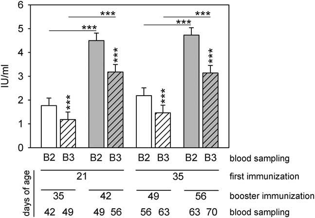
Before immunization, TT-specific antibody concentrations were below the detection limit of 0.09 IU/ml in all individuals (data not shown). In all immunization protocols, immunization induced an antibody response, yet with differential efficiency. Effects of the intervals between the primary and booster immunizations (P<0.001) as well as the intervals between the booster immunization and blood sampling (P<0.001) were found. Pairwise comparisons showed that administration of the booster immunization 3 weeks after the primary immunization yielded higher antibody concentrations than booster immunization 2 weeks after the primary immunization, independent of the piglets’ age at the time of first immunization. Indeed, for the primary immunization there was no age influence on antibody concentrations (Figure 2). Blood sampling revealed higher antibody concentrations 1 week after booster immunization than after 2 weeks (Figure 2). Gender did not affect TT-specific antibody production (P=0.934).
Tetanus toxoid (TT)-specific antibody plasma concentrations in response to different immunization protocols. TT-specific antibody plasma concentrations were highest when booster immunizations followed 3 weeks after primary immunizations and blood samples were taken 1 week after booster immunizations (B2). Horizontal asterisks indicate significant differences between booster immunizations 2 or 3 weeks following primary immunizations. Vertical asterisks indicate significant differences between blood samplings 1 (B2) or 2 weeks (B3) after booster immunizations (***P<0.001; n=23 to 25 per column).

Tetanus toxoid-specific proliferative response of lymphocytes
The proliferative response to TT was tested using in vitro proliferation tests with two different TT concentrations. We observed main effects of (i) gender×age at primary immunization (P=0.003), (ii) the time of blood sampling (P=0.011), (iii) gender×age at immunization×time of blood sampling (P=0.023), (iv) age at primary immunization×time of blood sampling (P=0.030), and (v) TT concentration using in vitro proliferation tests (P<0.001).
Pairwise comparisons by the Tukey–Kramer test revealed that the lymphocytes of males had a lower proliferative response to TT than those of females when immunization 1 was performed at 21 days of age (1.17±0.07 v. 1.37±0.08; P=0.039).
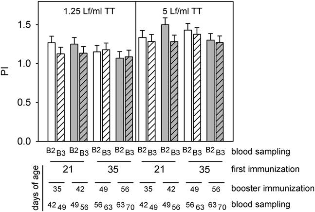
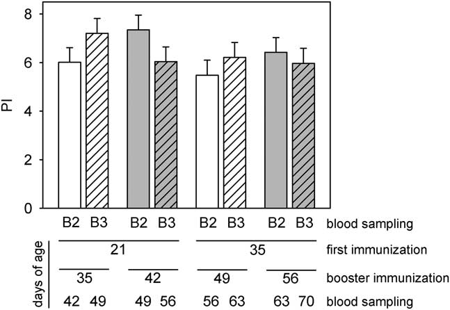
TT-specific proliferative responses increased with increasing TT concentrations (P<0.001; Figure 3) and were higher when blood sampling was performed 1 week compared with 2 weeks after booster immunization (P=0.011). When blood sampling was performed 1 week after booster immunization, the differences between the proliferative responses to 1.25 and 5 Lf/ml TT were significant in the group that had its primary and booster immunizations at 35 and 49 days of age, respectively (P=0.026). Proliferative responses did not show differences for the different immunization protocols (Figure 3).
Tetanus toxoid (TT)-specific proliferative responses of lymphocytes. The responses were independent of immunization protocols. B2 and B3 indicate blood sampling one and 2 weeks after booster immunizations, respectively. TT concentrations in cell culture were 1.25 and 5 Lf/ml (n=23 to 25 per column). PI=proliferation index.

Plasma IgG concentrations
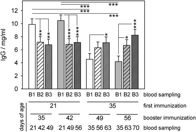
Before primary immunization at the age of 21 days, male piglets showed higher IgG concentrations than female piglets (9.8 v. 8.1 mg/ml; P=0.039). Plasma concentrations of IgG were highest at 21 days of age and decreased after that. Only in the piglets that had been first immunized at 35 days of age, there were significant increases in parallel with immunization. If these piglets were boosted 14 days later, IgG concentrations increased significantly at the third blood sampling; if they were boosted 21 days later, IgG concentrations increased significantly at the second and third blood samplings (Figure 4). Plasma IgG concentrations corresponded nicely with age at blood sampling, apparently independent of TT immunization (Figure 4). There was no main gender effect on plasma IgG (P=0.098).
Plasma IgG concentrations in relation to tetanus toxoid (TT) immunizations. Plasma IgG concentrations decreased in piglets immunized at 21 days of age but increased when piglets were first immunized at 35 days of age. TT immunization might have contributed to this increase. Horizontal asterisks indicate significant differences comparing the respective blood samples of the different vaccination protocols. Vertical asterisks indicate significant differences between the blood sample B1 (before immunizations) and the blood samples B2 and B3, which were taken 1 or 2 weeks after booster immunizations (*P<0.05; **P<0.01; ***P<0.001; n=23 to 25 per column).

Plasma IgM concentrations
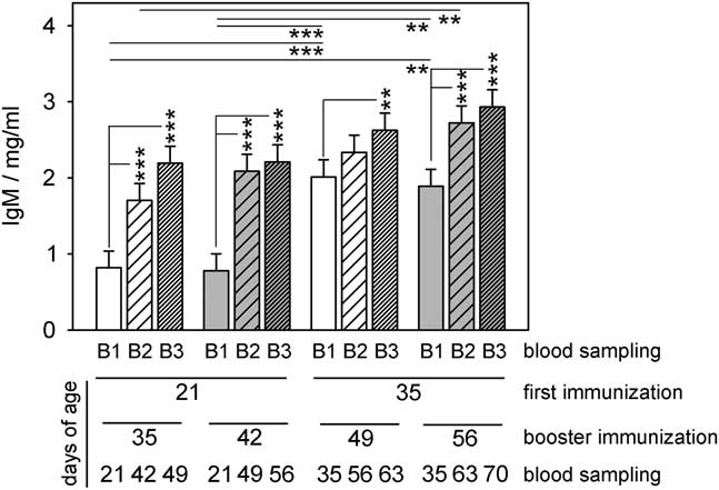
Plasma concentrations of IgM before immunization were significantly higher at 35 days of age than at 21 days of age (1.93 v. 0.80 mg/ml; P<0.001). There were increases in IgM concentrations paralleling immunization. However, these increases could also be age-related as the concentrations without immunization at 35 days of age were as high as with immunization at 49 and 56 days. When the first immunization was performed at 35 days, there were increases in IgM concentrations with age at blood sampling (Figure 5). There was no gender effect on plasma IgM (P=0.295).
Plasma IgM concentrations in relation to tetanus toxoid (TT) immunizations. Plasma IgM concentrations increased with age but independent of TT immunizations. Horizontal asterisks indicate significant differences comparing the respective blood samples of the different vaccination protocols. Vertical asterisks indicate significant differences between the blood sample B1 (before immunizations) and the blood samples B2 and B3, which were taken 1 or 2 weeks after booster immunizations (*P<0.05; **P<0.01; ***P<0.001; n=23 to 25 per column).

Mitogen-induced proliferation of lymphocytes
The proliferative responses of lymphocytes induced by the mitogen PHA were independent of the timing of primary and booster TT immunizations and thus independent of age as well as of the time of blood sampling (Figure 6). There was no gender effect on mitogen response (P=0.706).
Lymphocyte proliferative responses to phytohemagglutinin (PHA) were independent of tetanus toxoid (TT) immunizations and age. B2 and B3 indicate blood samples, which were taken 1 and 2 weeks after booster immunizations (n=23 to 25 per column). PI=proliferation index.

Discussion
In this study, we optimized TT vaccination as a tool to investigate immune competence in pigs. Different immunization and blood sampling protocols were tested to determine the optimal timing for maximum immune responses. In our setup, the age at primary immunization had no significant influence on the TT-specific antibody production. Booster immunizations induced higher antibody responses when performed three rather than two weeks after primary immunization. Antibody concentrations in blood samples were higher 1 week rather than 2 weeks after booster immunization. This can be explained by the fact that the ELISA recognizes both, IgG and IgM antibodies. The decrease in antibody titers 2 weeks after booster immunization could thus be due to a reduced production of IgM after an initial strong response and/or to a shorter half-life of IgM compared to IgG in the blood (Frazer and Capra, Reference Frazer and Capra1999).
TT-specific proliferative responses were independent of the immunization protocol, that is, the age at primary immunization and whether booster immunization was performed 2 or 3 weeks after primary immunization. By contrast, the time of blood sampling had a significant influence. Just as for antibody production, blood samples that were taken 1 week rather than 2 weeks after booster immunization showed higher proliferative responses.
The general proliferative ability of T cells was not influenced by TT immunization. Phytohemagglutinin-induced T cell proliferation did not show differences between animals that had undergone different immunization protocols. As expected, the time of blood sampling with respect to TT booster immunizations also did not influence the PHA-induced proliferation of T cells. Similar to TT-specific T cell responses, PHA-induced proliferation was independent of age. The proliferative responses to TT were lower than the proliferative responses to the mitogen PHA. This can be explained by the ability of PHA to induce proliferation in all T cells, while TT may only induce proliferation of TT-specific T cells.
Apparently, TT-specific antibody production was not reflected in general IgG and IgM plasma levels. Because our TT-specific antibody ELISA kit recognized both IgG and IgM antibodies and had IU/ml instead of mg/ml as readout, it is not easy to decide whether IgG, IgM or both levels were markedly increased by TT immunization. While IgG is the main porcine immunoglobulin, representing between 80% and 85% of all immunoglobulin in the blood, IgM is the second most abundant immunoglobulin, with 10% to 12%. For young piglets, it is well known that total IgG and IgM levels are strongly age dependent. Initially, IgG and IgM levels are high because suckling newborn piglets take up both immunoglobulins with colostrum during the first 24 to 36 h postpartum (Klobasa et al., Reference Klobasa, Werhahn and Butler1981; Le Dividich et al., Reference Le Dividich, Rooke and Herpin2005). IgG and IgM levels then decrease until the piglets’ own synthesis of immunoglobulin exceeds the depletion of the maternal antibodies (Klobasa et al., Reference Klobasa, Werhahn and Butler1981; Butler et al., Reference Butler, Zhao, Sinkora, Wertz and Kacskovics2009; Juul-Madsen et al., Reference Juul-Madsen, Jensen, Nielsen and Damgaard2010). Our own data show a decrease of IgG until the age of 56 days, followed by an increase, which probably reflects the synthesis of TT-specific IgG as well as IgG of other specificities. In contrast, we observed a perpetual increase for IgM. These dynamics may confound changes in antibody production due to external stimuli such as stress-induced immunosuppression. Specifically, a reduced antibody production in piglets due to weaning-related stress has been observed before (Blecha and Kelley, Reference Blecha and Kelley1981; Tuchscherer et al., Reference Tuchscherer, Kanitz, Puppe and Tuchscherer2006). Many studies on stress in domestic pigs are performed at an early age because stress sensitivity is stronger in piglets (Kanitz et al., Reference Kanitz, Puppe, Tuchscherer, Heberer, Viergutz and Tuchscherer2009). This suggests TT vaccination as an ideal surrogate test for immune competence because its effects are independent of age in piglets whose immune system is still developing. Moreover, stress may reduce vaccination responses in humans and animals (Pedersen et al., Reference Pedersen, Zachariae and Bovbjerg2009; Powell et al., Reference Powell, Allen, Hufnagle, Sheridan and Bailey2011; Segerstrom et al., Reference Segerstrom, Hardy, Evans and Greenberg2012). Thus, TT vaccination responses may reflect stress-induced reductions of immune competence in piglets.
The Equilis® TT vaccine has been used before in pigs although with aluminum hydroxide as an adjuvant. Studies with this vaccine reported antibody production and gene expression in a number of immune response-related pathways based on microarray analysis (Adler et al., Reference Adler, Murani, Brunner, Ponsuksili and Wimmers2013a and Reference Adler, Murani, Ponsuksili and Wimmers2013b; Reference Adler, Murani, Ponsuksili and Wimmers2015; Oster et al., Reference Oster, Scheel, Murani, Ponsuksili, Zebunke, Puppe and Wimmers2015). However, there are no published studies with our vaccine using ISCOM as an adjuvant. An upregulation of IL-1β, IL-18 and IL-10 messenger RNA (mRNA) as well as many other genes reflecting inflammation has been shown when an ISCOM adjuvant was injected into pigs without antigen (Ahlberg et al., Reference Ahlberg, Lovgren, Wallgren and Fossum2012). An earlier study showed that ISCOM adjuvants induced secretion of IL-12 (Smith et al., Reference Smith, Donachie, Grdic, Lycke and Mowat1999), which in combination with IL-18 is able to induce IFN-γ production in porcine NK and T cells even in the absence of antigen (Domeika et al., Reference Domeika, Berg, Eloranta and Alm2002). TT vaccination of pigs did not induce upregulation of the Th2 cytokine genes IL-4, IL-5 and IL-10, while IFN-γ mRNA was upregulated in response to booster vaccination (Adler et al., Reference Adler, Murani, Ponsuksili and Wimmers2013b). In TT vaccination studies with humans and mice, stronger IFN-γ than IL-4 responses have been observed (Robinson et al., Reference Robinson, Chamberlain, Lopez, Rush, Marcotte, Le Page and Wells2004; Livingston et al., Reference Livingston, Jiang and Stephensen2013). However, the determination of cytokines in plasma, in stimulated lymphocytes or in lymphocyte culture supernatants could be employed for a more complete assessment of immune competence.
For piglets, weaning stress is inevitable in standard husbandry. The process of weaning involves a number of stressors such as separation from the mother, change of nutrition and housing, and mixing with unfamiliar conspecifics (Puppe et al., Reference Puppe, Tuchscherer and Tuchscherer1997). To attenuate stress, we excluded mixing with unfamiliar conspecifics by litterwise weaning. The social support provided by familiar conspecifics has been shown to substantially reduce social stress (Kanitz et al., Reference Kanitz, Hameister, Tuchscherer, Tuchscherer and Puppe2014; Tuchscherer et al., Reference Tuchscherer, Kanitz, Tuchscherer and Puppe2016). This may be the reason why we did not observe any immunosuppressive effects of weaning.
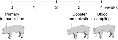
In pigs, tetanus is a rare disease (Wendt and Bickhardt, Reference Wendt and Bickhardt2001). As in other species, it is lethal for pigs, leaving no survivors with natural anti-TT antibodies. Despite this lethality, TT vaccination is not standard for pigs. For these reasons, pigs do not carry anti-TT antibodies in their blood unless they are immunized with TT. This ensures that the antibody synthesis and the TT-specific proliferative response were induced by our experimental immunization and exclusively reflected the immune response to TT vaccination. We see the specific immune responses to TT vaccination as a suitable tool to monitor the effects of stress or other challenges to immune competence. Our optimized immunization and blood sampling regime is depicted in Figure 7.
Optimized vaccination and blood sampling regime. Primary vaccination could be performed at 21 or 35 days of age with similar efficiency.

Acknowledgments
The authors wish to thank Birgit Sobczak and Regina Wal for excellent technical assistance.