Introduction
Kawasaki disease is a systemic vasculitis of unknown aetiology usually seen in children under 5 years of age. Its main complication is coronary artery aneurysms, and it is among the leading causes of acquired heart disease in children globally. Reference Agarwal and Agrawal1 Effective and validated indices to predict prognosis may guide treatment planning.
At present, hematological indices based on peripheral hemogram parameters are widely used as inflammatory prognostic markers. Reference Kawamura, Takeshita, Kanai, Yoshida and Nonoyama2,Reference Liu, Zhou and Hua3 The Systemic Immune-Inflammation Index is calculated using a combination of platelet, neutrophil, and lymphocyte counts, which may offer superiority over other simple parameters in demonstrating the severity of inflammation. Reference Yang, Wu and Hsu4
This study aimed to use the Systemic Immune-Inflammation Index to evaluate patients with Kawasaki disease and to investigate its possible role in predicting coronary artery involvement.
Methods
Patients who were followed up by the Paediatric Cardiology Clinic with the diagnosis of Kawasaki disease between January 2011 and December 2020 were retrospectively examined. The patients were diagnosed according to the clinical diagnostic criteria of the American Heart Association as follows. Patients with fever and an additional four clinical criteria were diagnosed with complete Kawasaki disease. Those with two or three of the clinical findings, three or more supportive laboratory findings, or less than three supportive laboratory findings and positive echocardiography findings were classified as incomplete Kawasaki disease. Reference McCrindle, Rowley and Newburger5 Supporting laboratory findings were anaemia for age; elevated transaminases; serum albumin level lower than 3 g/dL; platelet count higher than 450000/mm3 after day 7, white blood cell count higher than 15,000/mm3, and the presence of more than 10 white cells per high-power field in urine examination. Reference McCrindle, Rowley and Newburger5 During treatment, all patients received intravenous immunoglobulin and aspirin. The persistence or recurrence of fever 36 hours after the end of intravenous immunoglobulin treatment was defined as intravenous immunoglobulin resistance. Reference McCrindle, Rowley and Newburger5
Age- and sex-matched children who presented to the paediatric cardiology outpatient clinic due to a murmur and had no evidence of infection on examination or laboratory findings were included as a control group. Patients with multisystem inflammatory syndrome in children (MIS-C) were excluded from the study.
Epidemiological characteristics (e.g. age, sex) and laboratory findings including complete blood count parameters (white blood cell, neutrophil, lymphocyte, and platelet counts, neutrophil-to-lymphocyte ratio, mean platelet volume, erythrocyte sedimentation rate), C-reactive protein, and troponin-T levels were recorded upon admission for all patients and controls. Respiratory panel polymerase chain reaction test results, electrocardiography and echocardiography findings, and treatment responses were evaluated and recorded in the patient group. Systemic Immune-Inflammation Index was calculated as the neutrophil count multiplied by the platelet count, divided by the lymphocyte count. Reference Yang, Wu and Hsu4
Echocardiographic examination was performed using a GE Vingmed Vivid S6 device (GE Vingmed Ultrasound, Horten, Norway). On echocardiographic examination, meeting any of the following three conditions was considered positive: 1) A Z-score of ≥2.5 for the left anterior descending coronary artery (LAD) or right coronary artery, 2) detectable coronary artery aneurysm; 3) presence of at least three of the following together: LAD or right coronary artery Z-score of 2–2.5, decreased left ventricular function, mitral regurgitation, and pericardial effusion. Reference McCrindle, Rowley and Newburger5 Coronary arteries with a maximum Z-score ≥2 were evaluated as coronary artery lesions.
The studied parameters were compared between the Kawasaki disease and control groups, as well as between Kawasaki disease subgroups consisting of intravenous immunoglobulin-resistant and intravenous immunoglobulin-sensitive patients, and those with and without coronary artery lesions.
The study was approved by the University Faculty of Medicine Ethics Committee (approval no: 60116787-020/365442) and conducted in accordance with the principles of the Declaration of Helsinki.
Statistical analysis
Statistical analysis was performed using the IBM SPSS version 25.0 software (IBM Corp., Armonk, NY, USA). Continuous variables were presented as mean, standard deviation, range, and qualitative variables as number and percentage. Kolmogorov–Smirnov and Shapiro–Wilk tests were used for the determination of normal distribution. Comparisons between groups were made using an independent-samples t-test and one-way analysis of variance with post hoc Tukey test when parametric test assumptions were met, and Mann–Whitney U test and Kruskal–Wallis variance analysis with post hoc Mann–Whitney U test and Bonferroni correction when parametric test assumptions were not met. Categorical variables were compared using the chi-square test. Relationships between variables were evaluated using Spearman’s correlation coefficient. Statistical significance was set at p < 0.05.
Results
A total of 62 patients with acute Kawasaki disease were included in the study. The mean age was 44 ± 39 months (range: 3–146 months). Twenty-nine (47%) of the patients were female, and 33 (53%) were male. There was no statistical difference between the Kawasaki disease and control group in terms of age, sex, weight, height, or body mass index (p > 0.05). Patients presented most frequently in winter (n = 26, 41%) and spring (n = 13, 20%). The mean number of febrile days at admission was 7.8 ± 2.6 days (range: 4–15 days). At the time of admission, 54 (87%) of the patients had lip and oral changes, 53 (85%) had non-exudative conjunctivitis in the eyes, 52 (84%) had rash, 34 (58%) had swelling, redness, or peeling in the hands and feet, and 30 (48%) had lymphadenopathy. Nearly, all (n = 58, 94%) of the patients exhibited irritability. When additional clinical findings were evaluated, 11 patients (17%) had arthritis, 8 patients (13%) had erythema and induration at the Bacille Calmette-Guérin inoculation site. A respiratory panel polymerase chain reaction test was performed in 30 of the 62 patients, and the result was positive in 14 (46%). Of these patients, adenovirus was isolated in 5 (35%) and rhinovirus in 4 (28%). Troponin levels were evaluated in all patients; in only one patient, it was found to be three times higher than the normal range.
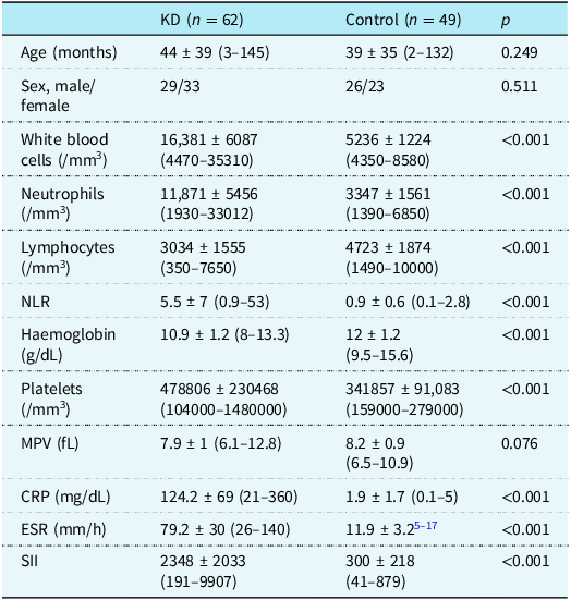
In Kawasaki disease patient group, the mean white blood cell count at the time of admission was 16,381 ± 6087/mm3 (range: 4470–35310/mm3), neutrophil count was 11,871 ± 5456/mm3 (range: 1930–33012/mm3), lymphocyte count was 3034 ± 1555/mm3 (range: 350–7650/mm3), neutrophil-to-lymphocyte ratio was 5.5 ± 7 (range: 0.9–53), haemoglobin was 10.9 ± 1.2 g/dL (range: 8–13.3 g/dL), platelet count was 478806 ± 230468/mm3 (range: 104000–1480000)/mm3), mean platelet volume was 7.9 ± 1 (range: 6.1–12.8) fL, C-reactive protein was 124.2 ± 69 mg/dL (range: 21–360 mg/dL), and erythrocyte sedimentation rate was 79.2 ± 30 mm/h (range: 26–140 mm/h). The mean Systemic Immune-Inflammation Index was calculated as 2348 ± 2033 × 109 (range: 191–9907 × 109) (Table 1).
Demographic characteristics and laboratory data of patients with Kawasaki disease and the control group

Data shown as mean ± standard deviation (range). KD = Kawasaki disease; NLR = Neutrophil-to-leukocyte ratio; MPV = Mean platelet volume; CRP = C-reactive protein; ESR = Erythrocyte sedimentation rate; SII = Systemic Immune-Inflammation Index.
White blood cell, neutrophil, and platelet counts, neutrophil-to-lymphocyte ratio, C-reactive protein level, and erythrocyte sedimentation rate were significantly higher in patients with Kawasaki disease compared to the control group, while haemoglobin level and lymphocyte count were significantly lower (p < 0.05). The mean Systemic Immune-Inflammation Index was also significantly higher in patients with Kawasaki disease than in the control group (2348 ± 2033 × 109 vs. 300 ± 218 × 109, p < 0.05). Although mean platelet volume was lower in the patient group compared to the control group, the difference was not statistically significant (Table 1).
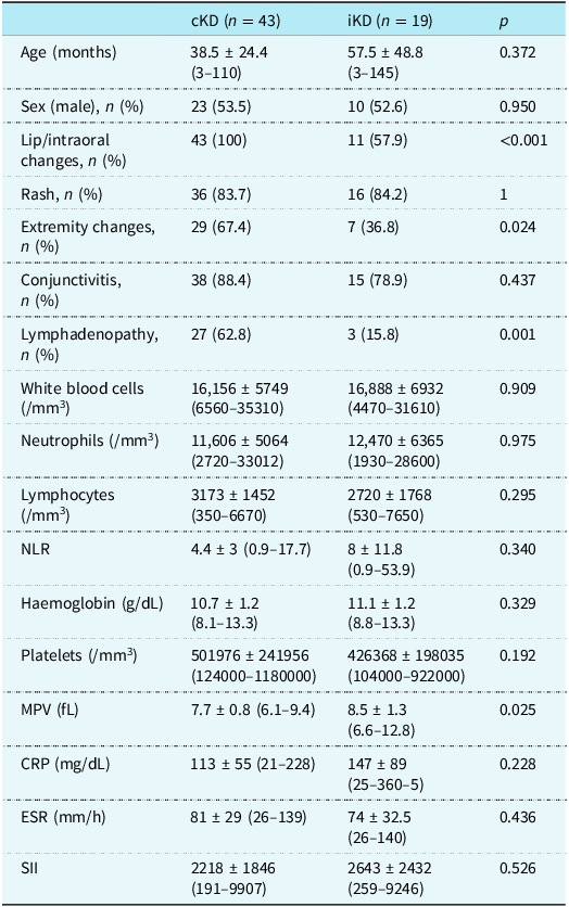
In the Kawasaki disease group, 43 (69%) of the patients had complete Kawasaki disease, and 19 (30%) had incomplete Kawasaki disease. Findings of lip and oral changes, swelling, redness, or peeling in the hands and feet, and lymphadenopathy were significantly more common in complete than in incomplete Kawasaki disease (p < 0.05). When the laboratory data were examined, mean platelet volume was significantly lower in the complete Kawasaki disease group (7.7 ± 0.8 fL, range: 6.1–9.4 fL) compared to the incomplete Kawasaki disease group (8.5 ± 1.3 fL, range: 6.6–12.8 fL) (p < 0.05). The mean Systemic Immune-Inflammation Index was 2348 ± 2033 × 109 (range: 191–9907 × 109) in patients with complete Kawasaki disease and 2643 ± 2432 × 109 (range: 259–9246 × 109) in patients with incomplete Kawasaki disease. Although Systemic Immune-Inflammation Index was higher in incomplete Kawasaki disease, the difference was not significant (p > 0.05) (Table 2).
Demographic characteristics and laboratory data of patients with complete and incomplete Kawasaki disease

Data shown as mean ± standard deviation (range) unless otherwise noted. cKD = Complete Kawasaki disease; iKD = Incomplete Kawasaki disease; NLR = Neutrophil-to-leukocyte ratio; MPV =Mean platelet volume; CRP = C-reactive protein; ESR = Erythrocyte sedimentation rate; SII = Systemic Immune-Inflammation Index.
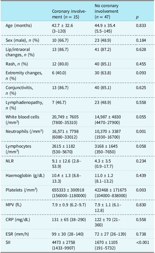
Echocardiographic evaluation revealed pericardial effusion in 13 patients (21%), mitral regurgitation in 6 patients (10%), and low ejection fraction in 1 patient. In the subacute period, coronary involvement was present in 15 patients (24%). Nine of the patients with coronary involvement had aneurysms in the coronary arteries. Neutrophil count, platelet count, and Systemic Immune-Inflammation Index were significantly higher at admission in patients with coronary involvement compared to those without (p < 0.05) (Table 3). Mean platelet count and Systemic Immune-Inflammation Index values, respectively, were 777333 ± 306872/mm3 (range: 356000–1180000/mm3) and 4688 ± 3081 × 109 (range: 1433–9907 × 109) in patients with aneurysm and 428622 ± 173604/mm3 (range: 104000–838000/mm3) and 1951 ± 1509 × 109 (range: 191–8418 × 109) in patients without aneurysm (p < 0.05 for both).
Demographic characteristics and laboratory data of patients with and without coronary involvement

Data shown as mean ± standard deviation (range) unless otherwise noted. NLR = Neutrophil-to-leukocyte ratio; MPV = Mean platelet volume; CRP = C-reactive protein; ESR = Erythrocyte sedimentation rate; SII = Systemic Immune-Inflammation Index.
All patients were treated with intravenous immunoglobulin and aspirin. Eight patients (13%) showed intravenous immunoglobulin resistance, all of whom had complete Kawasaki disease. The mean number of febrile days at admission was 7.8 ± 3 days (range: 4–12 days) among intravenous immunoglobulin-resistant patients and did not differ statistically from that of the intravenous immunoglobulin-sensitive group. Five (63%) of the intravenous immunoglobulin-resistant patients had coronary involvement. No significant differences in laboratory parameters at admission were observed between the intravenous immunoglobulin-resistant and sensitive groups (p > 0.05).
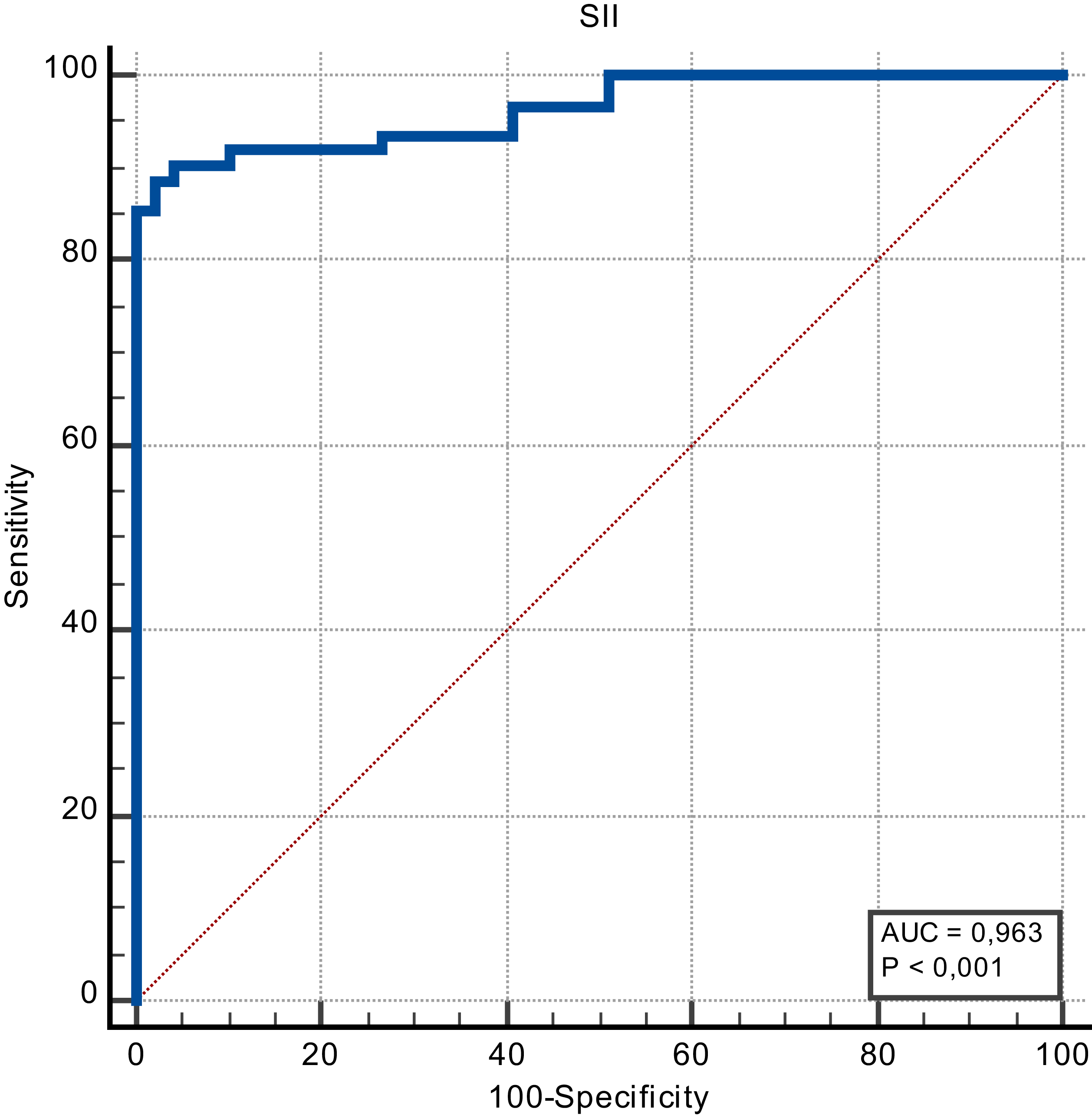
Systemic Immune-Inflammation Index was found to be significantly positively associated with the number of febrile days, white blood cell count, neutrophil count, platelet count, neutrophil-to-lymphocyte ratio, erythrocyte sedimentation rate, and C-reactive protein (p < 0.05; r values: 0.361, 0.452, 0.772, 0.55, 0.595, 0.658, 0.56, and 0.55, respectively). Systemic Immune-Inflammation Index was also negatively correlated with lymphocyte count, haemoglobin level, mean platelet volume, and coronary involvement (p < 0.05; r values: −0.46, −0.20, −0.42, and −0.59, respectively). According to the ROC Curve test result to determine the cut-off value of the Systemic Immune-Inflammation Index index, it was found that the Systemic Immune-Inflammation Index predicted the patient group with a cut-off value of >751.87, 96.3% AUC value, 88.71% sensitivity value, and 97.96% specificity value (Figure 1)
Examination of the predictability of the SII index in the patient group using the ROC curve test.

Discussion
Kawasaki disease is a vasculitis that can involve the medium-sized arteries throughout the body, especially the coronary arteries. The introduction of intravenous immunoglobulin therapy resulted in a significant decrease in rates of coronary artery involvement. The prevalence of coronary artery involvement is approximately 25% in untreated cases and can be reduced to the 5-10% range with treatment. Reference McCrindle, Rowley and Newburger5,Reference Türkuçar, Kaya and Çakmak6 Intravenous immunoglobulin resistance, defined as persistent or recurrent fever after completing the first intravenous immunoglobulin administration, occurs in 10–20% of patients. This group carries a higher risk of developing coronary artery involvement. Reference Newburger, Takahashi and Burns7 Consistent with the literature, coronary involvement was more frequent among the intravenous immunoglobulin-resistant patients in our study. Many scoring systems such as the Egami, Formosa, Kobayashi, and Sano scores have been developed to prevent coronary involvement and predict the risk of intravenous immunoglobulin resistance. Reference Chiang, Huang, Kuo, Huang and Chang8 However, their effectiveness needs to be verified taking into account regional and ethnic differences. Predicting a high risk of intravenous immunoglobulin resistance and/or coronary involvement at the time of admission can aid clinicians in identifying patients whose treatment should be revised. For this reason, they need predictors that are fast, low-cost, easy to use, and valid in their population. Studies on this subject are ongoing.
Neutrophils, a classic marker of inflammation, are known to play a critical role in the initial immune response in Kawasaki disease. This is followed by lymphocyte apoptosis, resulting in the release of various anti-inflammatory cytokines and immunosuppression. Reference Liu, Zhou and Hua3 Therefore, a high neutrophil and low lymphocyte counts may indicate intense inflammation and severe clinical course in patients with Kawasaki disease. Reference Kawamura, Takeshita, Kanai, Yoshida and Nonoyama2,Reference Takeshita, Kanai, Kawamura, Yoshida, Nonoyama and Bayry9 As in the Kobayashi and Sano scores, high neutrophil count has poor prognostic value in Kawasaki disease. Reference Chiang, Huang, Kuo, Huang and Chang8 In our study, the neutrophil counts were significantly higher in patients with coronary artery involvement, while lymphocyte counts were lower.
Platelet count reflects inflammation activity in Kawasaki disease, increasing due to megakaryocyte proliferation stimulated by proinflammatory cytokines. Reference Hwang, Shin and Jo10 High platelet counts and hypercoagulability have been associated with mortality and coronary involvement in Kawasaki disease. Reference Newburger, Takahashi and Gerber11,Reference Chen, Liu, Liu and Wu12 Studies testing the hypothesis that platelet volume may be a more sensitive marker than platelet count showed that increasing MPV levels were associated with myocardial infarction, acute ischaemic stroke, and diabetes, while decreasing mean platelet volume levels were associated with rheumatoid arthritis and ankylosing spondylitis. Reference Chen, Liu, Liu and Wu12,Reference Korniluk, Koper-Lenkiewicz, Kamińska, Kemona and Dymicka-Piekarska13,Reference Zheng, Wu, Zhang and Wu14 A decrease in mean platelet volume may occur as a result of the consumption or retention of large platelets in the vessels, or impaired thrombopoiesis. Reference Korniluk, Koper-Lenkiewicz, Kamińska, Kemona and Dymicka-Piekarska13 Low mean platelet volume levels have been demonstrated previously in patients with Kawasaki disease and their importance in early diagnosis has been emphasised. Reference Zheng, Wu, Zhang and Wu14 In another study evaluating the prognostic value of mean platelet volume in coronary artery involvement in 309 patients with Kawasaki disease, the authors reported that mean platelet volume was significantly lower in the patient group than the control group, but they were unable to demonstrate an association with coronary involvement. Reference Liu, Gao, Huo and Yi15 In contrast, we observed in our study that platelet counts were significantly higher in patients with coronary artery involvement compared to those without, while there was no significant difference in mean platelet volume.
As inflammatory markers can be easily affected by other factors, composite measures combining multiple inflammatory parameters may offer more reliable and powerful tools for assessing inflammation. Reference Okan, Doruk, Ozturk, Topaloglu, Dogdus and Yilmaz16 Neutrophil-to-lymphocyte ratio was reported to predict coronary aneurysm development and intravenous immunoglobulin resistance in patients with Kawasaki disease. Reference Liu, Zhou and Hua3,Reference Tsilimigras, Moris and Mehta17 In our study, neutrophil-to-lymphocyte ratio was significantly higher in patients with Kawasaki disease compared to the control group but was not associated with intravenous immunoglobulin resistance or coronary aneurysm development.
Systemic Immune-Inflammation Index is another composite parameter that combines neutrophil, lymphocyte, and platelet counts and has recently been evaluated as an indicator of poor outcomes in many diseases such as cancer, coronary artery disease, isolated coronary artery ectasia, saphenous vein graft disease, and rheumatological diseases. Reference Yang, Wu and Hsu4,Reference Tsilimigras, Moris and Mehta17–Reference Yalcinkaya, Öz and Durmuş21 There are only a few studies in the literature on Systemic Immune-Inflammation Index in Kawasaki disease. Reference Liu, Zhou and Hua3,Reference Dogdus, Dindas and Yenercag19,Reference Ermurat and Tezcan20
In one of those studies, Liu et al. Reference Liu, Zhou and Hua3 determined that Systemic Immune-Inflammation Index may serve as a reference for clinical management in predicting intravenous immunoglobulin resistance and various cardiovascular complications in patients with Kawasaki disease, but it was not found to be superior to neutrophil-to-lymphocyte ratio. The authors noted that Systemic Immune-Inflammation Index may not be applicable in Kawasaki disease patients with thrombocytopenia. However, they observed no difference in neutrophil-to-lymphocyte ratio or Systemic Immune-Inflammation Index between patients with and without coronary artery lesions. Yalçınkaya et al. determined that Systemic Immune-Inflammation Index was significantly lower in patients with coronary artery lesions compared to those without, while there was no difference in neutrophil-to-lymphocyte ratio. Reference Yalcinkaya, Öz and Durmuş21 Huang et al. observed a linear relationship between Systemic Immune-Inflammation Index at admission and the onset of coronary artery lesions and emphasised that high Systemic Immune-Inflammation Index values may correspond to a high risk for coronary artery lesion development. Reference Huang, Peng, Zhang, Zhu and Fan22
In the present study, neutrophil-to-lymphocyte ratio did not differ significantly in patients with coronary artery lesion or those with coronary artery aneurysms, whereas Systemic Immune-Inflammation Index was higher in these groups. When evaluating hematological indices, we believe that combining information on more inflammatory parameters may increase the power of the parameter. In addition, persistent and ongoing inflammation in Kawasaki disease is more associated with coronary artery involvement. Reference Liu, Zhou and Hua3 Calculating Systemic Immune-Inflammation Index at different times during the course of the disease and detecting dynamic variations may further increase its power in predicting coronary artery involvement.
Limitations of this study include its single-centre design, small sample size, and evaluation of coronary arteries only by echocardiography. However, the findings of this study demonstrate that Systemic Immune-Inflammation Index determined at the time of admission may predict coronary involvement in patients with Kawasaki disease. This composite parameter is a complementary adjunct to clinical and routine laboratory data and may have stronger prognostic value in future large-scale prospective studies.
Author Contributions
Merve KORKUT OĞUZ: Data curation, supervision, writing – original draft, writing – review and editing Dolunay GÜRSES: Conceptualization, data curation, writing – original draft Münevver YILMAZ: Clinical and imaging data curation, writing – review and editing Emine Sayın: Clinical and imaging data curation, writing – review and editing.
Conflicts of Interest
The authors declare no conflicts of interest.