Introduction
According to Diederich et al. (Reference Diederich, Lawrey and Ertz2018b), most species of lichenicolous fungi seem to be confined to specific host genera. Close examination of lichenicolous fungi growing on particular lichen taxa proved to be an effective approach to reveal the taxonomic diversity of these fungi. For example, during such case studies, two species of lichen-dwelling fungi were described as new from Arthrorhaphis (Hafellner & Obermayer Reference Hafellner and Obermayer1995), three from Baeomyces, Dibaeis and Icmadophila (Ihlen Reference Ihlen1998), three from Cladia (Zhurbenko & Pino-Bodas Reference Zhurbenko and Pino-Bodas2015), seven from Cladonia (Zhurbenko & Pino-Bodas Reference Zhurbenko and Pino-Bodas2017), ten from Graphidales (Diederich et al. Reference Diederich, Common, Braun, Heuchert, Millanes, Suija and Ertz2019), 13 from Lobariella (Flakus et al. Reference Flakus, Etayo, Miadlikowska, Lutzoni, Kukwa, Matura and Rodriguez-Flakus2019), four from Peltigera (Hawksworth Reference Hawksworth1980), one from Phlyctis (Muscavitch et al. Reference Muscavitch, Lendemer and Harris2017), one from Pilophorus (Zhurbenko & Triebel Reference Zhurbenko and Triebel2005), three from Placopsis (Brackel & Berger Reference Brackel and Berger2010), three from Stereocaulon (Zhurbenko Reference Zhurbenko2010a) and six from Thamnolia (Zhurbenko Reference Zhurbenko2012).
The families Baeomycetaceae and Icmadophilaceae sensu Lücking et al. (Reference Lücking, Hodkinson and Leavitt2016) have several morphologically similar genera in common and have sometimes been revised together (Burgaz Reference Burgaz2015). Lichenicolous fungi growing on species of Baeomyces (Baeomycetaceae), Dibaeis and Icmadophila (both belonging to Icmadophilaceae) in Norway have been studied by Ihlen (Reference Ihlen1998). We revisited these and also Phyllobaeis (Baeomycetaceae) and Pseudobaeomyces (Icmadophilaceae), which are morphologically similar to Baeomyces (baeomycetoid), from other regions of the world preserved in the herbarium of the National Museum of Nature and Science (TNS), Tsukuba, Japan. During this process we found 11 species of lichenicolous fungi, some of which were previously unknown to science.
The aims of this paper are to present the results of the revision, including the description of five new species, and to give an identification key to the species of lichenicolous fungi and lichens growing on baeomycetoid lichens and Icmadophila.
Materials and Methods
This study is based on 48 specimens of lichenicolous fungi mainly deposited in TNS (42 specimens) but also in several other herbaria: KRAM (1), LE (1), LPB (2) and the private collections of J. Etayo (1) and K. Kalb (2). A Stemi 2000-CS stereomicroscope and a Zeiss Axio Imager A1 compound microscope equipped with Nomarski differential interference contrast optics, fitted with an AxioCam MRc5 digital camera were used. Microscopic characters were studied using sections hand-cut with a razor blade and mounted in water, 10% potassium hydroxide (K), Lugol's iodine directly (I) or after a K pretreatment (K/I), brilliant cresyl blue (BCr), or concentrated nitric acid (N). Measurements were taken from water mounts. Where n >10, the length, width and length/width ratio (l/w) of the ascospores are given as (min−) (x̄ − SD) − (x̄ + SD) (−max), where ‘min’ and ‘max’ are the extreme values observed, x̄ the arithmetic mean and SD the corresponding standard deviation. Dimensions of ascospores were rounded to the nearest 0.5 μm. Colours were named according to Kornerup & Wanscher (Reference Kornerup and Wanscher1978).
The Species
Arthonia sp.
Ascomata apothecia, superficial, blackish, matt, epruinose, convex, immarginate, irregularly rounded in surface view, 60‒210 μm diam. (n = 10), aggregated to confluent. Epihymenium brown, c. 5 μm thick. Hymenium pale brown, not inspersed, 30‒35 μm tall, I+ red, K/I+ blue. Hypothecium brown, 40‒50 μm tall. Asci broadly clavate, c. 30 × 15 μm, 8-spored. Ascospores hyaline to brownish, clavate (club-shaped), irregularly arranged in the ascus, 12‒13.5 × 4.5‒5.5 μm (n = 6), 1-septate, halo not observed. Asexual morph not observed.
According to Diederich et al. (Reference Diederich, Lawrey and Ertz2018b), no Arthonia species have been reported on members of Baeomycetaceae and Icmadophilaceae. Keissler (Reference Keissler1930: 97) mentioned as ‘Celidium ericetorum Rehm apud Rabh.’ an Arthonia-like fungus growing on Baeomyces and Dibaeis, which has not been validly described. It differs markedly from the species that is mentioned above by its greenish epihymenium, I+ blue hymenium, colourless hypothecium, longer asci (60‒65 × 9‒10 μm) and hyaline, 1‒3-septate, narrower ascospores (10‒15 × 3 μm).
Rhymbocarpus ericetorum (Körb.) Etayo et al. growing on Dibaeis is somewhat reminiscent of an arthonioid fungus, but is quite distinct from the Arthonia species examined by having immersed apothecia, an olivaceous epihymenium and lateral exciple, a colourless to pale brown hypothecium, cylindrical to clavate asci with an only slightly thickened apical wall, and colourless, cylindrical-oblong, aseptate ascospores (Rambold & Triebel Reference Rambold and Triebel1990; Baral & Marson Reference Baral, Marson and Consiglio2001).
Specimen examined
Papua New Guinea: Morobe District: vicinity of Kasanombe, c. 30 km N of Lae, 06°22'S, 146°59'E, 1600–1700 m, on Pseudobaeomyces sp. (thallus, occasionally also on apothecia), 1975, S. Kurokawa 9560 (TNS, filed under the host).
Buelliella ohmurae Zhurb. & Diederich sp. nov.
MycoBank No.: MB 836571
Lichenicolous ascomycete. Distinguished from Buelliella minimula mainly by the somewhat narrower ascospores, 12.5‒17.5 × 5‒6.5 μm versus 14‒18 × 6‒8 μm, and a different host selection, Icmadophila versus Pertusaria.
Type: Japan, Honshu, Prov. Kai (Pref. Yamanashi), Mt Daibosatsu, 35°44'N, 138°50'E, on the thallus of Icmadophila ericetorum, 30 October 1955, Y. Kuwata (TNS-L-129845—holotype).
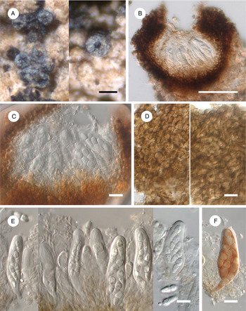
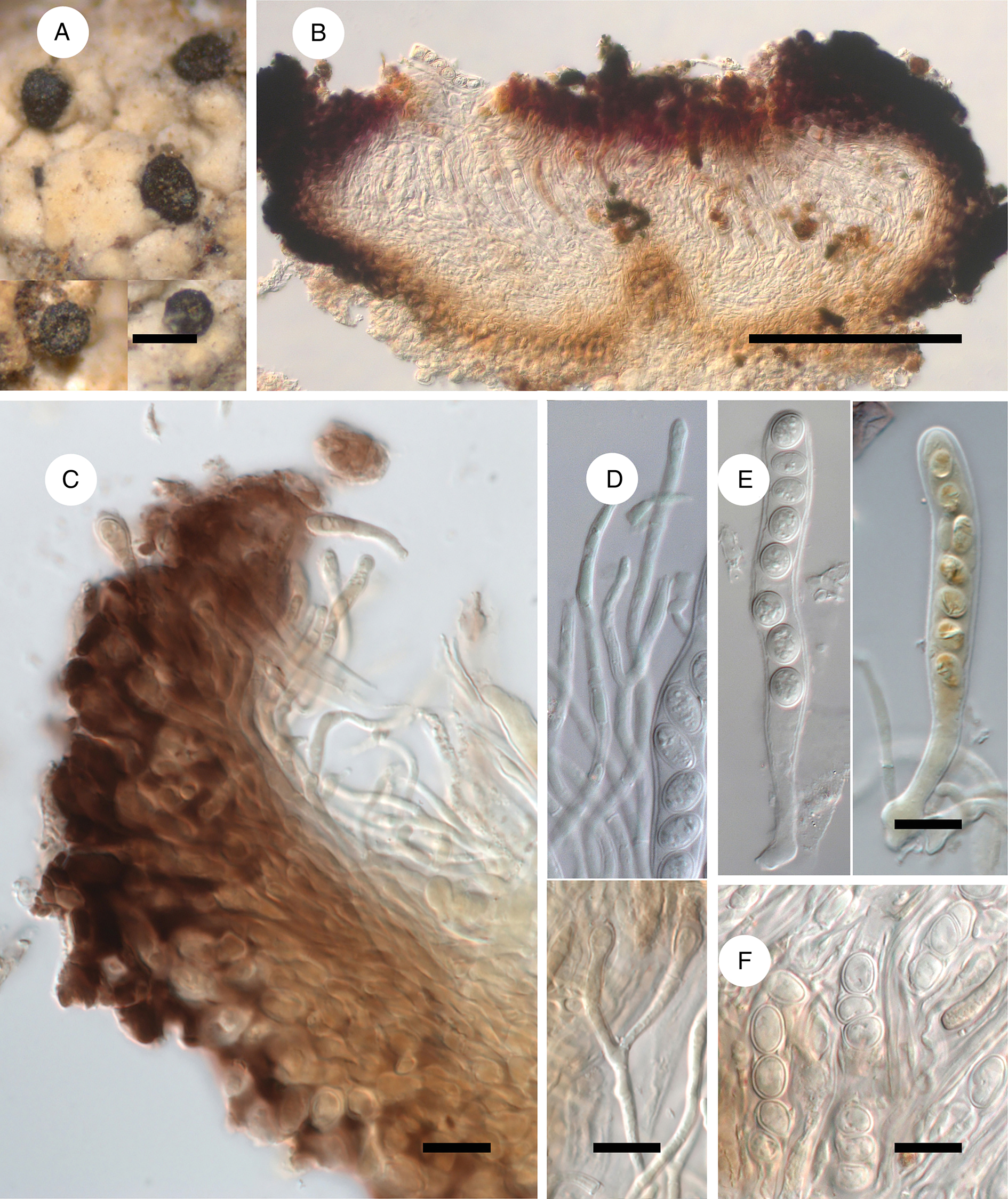
(Fig. 1)
Buelliella ohmurae (A, D–F, holotype; B, Shibuichi 4253; C, 1926, Okada (a)). A, habitus of ascomata on the thallus of Icmadophila ericetorum. B & C, ascoma in cross-section in water. D, different layers of the exciple in surface view in K. E, asci, ascospores and paraphysoids in K. F, ascus with spores in K/I. Scales: A = 100 μm; B = 50 μm; C–F = 10 μm. In colour online.

Ascomata initially almost closed, subspherical to flattened, partly immersed, eventually becoming urceolate, 80‒160 μm diam. (n = 14), erumpent, 1/2 to 3/4 exposed, blackish, matt, sometimes white pruinose, rough, with an irregularly rounded opening 10‒40 μm wide surrounded by radial splits, exposing part of the hymenium, aggregated to occasionally contiguous. Exciple brown, K− (light brown), 15‒30 μm thick, in surface view resembling textura epidermoidea and textura globulosa, in cross-section composed of circular or elongate cells up to 7 μm with walls 1‒2 μm thick. Periphyses not observed. Paraphysoids rather delicate and inconspicuous in water, clearly seen in K, 1‒1.5 μm diam., septate, flexuose, varying in thickness, sometimes slightly enlarged and pale brown at the apex, branched, occasionally anastomosed. Epihymenium pale brown, pigmentation not granulose, sometimes rather indistinct. Hymenium hyaline, 50‒60 μm tall, I−, K/I−. Asci bitunicate, elongate-clavate to narrowly clavate, with a distinct foot, with an ocular chamber 1‒2 μm long, 45‒55 × 13‒17 μm (n = 6), wall laterally rather thick, apically not to markedly thicker, I−, K/I−, 8-spored. Ascospores hyaline to occasionally medium brown, 1-septate, constricted at the septum, narrowly obovoid/soleiform with a broader upper cell, irregularly biseriate, partly overlapping diagonally in the ascus, (12.5‒)13.5‒15.5(‒17.5) × 5‒6(‒6.5) μm, l/w = (2.1‒)2.4‒2.8(‒3.0) (n = 22), sometimes disarticulating into semi-spores in squash mounts, wall c. 0.5 μm thick, smooth.
Asexual morph not observed.
Etymology
This species is dedicated to the Japanese lichenologist Yoshihito Ohmura, curator of the TNS lichen herbarium, in which material of the new species was discovered.
Distribution and host
The new species is known from four collections in Japan, growing on thalli and occasionally also on apothecia of Icmadophila ericetorum. Host tissues are bleached under severe infections.
Notes
The new species fits well with the concept of Buelliella Fink as presented in Hafellner (Reference Hafellner1979, Reference Hafellner, Nash, Ryan, Diederich, Gries and Bungartz2004), including diagnostic characters such as cleistohymenial ascomata, a hyaline, I−, K/I− hymenium, delicate, branched and anastomosed paraphysoids, bitunicate, I−, K/I− asci, and 1-septate ascospores, remaining hyaline for a long time. We did not see the short periphyses that occur in some Buelliella species (Ertz & Diederich Reference Ertz and Diederich2015) but neither were these observed in the generic type B. minimula (Tuck.) Fink.
The new species may be confused with other Buelliella species with ascospores similar in size. Among these, Buelliella eximia Kalb & Hafellner (on Pyxine) differs in the 1‒3-septate, shorter and wider ascospores, 12.5‒14 × 6‒8 μm (Kalb Reference Kalb1990); B. inops (Triebel & Rambold) Hafellner (on Caloplaca s. lat.) is distinct in the markedly enlarged, brown apical cells of paraphysoids and the 1(‒3)-septate, wider ascospores, 13‒18 × 6‒8.5 μm (Hafellner et al. Reference Hafellner, Triebel, Ryan and Nash2002); B. minimula (on Pertusaria) is distinguished by a distinct brown, granulose epihymenium and wider ascospores, 14‒18 × 6‒8 μm (Hafellner Reference Hafellner1979; specimens of B. minimula examined for comparison); B. physciicola Poelt & Hafellner (on Phaeophyscia and Physcia) has wider ascospores, 12‒17 × 6‒8.5 μm (Hafellner Reference Hafellner1979); B. protoparmeliopseos Etayo & Pérez-Ortega (on Protoparmeliopsis) differs in having lirelliform young ascomata, markedly enlarged, brown apical cells of paraphysoids and larger ascospores, 14‒21 × 4‒10 μm (Pérez-Ortega & Etayo Reference Pérez-Ortega and Etayo2010); and B. pusilla Hafellner (on Brigantiaea) differs in the I+ light blue then red-brown hymenium and smaller, verruculose ascospores, 11‒14 × 5‒6 μm (Hafellner Reference Hafellner1985).
Additional specimens examined (all on thalli and occasionally apothecia of Icmadophila ericetorum)
Japan: Honshu: Prov. Etchu (Pref. Toyama), Mt Yakushi, 36°28'N, 137°32'E, 1600 m, 1976, H. Kashiwadani 13480 (TNS); Prov. Shinano (Pref. Nagano), Yatsugatake Mts, 35°59'N, 138°21'E, 1926, Y. Okada (a) (TNS); Mt Kinpu, Minamisaku-gun, 35°52'N, 138°37'E, 2550 m, 1970, H. Shibuichi 4253 (TNS).
Specimens of Buelliella minimula used for comparison (both on Pertusaria spp.)
Brazil: São Paulo Municipality: zwischen São Lourenço und Juquií, etwa 65 km W von São Sebastião, am Rio Guaratuba, mangrove, 1980, K. Kalb (hb. Kalb).—Ecuador: Napo Province: zwischen Quito und Baeza, etwa 50 km NW von Baeza, in Paramovegetation, 4000 m, 1987, K. & A. Kalb 18669 (hb. Kalb).
Catillaria japonica Zhurb. & Hafellner sp. nov.
MycoBank No.: MB 836572
Lichenicolous ascomycete, not lichenized. Distinguished from Catillaria stereocaulorum by the rather frequently branched, anastomosed paraphyses, apically not or only slightly swollen, without a dark cap, medium reddish brown versus hyaline to pale yellowish brown hypothecium, and a different host selection, Dibaeis and Pseudobaeomyces versus Stereocaulon.
Type: Japan, Kyushu, Prov. Hyuga (Pref. Miyazaki), Ohkawauchi, Shiiba Research Forest, Kyushu University, Shiiba-son, Higashi-usuki-gun, 32°23′43"N, 131°11'10"E, 1140 m, on the thallus of Dibaeis sorediata, 13 November 2014, Y. Ohmura 11059 (TNS-L-129846—holotype).
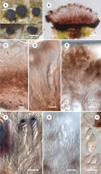
(Fig. 2)
Catillaria japonica (A & B, holotype; C & D, Kurokawa 62261; E–G, Kashiwadani 20272; H, Umezu 144). A, habitus of ascomata on the thallus of Dibaeis sorediata. B, ascoma in cross-section in water. C, exciple, hypothecium and hymenium in cross-section in water. D, hymenium and epihymenium in water. E, epihymenium in K. F, asci and paraphyses in K/I. G, paraphyses in K. H, ascospores in water. Scales: A = 200 μm; B = 50 μm; C & F = 20 μm; D, E, G & H = 10 μm. In colour online.
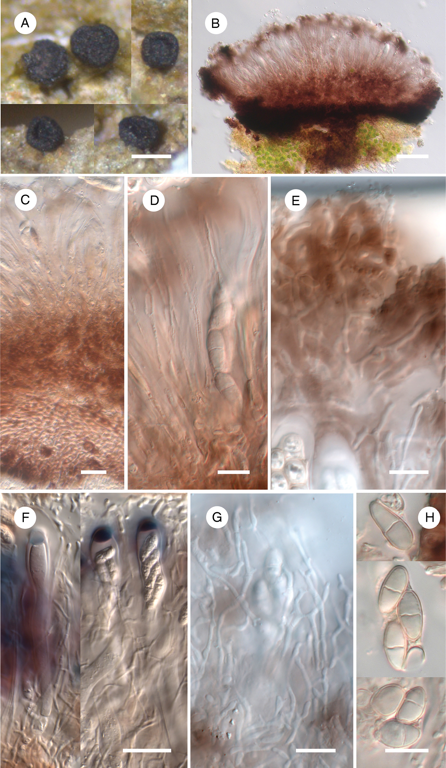
Ascomata apothecia, blackish, matt, epruinose, superficial, discoid, with slightly concave to slightly convex disc surrounded by an initially strongly elevated, later occasionally disappearing concolorous margin, constricted at the base, 150‒450(‒600) μm diam. (n = 20), arising singly or in small groups, occasionally contiguous. Exciple patchy medium to dark reddish brown, K+ slightly fading, 30‒50 μm laterally, 50‒250 μm basally where it sometimes forms a stipe immersed in the host thallus, in cross-section composed of circular or tangentially elongated cells up to 9 μm with walls 1.5‒2.5 μm thick. Hypothecium medium reddish brown, K+ slightly fading, 30‒70 μm tall, in cross-section of textura angularis. Epihymenium patchy medium to dark reddish brown, K+ slightly fading, pigmentation amorphous/non-granular, c. 10 μm tall. Reddish brown apothecial pigments N+ reddish orange to red. Hymenium hyaline to dull red, 55‒70 μm tall, entirely penetrated by a hymenial gel, without oil droplets, I+ blue, K/I+ blue with greyish red patches. Paraphyses 1‒2 μm diam., straight to sinuous (particularly above), extending beyond the asci, branched (sparingly below, rather frequently above), anastomosed, septate, with cells c. 3−10 μm long; tips not or slightly enlarged, without dark cap, embedded in reddish brown gelatinous gel. Asci of the Catillaria-type, subclavate, 50‒65 × 10‒12 μm (n = 10), tholus thickened, without any distinct apical apparatus, in K/I with a blue and partly reddish outer gelatinous coat and uniformly darker blue tholus, 8-spored. Ascospores hyaline to occasionally pale brownish orange or dull red, ellipsoid, narrowly ellipsoid, sometimes narrowly obovoid, with slightly wider upper cell, with rounded ends, (9.5‒)10.5‒13(‒15.5) × (4‒)4.5‒6(‒6.5) μm, l/w = (1.6‒)2.0‒2.6(‒3.0) (n = 78), (0‒)1(rarely 2‒3)-septate, not or only slightly constricted at the median septum, with smooth wall c. 0.5 μm thick throughout, non-halonate, usually without distinct guttules, (1‒)2(‒3)-seriate in the ascus.
In one specimen (Kashiwadani 20272) we observed semi-immersed, applanate, 30‒50 μm diam. pycnidia of unclear origin, occasionally growing on apothecial discs of Catillaria. They have a greenish grey wall and contain hyaline, short-bacilliform conidia, 3.5‒5 × 1‒1.5 μm.
Etymology
Named after Japan, where the type was collected.
Distribution and host
The new species is known from eight collections in Japan, growing on thalli of Dibaeis (mostly) and Pseudobaeomyces species, not visibly damaging the host.
Notes
To the best of our knowledge, the new species is most consistent with the broad concept of the large heterogeneous genus Catillaria A. Massal. (Hertel et al. Reference Hertel, Nash, Ryan, Nash, Gries and Bungartz2007). However, reddish brown apothecial pigments and a lichenicolous lifestyle are rare in that genus, and its paraphyses are typically simple or sparingly branched, abruptly swollen and have a dark brown cap at the apex. Its type species, C. chalybeia (Borrer) A. Massal., additionally differs in its carbonaceous black exciple and the presence of a green apothecial pigment (Hertel et al. Reference Hertel, Nash, Ryan, Nash, Gries and Bungartz2007). Compared to other lichenicolous species occurring in Catillaria (Diederich et al. Reference Diederich, Lawrey and Ertz2018b), C. japonica seems to be most similar to C. stereocaulorum (Th. Fr.) H. Olivier which also has reddish brown apothecial pigments but differs in its mostly simple paraphyses, apically abruptly swollen and with a dark reddish brown cap, and hyaline to pale yellowish brown hypothecium (Zhurbenko Reference Zhurbenko2010a).
Further genera comparable to the new species are Catinaria Vain. and Scutula Tul. The former differs in its ascospores having a compact gelatinous halo and the absence of a lichenicolous lifestyle (Gilbert Reference Gilbert, Smith, Aptroot, Coppins, Fletcher, Gilbert, James and Wolseley2009), and the latter in its asci with an amyloid tholus and diffuse non-amyloid axial body, and an epihymenium with a granular, greenish brown to greenish black pigment (Triebel & Kainz Reference Triebel, Kainz, Nash, Ryan, Diederich, Gries and Bungartz2004).
With its apothecial habitus, structure and pigmentation of the exciple and epihymenium, and shape and septation of the ascospores, Catillaria japonica resembles species of Sclerococcum Fr., particularly S. athallinum (Müll. Arg.) Ertz & Diederich growing on Baeomyces (Hafellner Reference Hafellner1979; present paper). However, species of that genus are easily distinguished by their asci with a K/I− tholus and brown ascospores (Hafellner Reference Hafellner, Nash, Ryan, Diederich, Gries and Bungartz2004).
Additional specimens examined
Japan: Hokkaido: Prov. Kitami, Bihoro Pass, Bihoro-cho, Abashiri-gun, 43°39′N, 144°15′E, 500 m, on Dibaeis arcuata (thallus), 1997, Y. Ohmura 3604 (TNS); Prov. Tokachi, along the trail from Me-akan Hot Spring to the top of Mt Me-akan, Ashoro-cho, Ashoro-gun, 43°23'N, 144°00′E, 920–1050 m, on D. arcuata (thallus), 1995, Y. Ohmura 1699 & H. Kashiwadani (TNS). Honshu: Prov. Rikuchu (Pref. Akita), around Sukawa Hot Spring, Mt Kurikoma, 38°57′N, 140°47'E, 1200 m, on D. arcuata (thallus), 1983, H. Kashiwadani 20272 (TNS); Prov. Shimotsukue (Pref. Tochigi), Nikko-city, 13 km NW of Nikko and 5 km ESE of the village Yumoto, at the river 150 m SE of the dirt road, 36°47'49.9"N, 139°28'50.0"E, 1570 m, on Dibaeis sp. (thallus) growing on Tsuga diversifolia log, 2015, G. Thor 32554 (TNS). Kyushu: Prov. Bungo (Pref. Ohita), Mt Taisen-zan, Kujyu-cho, Naoiri-gun, 33°05'N, 131°17′E, 1400 m, on D. arcuata (thallus), 1987, Y. Umezu 144 (TNS); Mt Yufu-dake, 33°16′N, 131°23′E, on Pseudobaeomyces pachycarpus (thallus), 1962, S. Kurokawa 62261 (TNS); Prov. Ohsumi (Pref. Kagoshima), Hananoego, Mt Kuromi-dake, Yakushima Island, 30°18'45"N, 130°30'38"E, 1550 m, on P. pachycarpus (thallus), 2005, K. Yoshida 13780 (TNS).
Cryptodiscus ihlenii Zhurb. sp. nov.
MycoBank No.: MB 836573
Lichenicolous ascomycete. Distinguished from Cryptodiscus galaninae mainly by the I− versus I+ blue then orange to red hymenium, narrowly obovate, 1-septate versus slightly fusiform, clavate or bacilliform, 0‒2-septate and larger ascospores (14‒20.5 × 5‒7.5 μm versus 7‒14.5 × 1.5‒2 μm), and a different host selection, Dibaeis versus Cladonia.
Type: Japan, Hokkaido, Prov. Tokachi, Mt Higashi-Nupukaushi, Kami-Shihoro-cho, Kato-gun, 43°14'N, 143°06'E, c. 1000 m, on the thallus of Dibaeis arcuata, 17 September 1979, H. Kashiwadani 15493 (TNS-L-129847—holotype).
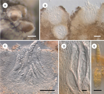
(Fig. 3)
Cryptodiscus ihlenii (holotype). A, habitus of ascomata on the thallus of Dibaeis arcuata. B & C, ascomata in cross-section in water. D, asci with spores in water. E, ascospores in I. Scales: A = 100 μm; B & C = 50 μm; D & E = 10 μm. In colour online.
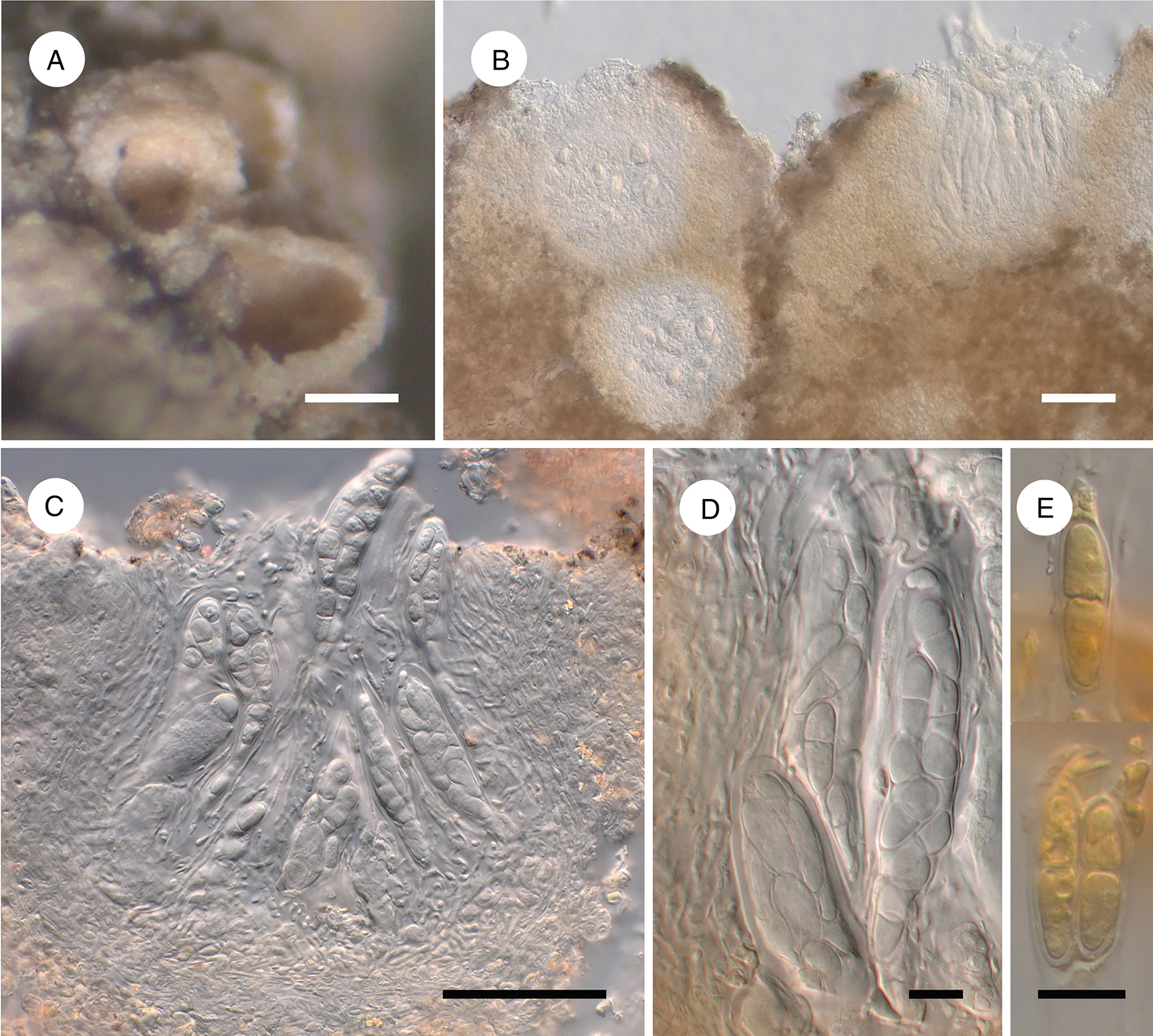
Ascomata cleistohymenial apothecia, aggregated, 80‒250 μm diam. (n = 7), persistently immersed in the host thallus, initially closed, subglobose, later opening by a small pore, finally urceolate or cupulate, with a widely exposed, concave, pale orange to orange-white, translucent, roundish to irregularly elongate (due to mutual compression) disc, inducing the formation of subglobose warts up to 0.4 mm diam. on the host thallus, containing one or several immersed apothecia. Exciple hyaline, without crystals, not differentiated into layers, 10‒20 μm thick throughout, in cross-section composed of tangentially somewhat elongated cells 2‒5 μm long, with walls 0.5‒1 μm thick, K−. Periphysoids absent. Epihymenium indistinct. Hymenium hyaline, 70‒100 μm tall, I−, K/I−. Subhymenium hyaline, c. 10 μm thick. Paraphyses hyaline, filiform, 1‒2 μm diam., septate, branched, apically not distinctly enlarged. Asci narrowly ellipsoid to elongate-clavate, wall 1‒3 μm thick, apically not or slightly thicker, apical structures not observed, 55‒80 × 13‒17 μm (n = 3), 8-spored, I−, K/I−. Ascospores hyaline, narrowly obovate, irregularly biseriate in the ascus, (14–)15.5‒19(‒20.5) × (5‒)5.5‒7(‒7.5) μm, l/w = (2.0‒)2.4‒3.2(‒3.7) (n = 29), 1-septate, not or slightly constricted at the septum, with smooth wall c. 0.5 μm thick, non-halonate, with many guttules.
Asexual morph not observed.
Etymology
The species is named after the Norwegian lichenologist Per Gerhard Ihlen who made a great contribution to the knowledge of lichenicolous fungi growing on baeomycetoid lichens.
Distribution and host
The new species is known from the type collection in Japan, growing on the thallus of Dibaeis arcuata, inducing gall-like warts on the host thallus; otherwise pathogenicity was not observed.
Notes
The new species fits the concept of Cryptodiscus Corda presented in Baloch et al. (Reference Baloch, Gilenstam and Wedin2009) and Pino-Bodas et al. (Reference Pino-Bodas, Zhurbenko and Stenroos2017). Most species of this genus are characterized by the K/I+ blue hymenium, K/I+ faintly blue ascus wall, simple, sometimes slightly forked above and apically often enlarged paraphyses, cylindrical phragmosporous ascospores and saprobic lifestyle on wood. However, three previously known species of Cryptodiscus are obligately lichenicolous (Diederich et al. Reference Diederich, Lawrey and Ertz2018b), the hymenium and ascus wall of C. epicladonia Zhurb. & Pino-Bodas do not change colour with K/I, the paraphyses of C. cladoniicola (D. Hawksw. & R. Sant.) Pino-Bodas et al. are regularly branched (Pino-Bodas et al. Reference Pino-Bodas, Zhurbenko and Stenroos2017), and both C. pini (Romell) Baloch et al. and C. foveolaris (Rehm) Rehm have 1-septate ascospores (Baloch et al. Reference Baloch, Gilenstam and Wedin2009).
The other lichenicolous species of Cryptodiscus, viz. C. cladoniicola, C. epicladonia and C. galaninae Zhurb. & Pino-Bodas, all growing on Cladonia, are readily distinguished from C. ihlenii by their eventually superficial ascomata and much narrower ascospores, up to 3 μm wide (Pino-Bodas et al. Reference Pino-Bodas, Zhurbenko and Stenroos2017). Additionally, C. cladoniicola is distinct, for example, in the I+ fleetingly blue hymenium and cylindrical to slightly fusiform, 2‒4-septate ascospores; C. epicladonia differs in its apothecia with a crystalline rim and filiform to cylindrical, 5‒11-septate ascospores; and C. galaninae differs in the I+ blue then orange to red hymenium and slightly fusiform, clavate or bacilliform, 0‒2-septate ascospores.
Cryptodiscus ihlenii is morphologically and anatomically comparable to species of Aabaarnia Diederich, Absconditella Vězda, Dimerella Trevis., Gyalecta Ach., Lettauia D. Hawksw. & R. Sant. s. lat., Nanostictis M. S. Christ. and Stictis Pers. Aabaarnia can be distinguished by its K/I+ blue hymenium, subcylindrical, 4‒6-spored asci and 3-septate ascospores with a perispore (Diederich Reference Diederich2014). Absconditella, which is phylogenetically very close to Cryptodiscus (Baloch et al. Reference Baloch, Gilenstam and Wedin2009), mainly differs in its lichenized not lichenicolous lifestyle and asci with a distinct apical dome (Coppins Reference Coppins, Smith, Aptroot, Coppins, Fletcher, Gilbert, James and Wolseley2009). Dimerella differs in its lichenized not lichenicolous lifestyle, sessile apothecia, I+ blue hymenium, paraphyses with a pair of swollen apical cells and asci with a K/I+ blue wall (Benfield et al. Reference Benfield, Purvis, Coppins, Smith, Aptroot, Coppins, Fletcher, Gilbert, James and Wolseley2009). Gyalecta differs in its lichenized not lichenicolous lifestyle and trans-septate to muriform ascospores (Gilbert et al. Reference Gilbert, James, Woods, Smith, Aptroot, Coppins, Fletcher, Gilbert, James and Wolseley2009). The type of Lettauia belongs to Cryptodiscus (Pino-Bodas et al. Reference Pino-Bodas, Zhurbenko and Stenroos2017); the phylogenetic positions of the other species of this genus, viz. L. hypotrachynae Etayo, L. santessonii Ihlen & Tønsberg and L. usneae Etayo are still unclear but they differ in having eventually superficial apothecia with a plane disc and a K/I+ blue hymenium (Ihlen & Tønsberg Reference Ihlen and Tønsberg1996; Etayo Reference Etayo2002, Reference Etayo2017). Nanostictis mainly differs in its filiform, cylindrical or acicular, exceptionally ellipsoid, ascospores with few or many trans-septa (Christiansen Reference Christiansen1954; Etayo & Diederich Reference Etayo and Diederich1996; Etayo Reference Etayo2002, Reference Etayo2017; Etayo & Sancho Reference Etayo and Sancho2008; Zhurbenko Reference Zhurbenko2017). Typically, Stictis includes saprobic or lichenized species with the ascomatal margin lined by periphysoids and filiform ascospores (Sherwood Reference Sherwood1977; Wedin et al. Reference Wedin, Döring and Gilsenstam2006). The generic position of the only lichenicolous species placed in this genus, S. cladoniae (Rehm) Sacc., is unclear; it lacks periphysoids but is well distinguished by the brown-black apothecia with blackish disc and an I+ red, K/I+ blue hymenium (Pino-Bodas et al. Reference Pino-Bodas, Zhurbenko and Stenroos2017).
Llimoniella chilensis Zhurb. sp. nov.
MycoBank No.: MB 836574
Lichenicolous ascomycete. Distinguished from the formerly known Llimoniella species by the combination of the K+ green reaction of the exciple and epihymenium, aseptate, broadly ellipsoid ascospores, and the host selection (growing on Dibaeis).
Type: Chile, Region X, Prov. Valdivia, Fundo San Martín (Universidad Austral de Chile), c. 30 km S of San José de la Mariquina, 40°38'S, 73°05'W, 10 m, mixed forest with Nothofagus obliqua, on the thallus of terricolous Dibaeis sp., 18 November 1987, H. Kashiwadani 35425a (TNS-L-129848—holotype).
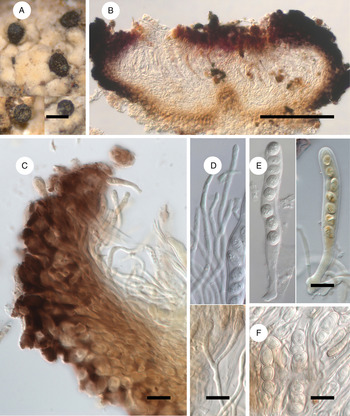
(Fig. 4)
Llimoniella chilensis (A, C, D (below) & E (on the right), holotype; B, D (above), E (on the left) & F, Imshaug 36984). A, habitus of ascomata on the thallus of Dibaeis sp. B, ascoma in cross-section in water. C, exciple in cross-section in K/I. D, paraphyses in K (above) and in K/I (below). E, asci with spores in K (on the left) and K/I (on the right). F, ascospores in water. Scales: A = 200 μm; B = 100 μm; C–F = 10 μm. In colour online.
Ascomata apothecia, loosely aggregated, rarely confluent, erumpent, eventually 1/4 to 1/2 immersed, urceolate to cupulate, constricted at the base, roundish in surface view, 70‒250 μm diam. (n = 20); margin prominent, often partially covering the disc, blackish, matt, rough, occasionally with radial fissures, without hairs; disc initially almost enclosed, later widely exposed, blackish, not translucent, slightly concave to plane. Exciple in cross-section composed of circular to elongate cells up to 10 μm, with walls 1‒2.5 μm thick, innermost cells usually tangentially elongated, thinner, with paler and thinner walls; laterally 20‒50 μm thick, dark violet-brown, K+ greyish green to deep green, N+ reddish brown; basally 20‒70 μm thick, occasionally prolonged into a short immersed stipe, pale to medium greyish or brownish orange, K−, N+ brownish orange. Epihymenium reddish brown and partly violet-brown (pigmentation ill-defined), K+ greyish green to deep green, K/I+ greyish red, N+ brownish orange, 10‒15 μm tall. Hymenium pale reddish to violet-brown above, colourless below, not inspersed, 60‒70 μm tall, I−, K/I−, partly K+ greenish. Subhymenium colourless to orangish. Paraphyses 2‒3.5 μm thick, apically sometimes slightly swollen (to 4 μm), scarcely septate, occasionally branched, not pigmented. Asci subcylindrical, apically rounded to somewhat applanate, with a long foot, croziers not observed, wall 0.5‒1 μm thick, not thickened at the apex, without ocular chamber, 55‒75 × 7.5‒10 μm (n = 13), 8-spored, I−, K/I−. Ascospores hyaline, mostly broadly ellipsoid, occasionally ellipsoid or subglobose, uniseriate in the ascus, (5‒)6.5‒8.5(‒10.5) × (4‒)4.5‒5.5(‒7) μm, l/w = (1.0‒)1.3‒1.7(‒2.3) (n = 100), aseptate, wall c. 0.5 μm thick, smooth, without perispore, indistinctly guttulate.
Asexual morph not observed.
Etymology
Named after Chile, where the type was collected.
Distribution and host
The new species is known from two collections in Chile. It grows on the thalli of terricolous Dibaeis sp. and a sterile microsquamulose lichen, partly associated with discolorations.
Notes
The new species fits well with the generic concept of Llimoniella Hafellner & Nav.-Ros. in its broad sense (Hafellner & Navarro-Rosinés Reference Hafellner and Navarro-Rosinés1993; Diederich & Etayo Reference Diederich and Etayo2000; Diederich et al. Reference Diederich, Ertz and Etayo2010) and is distinct from all its species keyed out in Diederich et al. (Reference Diederich, Ertz and Etayo2010) and described in subsequent publications (Pérez Ortega et al. Reference Pérez, Etayo and Spribille2011; Vondrák et al. Reference Vondrák, Palice, Mares and Kocourková2013; Zhurbenko Reference Zhurbenko2013; Etayo Reference Etayo2017) by the combination of the K+ green reaction of the exciple and epihymenium and aseptate, broadly ellipsoid ascospores. Llimoniella pertusariae Diederich & Etayo (on Lepra ophthalmiza and Pertusaria glaucomela) and L. pyrenulae Diederich & Etayo (on Pyrenula) are also characterized by the K+ green reaction and aseptate ascospores, but the latter are narrowly ellipsoid to fusiform (Diederich & Etayo Reference Diederich and Etayo2000; Diederich et al. Reference Diederich, Ertz and Etayo2010, Reference Diederich, Lawrey and Ertz2018b). In its broadly ellipsoid, aseptate ascospores the new species is similar to Llimoniella fuscatae Hafellner & Obermayer (on Acarospora fuscata), L. gregorellae Kocourk. & Vondrák (on Gregorella humida) and L. terricola (Arnold) M. Schultz et al. (on Leptogium byssinum) (Ertz & Diederich Reference Ertz and Diederich2006; Hafellner & Obermayer Reference Hafellner and Obermayer2007; Vondrák et al. Reference Vondrák, Palice, Mares and Kocourková2013). However, Llimoniella fuscatae is distinct in its red-brown, K+ purple-violet later dark brown exciple and larger ascospores, 9‒12 × 5.5‒7.5 μm; L. gregorellae differs in its orange-brown, K+ brown-purple exciple; and L. terricola differs in its orange-brown, K+ slightly darker reddish brown exciple.
In its urceolate to cupulate apothecia and hyaline, broadly ellipsoid, aseptate, uniseriate ascospores of similar size, the new species resembles Geltingia associata (Th. Fr.) Alstrup & D. Hawksw. However, that species, usually growing on Ochrolechia, differs in its more or less immersed apothecia, orange-brown, K−, N− exciple, and asci with an apically thickened wall and distinct ocular chamber (Diederich et al. Reference Diederich, Ertz and Etayo2010).
Additional specimen examined
Chile: Valparaíso Region: Juan Fernandez Islands, Más Afuera Island, Campo Correspondencia, 1000 m, on terricolous sterile microsquamulose lichen (thallus), 1965, H. A. Imshaug 36984 (TNS).
Micarea inquinans (Tul.) Coppins
This species is known from scattered reports throughout Europe (Brackel Reference Brackel2014) and single finds in North America (Alaska, USA; Fryday Reference Fryday2017) and Asia (Kai Province, Japan; Zhurbenko et al. Reference Zhurbenko, Frisch, Ohmura and Thor2015). Dibaeis arcuata is a new host species.
Specimens examined
Japan: Honshu: Prov. Shinano (Pref. Nagano), Mt Neko-dake, Ueda-city, 36°32'54.7"N, 138°23'34.5"E, 2150 m, mountain heath, on Dibaeis baeomyces (thallus), 2012, A. Frisch 12/Jp398 (TNS, filed under the host). Hokkaido: Prov. Kushiro, along the trail from Taro-ko to the top of Mt O-akan, Akan-gun, 43°27′N, 144°09′E, 1180–1370 m, on D. arcuata (thallus), 1995, Y. Ohmura 1619 & H. Kashiwadani (TNS, filed under the host).
Pyrenidium actinellum Nyl. s. lat.
This species was described from Scytinium teretiusculum (Nylander Reference Nylander1865) and subsequently reported from a wide range of lichen genera (see e.g. Brackel Reference Brackel2014). In its broad sense it probably includes a number of host-specific, yet undescribed species (Huanraluek et al. Reference Huanraluek, Ertz, Phukhamsakda, Hongsanan, Jayawardena and Hyde2019). The specimens examined for this study are characterized by blue-green flecks occurring in the ostiolar region, asci with four mature spores, and 3-septate ascospores measuring (17.5–)20.5–26.5(–31) × (7–)8–9.5 μm, l/w = (2–)2.4–3.2(–4.2) (n = 48; on Baeomyces) and (20–)22–26.5(–30) × (7.5–)8–9(–9.5) μm, l/w = (2.2–)2.5–3.1(–3.3) (n = 19; on Dibaeis).
Specimens examined
Japan: Hokkaido: Prov. Kushiro, along the trail from Taro-ko to the top of Mt O-akan, Akan-gun, 43°27′N, 144°09′E, 700–1180 m, on Dibaeis baeomyces (thallus), 1995, Y. Ohmura 1550 & H. Kashiwadani (TNS, filed under the host); on Baeomyces placophyllus (thallus), 1995, Y. Ohmura 1551 & H. Kashiwadani (TNS, filed under the host); around Hyotan-numa, Akan-cho, Akan-gun, 43°25′N, 144°11′E, 460 m, on B. rufus (thallus), 1995, Y. Ohmura 2052 & H. Kashiwadani (TNS, filed under the host). Honshu: Prov. Shimotsuke (Pref. Tochigi), Nikko-city, 13 km NW of Nikko and 5 km ESE of the village Yumoto, at the river 150 m SE of the dirt road, 36°47'49.9"N, 139°28'50.0"E, 1570 m, on Dibaeis sp. (thallus) growing on Tsuga diversifolia, 2015, G. Thor 32553 (TNS, filed under the host); Prov. Shinano (Pref. Nagano), Yatsugatake Mts, 35°59′N, 138°21′E, on Baeomyces rufus (thallus), 1959, M. Togashi & S. Kurokawa (TNS, filed under the host); Shibunoyu Hot Spring, Yatsugatake Mts, 36°02′10"N, 138°19′43"E, 1800 m, on B. rufus (thallus), 1958, S. Kurokawa 58262 (TNS, filed under the host); Pref. Gunma, Katashina-mura, 25 km WNW of Nikko, 5 km ENE of Marunuma-kogen ski resort, 36°49'15.1"N, 139°22'35.1"E, 1780 m, on B. placophyllus (thallus) on log, 2015, G. Thor 32439 (TNS, filed under the host).—Chile: Region X: Prov. Valdivia, Fundo San Martín (Universidad Austral de Chile), c. 30 km S of San José de la Mariquina, 40°38′S, 5°50′W, 10 m, mixed forest with Nothofagus obliqua, on Dibaeis sp. (thallus), 1987, H. Kashiwadani 35425b (TNS, filed under the host).
Sclerococcum athallinum (Müll. Arg.) Ertz & Diederich
Exciple and epihymenium reddish brown, hymenium hyaline to dull red, pigmentation slightly fading in K, the K+ purplish tinge of epihymenium reported in Hafellner (Reference Hafellner1979) not observed. Ascospores (rarely 0‒)1-septate, somewhat smaller than reported in Hafellner (Reference Hafellner1979) and Ihlen (Reference Ihlen1998), (6‒)7‒9(‒10.5) × (3.5‒)4‒4.5(‒5) μm, l/w = (1.3‒)1.7‒2.1(‒2.4) (n = 49) versus 9–14.5 × 4–7 μm and (7.5‒)9‒11 × 5‒6 μm respectively. In the Southern Hemisphere, Sclerococcum athallinum was until now known from a single report in the state of Victoria, Australia (Staatliche Naturwissenschaftliche Sammlungen Bayerns 2020) and is newly reported here from two other Australian states, New South Wales and Tasmania.
Specimens examined (both on thalli of Baeomyces heteromorphus)
Australia: New South Wales: Blue Mountains, Mt Wilson, 33°30′S, 150°22′E, 1000 m, 1965, S. Kurokawa 5025 (TNS, filed under the host). Tasmania: SW of Hobart, Old Huon Road, 1887(?), R. A. Bastow (TNS, filed under the host).
Specimen of Sclerococcum athallinum used for comparison
Australia: Victoria: 12 km NW of Castlemaine, Harcourt-Sutton Grange Road, 36°58'S, 144°18'E, 450 m, on Baeomyces heteromorphus (thallus), 1994, H. T. Lumbsch, A. Dickhäuser & H. Streimann (D. Triebel, Microfungi Exsiccati, no. 129; LE 200943).
Sclerococcum attendendum (Nyl.) Ertz & Diederich
Apothecia black, sessile, discoid, with a thick, rough, elevated margin, constricted at the base, 100‒310 μm diam. (n = 41), growing on thalli of Icmadophila species and occasionally on adjacent rotten wood. Exciple reddish brown, 20‒50 μm laterally, basally sometimes forming a stipe up to 180 μm tall immersed in the host thallus. Hypothecium brown to reddish brown. Epihymenium reddish brown, c. 5 μm tall. Hymenium hyaline to pale reddish brown below, 30‒50 μm tall. Asci 8-spored. Ascospores brown, broadly to narrowly ellipsoid or oblong, biseriate in the ascus, (6.5‒)8.5‒11(‒13) × (3.5‒)4‒5(‒6) μm, l/w = (1.4‒)1.9‒2.5(‒3.0) (n = 132), (0‒)1‒3-septate, often slightly constricted at septa, smooth, non-halonate.
This species was described from Pilophorus (Lecanorales) and subsequently reported from a number of distantly related genera, viz. Amygdalaria (Lecideales), Icmadophila (Pertusariales) and Porpidia (Lecideales) (Triebel Reference Triebel1989; Ihlen Reference Ihlen1998). According to Triebel (Reference Triebel1989), ascospores of this species are somewhat longer than in our material ((10‒)11–15(‒17) × (4‒)4.5‒5.5(‒6.5) μm), which might suggest that the specimens growing on Icmadophila are not conspecific with those on the other diverse host genera. However, the ascospore sizes of the additionally examined specimen growing on the type host Pilophorus cereolus (LE 207747), (6.5‒)8.5‒11(‒13) × (3.5‒)4.5‒5(‒6) μm, l/w = (1.4‒)1.8‒2.4(‒2.8) (n = 102), perfectly match our material on Icmadophila. The species was previously known in Japan from a single collection in Honshu Province growing on Pilophorus clavatus (Triebel Reference Triebel1989); it is newly reported here for the Sakhalin Region of Russia.
Specimens examined
Japan: Hokkaido: Prov. Hidaka, foot of Mt Petegari, 42°59′N, 142°52′E, 750 m, on Icmadophila japonica (thallus), 1970, S. Kurokawa 70226 (TNS, filed under the host); Prov. Kushiro, Furebetsu Bokke, N slope of Mt Furebetsu, Akan-cho, Akan-gun, 43°24′N, 144°05′E, 780 m, on I. japonica (thallus), 1995, Y. Ohmura 1944 & H. Kashiwadani (TNS, filed under the host); Prov. Tokachi, Mt Higashi-Nupukaushi, Shikaoi-mura, Kato-gun, 43°14′N, 143°06'E, 1000 m, on I. japonica (thallus), 1979, H. Kashiwadani 15363 (TNS, filed under the host); foot of Mt Hakuun, Lake Shikaribetsu, Kato-gun, 43°15′N, 143°06′E, 900 m, on I. japonica (thallus), 1970, M. Togashi [Y. Ohmura, Lichenes Minus Cogniti Exsiccati, no. 381] (TNS, filed under the host). Honshu: Prov. Echigo (Pref. Niigata), on trail from the summit to Akayu Hot Spring, Mt Naeba, 36°49′N, 138°43′E, on I. ericetorum (thallus), 1957, S. Kurokawa 57199 (TNS, filed under the host); Prov. Kai (Pref. Yamanashi), Mt Kimpu, 35°52′N, 138°37′E, on I. ericetorum (thallus), 1953, S. Kurokawa 521163 (TNS, filed under the host); Prov. Kozuke (Pref. Gunma), Mt Shibutsu, Oze, 36°54′N, 139°10′E, on I. japonica (thallus), 1950, Y. Asahina 5044 (TNS, filed under the host); Prov. Shinano (Pref. Nagano), Yatsugatake Mts, 35°59'N, 138°21'E, on I. ericetorum (thallus), 1926, Y. Okada (b) (TNS, filed under the host); Mt Sannosawa-dake, Kiso-Komagatake Mts, 35°46′N, 137°47′E, 2700 m, on I. ericetorum (thallus), 1969, S. Kurokawa 69068 (TNS, filed under the host); Prov. Iwashiro (Pref. Fukushima), Oze, 36°56′N, 139°14′E, on I. japonica (thallus) and occasionally on neighbouring rotten wood, 1933, S. Asahina (TNS, filed under the host).—Russia: Sakhalin Region: Sakhalin Island, Mt Tosso, 48°03'N, 142°32′E, on I. ericetorum (thallus) and occasionally on neighbouring rotten wood, 1932, M. Sato (TNS, filed under the host).
Specimen of Sclerococcum attendendum used for comparison
Russia: Komi Republic: headwaters of the Pechora River, valley between Yanypupuner Range and Mt Medvezh'ya, 62°04'N, 59°08'E, 500 m, on Pilophorus cereolus (thallus), 1997, M. P. Zhurbenko 97166 (LE 207747).
Sphaerellothecium coniodes (Nyl.) Cl. Roux & Diederich
The range of ascospore sizes in the material we studied was (11–)12–14.5(–16.5) × (3.5–)4.5–5.5(–6) μm, l/w = (2.2–)2.4–3.0(–3.8) (n = 26) which is slightly greater than those presented in Ihlen (Reference Ihlen1998) and Roux & Triebel (Reference Roux and Triebel1994), viz 11–14 × 4–5 μm and 11–13 × 3.5–5 μm respectively. To date, the species was known from Europe and North America (Hodkinson et al. Reference Hodkinson, Harris and Case2009; Brackel Reference Brackel2014) and is newly reported here for Asia and Japan.
Specimens examined (both on thalli of Baeomyces rufus)
Japan: Hokkaido: Prov. Tokachi, along the trail from Me-akan Hot Spring to the top of Mt Me-akan, Ashoro-cho, Ashoro-gun, 43°24′N, 143°59′E, 710–920 m, 1995, Y. Ohmura 1682 & H. Kashiwadani (TNS, filed under the host). Kyushu: Prov. Ohsumi (Pref. Kagoshima), Hananoego, Yakushima Island, 30°18'45"N, 130°30'38"E, 1933, F. Fujikawa (TNS, filed under the host).
Stigmidium phyllobaeidis Zhurb., Etayo & Flakus sp. nov.
MycoBank No.: MB 836575
Lichenicolous ascomycete growing on Phyllobaeis. Vegetative hyphae reduced; ascomata perithecioid, ostiolate, brownish black, glabrous, immersed to semi-immersed; exciple dark brown, K−; interascal hyphae lacking; interascal gel I+ red, K/I+ blue; periphysoids 1‒2(‒4)-celled; asci bitunicate, 8-spored, I−, K/I−; ascospores hyaline, 1-septate, with slightly wider upper cell, (6.5‒)8‒10.5(‒13) × (2.5‒)3‒4(‒5) μm.
Type: Peru, Dept. Cuzco, Prov. Paucartambo, Abra Acjanaco, near Paucartambo, 3400‒3500 m, upper part of ‘Ceja de Selva’ zone, on apothecial discs of Phyllobaeis erythrella growing on soil, 17 September 1984, H. Kashiwadani 21946 (TNS-L-129849—holotype).
(Fig. 5)
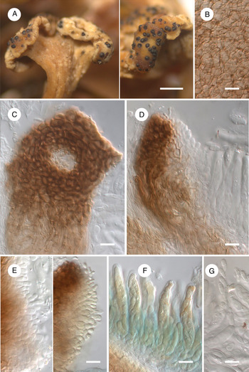
Stigmidium phyllobaeidis (holotype). A, habitus of ascomata on apothecial discs of Phyllobaeis erythrella. B, basal exciple in surface view in K. C, exciple around ostiole in surface view in K. D, exciple in cross-section in K. E, periphysoids in K (on the left) and in K/I (on the right). F, asci and interascal gel in K/I. G, ascospores in K. Scales: A = 500 μm; B–G = 10 μm. In colour online.
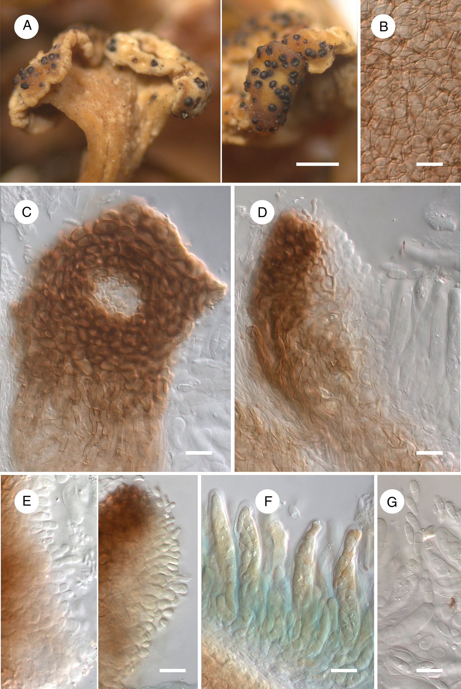
Vegetative hyphae brown, immersed, reduced. Ascomata perithecioid, mostly completely immersed to semi-immersed, occasionally 1/3 protruding, brownish black, somewhat glossy, glabrous, epruinose, subglobose, 60‒130 μm diam. (n = 20), with an ostiole 10‒15 μm diam., aggregated. Exciple 10‒20 μm thick, exposed upper part medium to dark brown, K−, in surface view resembling textura angularis or textura globulosa, composed of cells up to 7 μm with walls 1‒1.5 μm thick; immersed lower part pale brown, in surface view resembling textura angularis or textura porrecta, mainly composed of cells up to 12 μm with walls c. 0.5 μm thick; in cross-section composed of c. 5 layers of tangentially strongly elongated cells, the innermost cells thinner, with paler and thinner walls. Periphysoids resemble internal periphyses (above) and pseudoparaphyses (below) sensu Roux & Triebel (Reference Roux and Triebel1994: fig. 2), well-developed throughout the upper half of the inner exciple, hyaline, 1‒2(‒4)-celled, up to 12 μm long; external, brown periphyses absent. Interascal hyphae absent. Interascal gel I+ red, K/I+ blue and partly red (staining does not fade). Asci bitunicate, subcylindrical to narrowly ellipsoid, slightly wider in the central or lower part, rounded at the apex, with a distinct foot, 35‒50 × 7‒11 μm (n = 12), wall apically thickened, sometimes with a small ocular chamber, I−, K/I−, plasma BCr+ blue to violet, 8-spored. Ascospores hyaline, narrowly obovate, with slightly wider upper cell, (6.5‒)8‒10.5(‒13) × (2.5‒)3‒4(‒5) μm, l/w = (1.7‒)2.2‒3.0(‒3.4) (n = 55), diagonally uniseriate to irregularly biseriate in the ascus, 1-septate, not constricted at the septum, with a smooth wall c. 0.3 μm thick, non-halonate, usually without distinct guttules.
Etymology
Referring to its growth on Phyllobaeis.
Distribution and host
The new species is known from three collections in Bolivia and Peru, growing on apothecial discs of Phyllobaeis erythrella and P. imbricata. Host tissues are bleached under severe infections.
Notes
The new species does not perfectly match any of the genera known to us and is tentatively placed in the large, almost exclusively lichenicolous genus Stigmidium Trevis. Characteristics of Stigmidium fit the material examined except for the hemiamyloid reaction of the interascal gel never reported in the genus (Triebel & Cáceres Reference Triebel, Cáceres, Nash, Ryan, Diederich, Gries and Bungartz2004). However, some Stigmidium species, such as S. cartilagineae Calatayud & Triebel, S. psorae (Anzi) Hafellner, S. squamarinicola Calatayud & Triebel and S. triebeliae Etayo display an amyloid reaction of the interascal gel (Etayo Reference Etayo2000; Calatayud & Triebel Reference Calatayud and Triebel2003). A number of Stigmidium species, such as S. buelliae Zhurb. & Himelbrant, S. cerinae Cl. Roux & Triebel, S. collematis Cl. Roux & Triebel, S. congestum (Körb.) Triebel, S. lecidellae Triebel et al. and S. disconephromeum Etayo, are also confined to the apothecial discs of their host lichens (Roux & Triebel Reference Roux and Triebel1994; Roux et al. Reference Roux, Triebel, Bricaud and Le1995; Etayo & Sancho Reference Etayo and Sancho2008; Zhurbenko et al. Reference Zhurbenko, Himelbrant, Kuznetsova and Stepanchikova2012). With its well-developed periphysoids, hemiamyloid hymenial gel and elongate asci, the new species might fit the broad concept of Pseudostigmidium Etayo (Etayo & Sancho Reference Etayo and Sancho2008). However, this genus is known to grow on foliose macrolichens of the suborder Peltigerineae and is characterized by almost exclusively 2‒3-septate ascospores, though 1-septate ascospores also occur in Pseudostigmidium disparatum Etayo (Etayo & Sancho Reference Etayo and Sancho2008). The generic type, P. nephromiarium (Linds.) Etayo, differs in having a distinct net of brown vegetative hyphae, hemispherical to subconical ascomata, I+, K/I+ light purple interascal gel and pycnidia with hyaline, bacilliform conidia (Etayo & Sancho Reference Etayo and Sancho2008). In most respects the new species is also similar to species of Austrostigmidium Pérez-Ortega & Garrido-Benavent, Clauzadella Nav.-Ros. & Cl. Roux, Endococcus Nyl., Epibryon Döbbeler and Sphaerellothecium Zopf. However, Austrostigmidium is distinct in its I−, K/I− interascal gel and 3-septate ascospores (Pérez-Ortega et al. Reference Pérez-Ortega, Garrido-Benavent and De Los2015), Clauzadella is distinguished by its comparatively thick, violet-tinged ascomatal wall and 3-septate ascospores (Navarro-Rosinés & Roux Reference Navarro-Rosinés and Roux1996), Endococcus is distinguished by its more or less pigmented ascospores (Kainz & Triebel Reference Kainz, Triebel, Nash, Ryan, Diederich, Gries and Bungartz2004; Etayo & Sancho Reference Etayo and Sancho2008), Epibryon differs in its typically setose ascomata and poorly developed periphyses (Döbbeler Reference Döbbeler1978; Hoffmann & Hafellner Reference Hoffmann and Hafellner2000), and Sphaerellothecium has an I−, K/I− interascal gel and typically produces a conspicuous net of dark vegetative hyphae as well as paraphysoids (Cáceres & Triebel Reference Cáceres, Triebel, Nash, Ryan, Diederich, Gries and Bungartz2004). The only species of the compared genera growing on members of the same host family Icmadophilaceae is Sphaerellothecium icmadophilae (R. Sant.) Zhurb. It grows on thalli of Icmadophila ericetorum and is clearly different from the new species, apart from the features noted above, in its 1(‒3)-septate, hyaline to eventually brown, larger ascospores, 15‒21 × 5–7 μm (Zhurbenko Reference Zhurbenko2008).
In one specimen (Etayo 27749), perithecia of Stigmidium phyllobaeidis were intermixed with brown sporodochia 20‒40 μm diam., originating from subhyaline to pale brownish hyphae deeply immersed in the host hymenium. They are characterized by prismatic to ellipsoid conidiophores and conidiogenous cells measuring 4‒6 × 2.5‒4.5 μm, producing acrogenous, simple, obpyriform, thin-walled, light brown conidia with a pointed end and truncate base, 11‒14 × 2.5‒3.5 μm. To the best of our knowledge, such an anamorph has not been recorded in any lichenicolous genus.
Additional specimens examined
Bolivia: Dept. Cochabamba: Carrasco Parque Nacional Carrasco, Wayra Mayu close to Monte Punku, 17°32'27"S, 65°16'14"W, 2553 m, lower montane Yungas cloud forest, on Phyllobaeis imbricata (discs of apothecia), 2014, A. Flakus 25830 (KRAM, LPB). Dept. La Paz: Prov. Nor Yungas, Parque Nacional y Área Natural de Manejo Integrado Cotapata, between Tunkini and Chairo villages, above Tunkini, 16°11′S, 67°52′W, 1300–1600 m, Yungas montane forest, on P. erythrella (discs of apothecia), 2011, J. Etayo 27749 (hb. Etayo, LPB).
A key to the species of lichenicolous fungi and lichens growing on baeomycetoid lichens and Icmadophila
This key is based on data from Keissler (Reference Keissler1930), Hafellner (Reference Hafellner1979), Hawksworth (Reference Hawksworth1979), Triebel (Reference Triebel1989), Alstrup & Hawksworth (Reference Alstrup and Hawksworth1990), Rambold & Triebel (Reference Rambold and Triebel1990), Gierl & Kalb (Reference Gierl and Kalb1993), Obermayer (Reference Obermayer1994), Roux & Triebel (Reference Roux and Triebel1994), Aptroot et al. (Reference Aptroot, Diederich, Sérusiaux and Sipman1997), Ihlen (Reference Ihlen1998), Alstrup et al. (Reference Alstrup, Hansen and Daniels2000), Baral & Marson (Reference Baral, Marson and Consiglio2001), Hafellner et al. (Reference Hafellner, Triebel, Ryan and Nash2002), Punithalingam (Reference Punithalingam2003), Zhurbenko (Reference Zhurbenko2008), Duke & Purvis (Reference Duke, Purvis, Smith, Aptroot, Coppins, Fletcher, Gilbert, James and Wolseley2009), Orange et al. (Reference Orange, Watson, James, Moore, Smith, Aptroot, Coppins, Fletcher, Gilbert, James and Wolseley2009), Ertz et al. (Reference Ertz, Lawrey, Common and Diederich2014), Heuchert et al. (Reference Heuchert, Braun, Diederich and Ertz2018), Diederich et al. (Reference Diederich, Common, Braun, Heuchert, Millanes, Suija and Ertz2019) and the present contribution.
1 Spores (conidia) produced on conidiogenous cells ……… 2
Spores produced in asci ……… 6
2(1) Conidiomata pycnidial, conidia hyaline ……… 3
Conidiomata not pycnidial, conidia brown ……… 5
3(2) Conidia Y-shaped, consisting of main body 9.5–11.5 × 2–2.5 μm (including basal appendage) and two divergent arms; on various host genera ……… Spirographa ciliata s. lat.
Conidia not Y-shaped ……… 4
4(3) Conidia cylindrical, 12–20 × 2.5–3.5 μm, with filiform lateral appendages, 18–36 × 1–2 μm, often in chains; on Icmadophila……… ……… Acarosporium lichenicola
Conidia narrowly ellipsoid to bacilliform, 5–6(–7) × 2–2.5 μm, without appendages, not in chains; on Dibaeis ……… ……… Phoma maculiformans
5(2) Conidiomata stromatic; conidia composed of 4–22 cells, subspherical to ellipsoid, (10–)11–13.5(–15) × (7–)8.5–10.5(–12) μm, not in chains; on various host genera ……… Lichenostigma alpinum
Conidiomata not stromatic (hyphomycete); conidia (0–)1–2(–3)-septate, subcylindrical, doliiform, pyriform, ellipsoid or limoniform, 4–17 × 3–8 μm, mostly in chains; on various host genera ……… Taeniolella delicata
6(1) Ascomata yellow; asci multispored; ascospores hyaline, aseptate ……… 7
Ascomata blackish; asci up to 8-spored; ascospores various ……… 9
7(6) Ascomata with at least slightly exposed discs, shortly cylindrical, obconical or dish-shaped; asci cylindrical clavate; ascospores (4‒)5‒7.5 × 1.5‒2.5 μm; on various host genera ……… Thelocarpon lichenicola
Ascomata without exposed discs, ovoid or conical; asci flask-shaped, tapering to a slender neck above ……… 8
8(7) Ascospores 4‒6 × 1.5‒2 μm; on various host genera ……… Thelocarpon epibolum var. epibolum
Ascospores (6‒)8‒10(‒12) × 2‒3 μm; on various host genera ……… Thelocarpon epibolum var. epithallinum
9(6) Ascomata apothecioid ……… 10
Ascomata perithecioid ……… 26
10(9) Ascospores consistently hyaline ……… 11
Ascospores at least partly pigmented ……… 20
11(10) Ascospores aseptate ……… 12
Ascospores septate ……… 14
12(11) Apothecia completely immersed, disc brownish with black dots; ascospores (9.5–)10.5–14(–16.5) × (2.5–)3–3.5 μm; on Dibaeis ………Rhymbocarpus ericetorum
Apothecia more or less superficial, disc blackish; ascospores shorter and wider ……… 13
13(12) Apothecia urceolate to cupulate, with prominent margin; ascospores (5‒)6.5‒8.5(‒10.5) × (4‒)4.5‒5.5(‒7) μm, uniseriate in the ascus; on Dibaeis and a sterile microsquamulose lichen ……… Llimoniella chilensis
Apothecia convex, immarginate; ascospores 7–12 × 5–6 μm, irregularly arranged in the ascus; on Dibaeis ……… ……… Micarea inquinans
14(11) Apothecia pale orange to orange-white, urceolate or cupulate; ascospores 1-septate, narrowly obovate, (14‒)15.5‒19(‒20.5) × (5‒)5.5‒7(‒7.5) μm; on Dibaeis ……… Cryptodiscus ihlenii
Apothecia black, not cupulate; at least some ascospores with more than one septum ……… 15
15(14) Ascospores rod-shaped, 1‒3-septate, 10‒15 × 3 μm; on Baeomyces and Dibaeis ……… ………‘Celidium ericetorum Rehm apud Rabh.’ sensu Keissler (Reference Keissler1930: 97)
Ascospores cylindrical to acicular, 3‒16-septate, 15‒110 × 2‒5 μm ……… 16
16(15) Not lichenized; on Dibaeis ……… Arthrorhaphis muddii
Producing lichenized thallus ……… 17
17(16) Lichenized thallus greyish, rather indistinct; on Baeomyces ……… Arthrorhaphis grisea
Lichenized thallus greenish yellow, distinct ……… 18
18(17) Ascospores 3(‒5)-septate; on Baeomyces ……… Arthrorhaphis vacillans
Ascospores 4‒16-septate ……… 19
19(18) Lichenized thallus of strongly convex areoles, not sorediate; ascospores mainly 25‒45 μm long; on Baeomyces ……… ……… Arthrorhaphis alpina
Lichenized thallus of flat to slightly convex areoles, sorediate; ascospores mainly 55‒80 μm long; on Baeomyces ……… ……… Arthrorhaphis citrinella
20(10) Ascospores hyaline to occasionally pigmented ……… 21
Ascospores consistently pigmented (brown) ……… 23
21(20) Apothecia eventually urceolate; ascospores 1-septate, (12.5‒)13.5‒15.5(‒17.5) × 5‒6(‒6.5) μm; on Icmadophila ……… ……… Buelliella ohmurae
Apothecia not urceolate; ascospores various ……… 22
22(21) Apothecia convex, immarginate; hymenium I+ red; ascospores 1-septate, clavate, 12‒13.5 × 4.5‒5.5 μm; on Pseudobaeomyces ……… Arthonia sp. (characterized in the present paper)
Apothecia discoid, usually marginate; hymenium I+ blue; ascospores (0‒)1(‒3)-septate, mainly ellipsoid, (9.5‒)10.5‒13(‒15.5) × (4‒)4.5‒6(‒6.5) μm; on Dibaeis and Pseudobaeomyces ……… Catillaria japonica
23(20) Producing yellow to yellowish green lichenized thallus; apothecia convex, immarginate; ascospores 1-septate, (10–)11.5–14(–18) × (4–)5.5–7.5(–9) μm; on Baeomyces ……… Epilichen scabrosus
Lichenized thallus absent; apothecia discoid, marginate; ascospores 1- or more septate ……… 24
24(23) Ascospores 1‒3-septate, (6.5‒)8.5‒11(‒13) × (3.5‒)4‒5(‒6) μm; on various host genera ……… Sclerococcum attendendum
Ascospores 1-septate ……… 25
25(24) Epihymenium reddish brown; ascospores (6‒)7‒9(‒10.5) × (3.5‒)4‒4.5(‒5) μm; on Baeomyces ……… ……… Sclerococcum athallinum
Epihymenium brown; ascospores (7–)10.5–14.5(–17) × (5–)6–9(–10) μm; on Baeomyces ……… Epilichen glauconigellus
26(9) Ascomata densely setose when young; ascospores hyaline to pale brown, 1-septate, 8‒10 × 3.5‒4 μm; on Baeomyces ……… ……… Capronia baeomycetis
Ascomata without setae; ascospores various ……… 27
27(26) Exciple greenish above, hyaline below; ascospores hyaline, 1-septate, 12–15(–16) × 4–5(–6) μm; on Baeomyces ……… ……… Cercidospora parva
Exciple brown; ascospores various ……… 28
28(27) Ostiolar region with blue-green flecks; asci 4-spored; ascospores brown, sometimes with paler end cells, 2‒4-septate, (17.5–)21–26.5(–31) × (7–)8–9.5 μm; on various host genera ……… Pyrenidium actinellum s. lat.
Ostiolar region without blue-green flecks; asci 8-spored; ascospores various ……… 29
29(28) Ascospores submuriform to muriform, brown, (14‒)15‒24.5(‒32) × (6.5‒)8‒12(‒15) μm; on various host genera ……… ……… Merismatium nigritellum
Ascospores trans-septate, mostly or consistently hyaline ……… 30
30(29) Ascospores (6.5‒)8‒10.5(‒13) × (2.5‒)3‒4(‒5) μm, hyaline, 1-septate; on Phyllobaeis ……… Stigmidium phyllobaeidis
Ascospores larger, hyaline to brown when overmature ……… 31
31(30) Ascospores (11–)12–14.5(–16.5) × (3.5–)4.5–5.5(–6) μm, 1-septate; on Baeomyces ……… Sphaerellothecium coniodes
Ascospores (15–)16.5–19(–21) × 5–6.5(–7) μm, 1(–3)-septate; on Icmadophila ……… Sphaerellothecium icmadophilae
Discussion
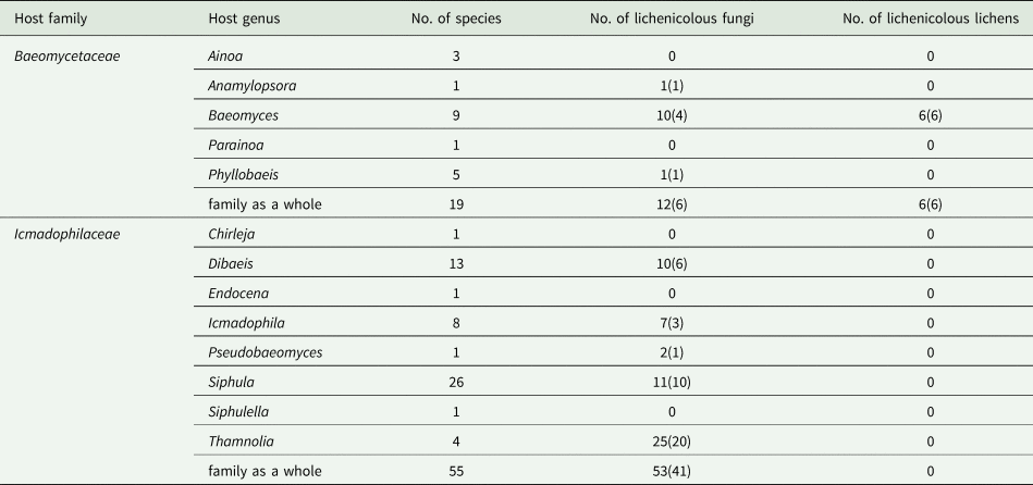
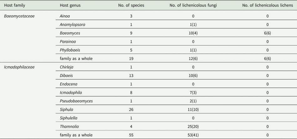
The present study of lichenicolous fungi on baeomycetoid lichens and Icmadophila, coupled with recent revisions of these fungi growing on Thamnolia (Icmadophilaceae; Zhurbenko Reference Zhurbenko2012) and Siphula (Icmadophilaceae; Motiejūnaitė et al. Reference Motiejūnaitė, Zhurbenko, Suija and Kantvilas2019), gives a fairly complete picture of the diversity of lichenicolous fungi growing on members of the Baeomycetaceae (12 species) and Icmadophilaceae (53 species) (Table 1). These fungi are known to grow on species of three of the five genera of Baeomycetaceae and on five of the eight genera of Icmadophilaceae. The ratio of the number of parasite species to the number of host species is 0.6 for Baeomycetaceae and 1.0 for Icmadophilaceae; at the generic level it ranges from 6.3 for Thamnolia to 0.2 for Phyllobaeis. In total, 62 species of lichenicolous fungi from 40 genera were recorded on lichens of these families, of which three species (‘Celidium ericetorum Rehm apud Rabh.’, Pyrenidium actinellum s. lat. and Thelocarpon lichenicola) and eight genera (Capronia Sacc., Cercidospora Körb., Polycoccum Körb., Pyrenidium Nyl., Sclerococcum, Sphaerellothecium, Stigmidium and Thelocarpon Nyl.) colonize hosts from both families. The proportion of parasite species specific to one host genus varies from 0.4 (for Icmadophila) to 1.0 (for Anamylopsora and Phyllobaeis). The largest numbers of associated lichenicolous fungi species are known for Thamnolia (25), Siphula (11) and Baeomyces (10). These numbers are relatively small, compared to those for the most ‘hospitable’ lichen genera, such as Cladonia, supporting no less than 130–140 species (Zhurbenko & Pino-Bodas Reference Zhurbenko and Pino-Bodas2017). Lichenicolous lichens occur within these families only on species of Baeomyces.
Number of species of lichenicolous fungi and lichens growing on genera of the Baeomycetaceae and Icmadophilaceae (sensu Lücking et al. Reference Lücking, Hodkinson and Leavitt2016). Numbers in parentheses indicate the number of species known to occur only on this genus. Based on data from Etayo (Reference Etayo2010), Zhurbenko (Reference Zhurbenko2010b, Reference Zhurbenko2012), Diederich et al. (Reference Diederich, Zimmermann, Sikaroodi, Ghobad-Nejhad and Lawrey2018a), Motiejūnaitė et al. (Reference Motiejūnaitė, Zhurbenko, Suija and Kantvilas2019), Zhurbenko et al. (Reference Zhurbenko, Enkhtuya and Javkhlan2019) and the present paper. Total counts for the ‘family as a whole’ may not reflect numbers in the columns as different species of lichenicolous fungi may occur on more than one host genus.
Acknowledgements
The research of MPZ was carried out within the framework of the research project of the V. L. Komarov Botanical Institute of the Russian Academy of Sciences ‘Biodiversity, ecology and structural and functional features of fungi and fungus-like protists’ (АААА-А19-119020890079-6) using the equipment of its Core Facility Center ‘Cell and Molecular Technologies in Plant Science’; his visit to TNS in 2017 was supported by a JSPS Invitation Fellowship for Research in Japan (no. S16173). We are indebted to A. Flakus and J. Etayo for the provision of specimens and to all staff of the Herbario Nacional de Bolivia, Instituto de Ecología, Universidad Mayor de San Andrés, La Paz, for their generous cooperation, and to SERNAP (http://sernap.gob.bo), and all protected areas staff, for providing permits for scientific studies, as well as for their assistance during fieldwork, which resulted in the description of Stigmidium phyllobaeidis. Paul Diederich provided valuable remarks on the manuscript. Rod Seppelt kindly revised the English.
Author ORCIDs
Milhail P. Zhurbenko, 0000-0002-9839-4698; Yoshihito Ohmura, 0000-0003-2557-2761.