Introduction
Sinonasal inverted papillomas are benign tumours of the nasal cavity of ectodermal origin, described for the first time by Ward in 1854.Reference Ward1 The low incidence of 0.2 to 1.5 new cases annually per 100 000 people contributes less than 5 per cent of sinonasal tumours, making them some of the rarer neoplasms of the nose and paranasal sinuses.Reference Re, Gioacchini, Bajraktari, Tomasetti, Kaleci and Rubini2 Inverted papillomas tend to expand locally, sometimes aggressively,Reference Lund, Stammberger, Nicolai, Castelnuovo, Beal and Beham3 and have considerable recurrence rates after surgical excision, ranging between 12 and 20 per cent.Reference Peng, Thamboo, Choby, Ma, Zhou and Hwang4
The introduction of endoscopic techniques has resulted in a reduction of recurrence rates, as indicated by two meta-analyses.Reference Peng, Thamboo, Choby, Ma, Zhou and Hwang4,Reference Busquets and Hwang5 It is believed that although intrinsic factors associated with the papillomas may play a role, recurrence is mostly the result of incomplete excision.Reference Attlmayr, Derbyshire, Kasbekar and Swift6 These features of inverted papillomas, coupled with a high risk of malignancy (either synchronous or metachronous) of between 3.4 and 9.7 per cent, render definitive management imperative.Reference Grayson, Khichi, Cho, Riley and Woodworth7,Reference Wood and Casiano8 However, this can be challenging, especially in the case of inverted papillomas invading structures such as the orbit and the skull base.Reference Georgalas and Wytske9 Such extensive papillomas pose significant challenges and may demand the use of multiple, novel techniques.Reference Bathma, Harvinder, Philip, Rosalind and Gurdeep10,Reference Castelnuovo, Dallan, Battaglia and Bignami11
We present prospectively collected data on a group of patients with inverted papillomas extending intracranially or intraorbitally and also identifying all literature cases of patients with such tumours.
Materials and methods
Demographic data were recorded upon admission. Date of the operation, details of the surgical technique and anatomical structures resected, time from initial diagnosis, follow ups, and revisions if there were any were prospectively collected. All operations were carried out by the senior author (C Georgalas).
We reported all cases of inverted papillomas invading the skull base (anterior cranial fossa, carotid-optic canal, dura) as ‘intracranial invasion’, and we reported all cases of inverted papillomas invading the orbit (lamina papyracea, periorbita) as ‘intraorbital invasion’.
We excluded all cases of inverted papillomas with intraorbital invasion or skull base extension with no radiological, endoscopic or intra-operative invasion evidence (bony erosion, dura–brain parenchyma, orbit-periorbita infiltration), in situ carcinoma, or frank malignant transformation. All the resected structures that were radiographically or intra-operatively suspected for disease infiltration were sent for histology. A senior pathologist examined the whole specimen in all our cases, excluding any underlying malignant foci.
Computed tomography (CT) and magnetic resonance imaging (MRI) were used in all cases and magnetic resonance angiography was used only in carotid artery involvement. Visual acuity and fields were pre-operatively assessed in patients with optic nerve or orbital involvement.
Consent was received from the patients, and the Committee of the National and Kapodistrian University of Athens reviewed and approved the collection of their data and its use in this case series.
Results
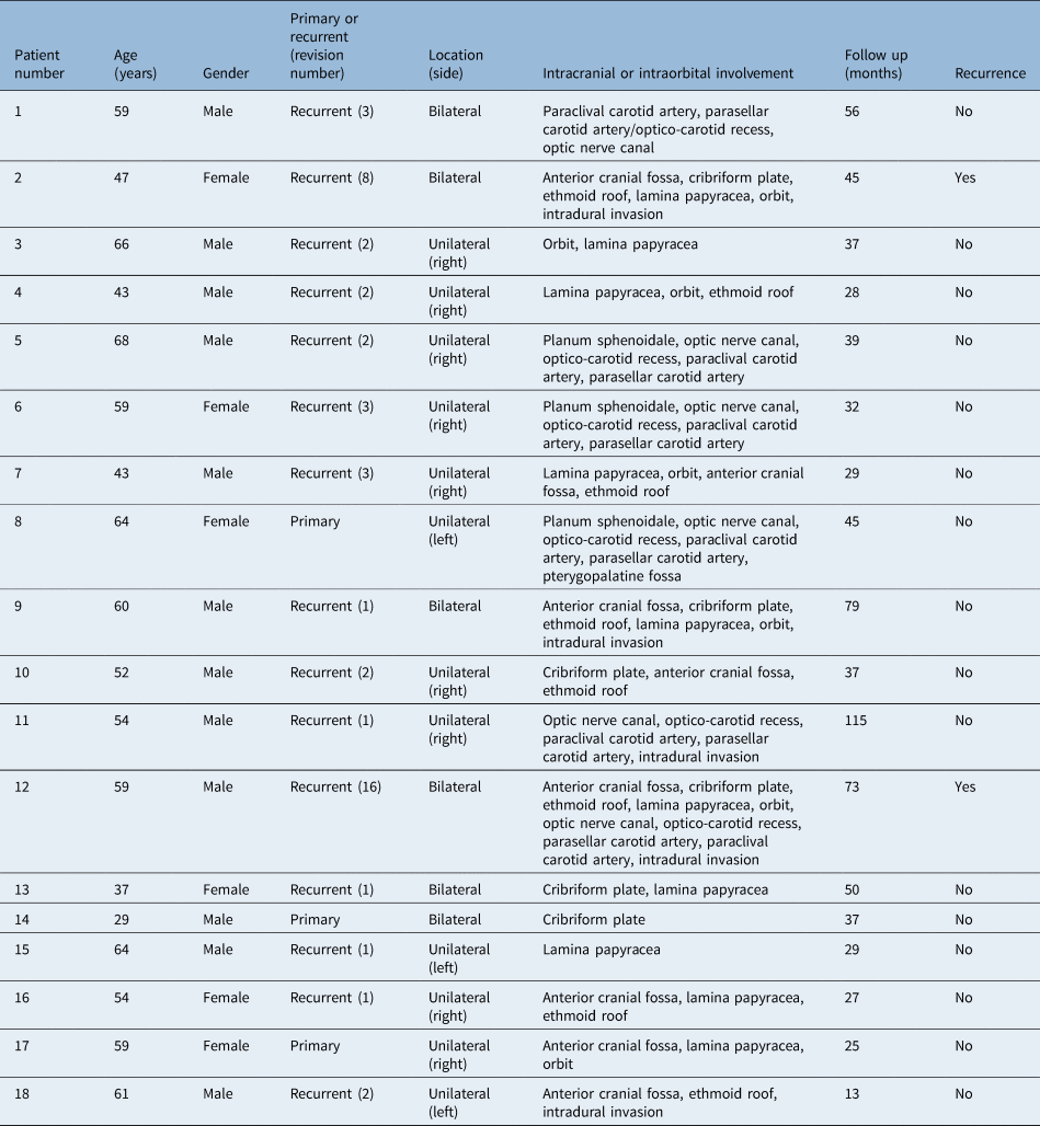
Individual patient data are presented in Table 1. Out of a total of 65 patients with inverted papilloma who underwent operations performed by the senior author between 2011 and 2019, 18 patients met inclusion criteria. There were 13 males and 5 females with an average age of 54.3 and a range of 29 to 68 years at the time of surgery. The average time from initial diagnosis to surgery was 110 months (range, 5 to 468 months). Fifteen patients had been previously operated on, five of whom had undergone more than two revisions. The median number of previous operations was 2 (range, 0–16). Median follow-up time was 37 months and ranged between 13 to 115 months.
Patient demographics and structures involved

The follow-up protocol included endoscopy monthly for the first three months, then every two months for the next six months, then every six months for years two to five and yearly thereafter. Imaging studies, usually MRI, were requested in cases of suspected recurrence or when the anatomical area of initial involvement could not be endoscopically assessed.Reference Lund, Stammberger, Nicolai, Castelnuovo, Beal and Beham3
Overall, intracranial invasion involvement (anterior cranial fossa with or without intradural or intraparenchymal involvement, optico-carotid recess or parasellar or paraclival carotid artery) was identified in 16 out of the 18 patients (88.9 per cent), orbital extension was identified in 10 out of 18 patients (55.5 per cent) and both were identified in 8 patients (44.4 per cent of total cases) who presented with multiple sites of origin of the disease.
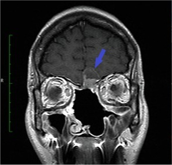
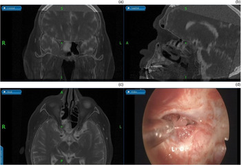
Erosion was found in six patients with paraclival or parasellar carotid artery and in five patients with anterior cranial fossa with intradural invasion (Figure 1). Erosion was also seen at the attachment at the optico-carotid recess in six patients (Figure 2), and the frontal sinus posterior wall was eroded in six patients. Four patients had attachment to the sphenoid sinus walls related to the skull base, either planum sphenoidale or lateral wall or sella, without any attachment to the carotid artery.
(Patient 2). Coronal plane magnetic resonance image (T1-weighted with contrast) of an inverted papilloma with anterior skull base site of attachment. The inverted papilloma occupied the frontal sinuses and had invaded intracranially (blue arrow). R = right

(Patient 5). Intra-operative neuronavigation image of inverted papilloma invading the medial optico-carotid recess and associated with a secondary sphenoid mucocele. (a) Coronal plane fusion-merged magnetic resonance image (MRI) and computed tomography (CT) image showing an inverted papilloma invading the medial optico-carotid recess and associated with a secondary sphenoid mucocele. (b) Sagittal plane fusion-merged MRI and CT image showing the same pathology. (c) Axial plane fusion-merged MRI and CT image showing the same pathology. (d) Intra-operative neuronavigation image showing the same pathology with the suction tip at the right optico-carotid recess. R = right; L = left; A = anterior; S = superior; I = inferior; P = posterior

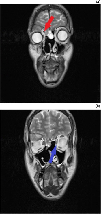
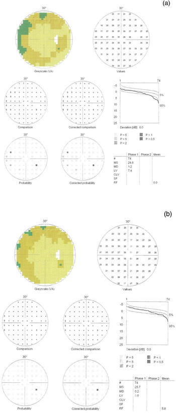
Ten of our patients presented with extended disease invading the orbit, amongst whom four had either diplopia or decreased visual acuity or impaired visual fields because of the entrapment of the optic nerve or artery or orbital involvement (Figure 3). Pre-operative and post-operative visual fields were assessed in all these cases (Figure 4). In four of our patients, disease involved the bony carotid canal within the sphenoid or the clivus; in three patients the inverted papilloma was attached to it, whereas in one patient it had eroded the bony carotid canal, encroaching but not invading the artery itself.
T2-weighted coronal magnetic resonance image demonstrating (a) the presence of the inverted papilloma at the area of the right lamina papyracea (red arrow) and (b) the fat pad medially to the medial rectus muscle is absent while present at the normal left side (blue arrow). The mucocele invades the orbit displacing the eye inferiorly and anteriorly. The desiccated and high protein content make it appear hypointense in T2-weighted images. Two years later the orbit has returned to its normal position and there are no signs of recurrence. R = right; L = left; I = inferior

Pre- (a) and post-operative (b) visual field examination of patient 5 with inverted papilloma originating from optico-carotid recess extending into the optic canal. VA = visual acuity; MS = mean sensitivity; MD = mean defect; LV = loss variance; CLV = corrected loss variance; SF = short-term fluctuations; RF = reliability factor

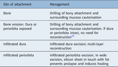
All patients were treated via endoscopic endonasal resection, using CT and MRI fusion neuronavigation. We followed the pedicle targeted surgery,Reference Pagella, Pusateri, Giourgos, Tinelli and Matti12 which allowed us to undertake a far less invasive dissection with greater radicality of the areas the inverted papilloma had invaded. Access was maximised with the use of the Draf III procedure in anterior cranial fossa or frontal sinus involvement, rostrum removal and bilateral extensive sphenoidotomies in sphenoid, optico-carotid recess or carotid artery involvement. A bi-nostril approach and the three or four hands technique were applied in most cases.Reference Castelnuovo, Pistochini and Locatelli13 Extended biopsies from multiple locations of the tumour were undertaken pre-operatively in order to confirm that there was no focal malignant transformation.Reference Landsberg, Cavel, Segev, Khafif and Fliss14 Strategy management according to tumour extension is shown in Table 2.
Treatment strategies depending on structures involved

Intra-operatively, the pedicle of the inverted papilloma was identified and, if required for better exposure, the mass of papilloma was debulked, without removing the mucosa, in order to identify the site of origin. Upon identification, the involved bone was drilled with rough diamond burr and removed. Specifically, the cribriform plate and the periorbita were completely excised when the inverted papilloma invaded them. The involved mucosa was excised with macroscopically clear margins, while frozen sections were sent to the pathology laboratory to exclude microscopic infiltration. When the tumour was compressing neighbouring structures as a space-occupying lesion, without truly invading structures, the involved mucosa that was in contact but not invaded by the papilloma was preserved. We used a variety of drills, including 15-, 45- and 70-degree drills that were mostly rough diamond (non-cutting) while also using a 30° rigid endoscope supplemented by 45° and 70° rigid endoscopes. Nasal packing was not used, although a temporary rolled silastic sheet was used in cases with Draf III procedures.
No major post-operative complications (cerebrospinal fluid leak, orbital injury or bleeding, major haemorrhage, meningitis) were encountered. One of our patients developed a post-operative mucocele in the right orbit that was subsequently drained as an out-patient procedure under local anaesthesia.
During the follow-up period, we report two recurrences: patients number 2 and 12 (Table 1). Patient 2 was previously operated on a total of 8 times over a period of 20 years. The inverted papilloma was invading the intradural space and an extended dissection of this area in the form of a combined approach (craniotomy and endoscopy) was required, which the patient declined. Patient number 12 had undergone 16 previous operations, and disease had undergone intraorbital and anterior cranial fossa invasion, making radical surgery not a viable option.
Discussion
The current strategy for treating inverted papillomas is focused on tumour pedicle removal. This means that simple extension, without invasion, does not affect surgical decision making.Reference Pagella, Pusateri, Giourgos, Tinelli and Matti12 In fact, the term ‘extent of disease’ may be misleading, as it is in fact space occupation instead of frank invasion. Although the Krouse staging systemReference Krouse15 is widely accepted, it provides little assistance in planning a complete excision of inverted papilloma. Like most staging systems, it refers to the anatomical subsites (frontal sinus, sphenoid sinus) occupied by the papilloma; however, primary papillomas usually grow in an exophytic, non-invasive way, and as a result their site of attachment is what needs to be resected with oncological principles. Once this has been dealt with, the remainder of the papilloma can be removed and has minimal impact on the extent of surgery. The pedicle-oriented surgical excision technique is the current standard in inverted papilloma excision, allowing residual foci removal with minimally invasive surgery.
In most cases, the inadequate surgical access leads to recurrence and more aggressive behavior.Reference Adriaensen, Lim, Georgalas, Reinartz and Fokkens16 Indeed, just 3 of our 18 patients had primary disease, while all the remaining patients had been operated upon multiple (as many as 16) times, thus highlighting the importance of ‘getting it right the first time’ or risk having to deal with difficult to treat areas later. A common pattern observed in some of our patients who had undergone previous operations in the area of the sphenoid was inadequate exposure (unilateral approach).
The importance of the bi-nostril approach cannot be understated in these cases as a way to ensure that we can reach all the walls of the sphenoid and can perform all the required manoeuvres for the excision using the three or four hands technique. The same wide exposure can be achieved in the frontal sinus, skull base and orbit by a Draf III procedure. In cases of inverted papillomas involving the sphenoid, a posterior septectomy and wide bilateral sphenoidectomy was performed to allow for wide visualisation and contralateral bimanual access. For the same reasons, in all the cases of inverted papillomas involving the lateral part of the frontal sinus, the posterior table or posterior wall of frontal sinus, a Draf III technique was employed with use of the contralateral nostril for bimanual lateral access.
Pre-operative imaging is also of paramount importance when treating inverted papillomas, with CT and MRI being complementary. We use CT imaging to identify the presence of osteitis, thus predicting the site of attachment of the inverted papilloma. Nevertheless, CT cannot distinguish the inverted papilloma from adjacent soft tissue, such as mucosa and retained secretions. MRI on the other hand cannot provide detailed data on sinus wall destruction, but tumour invasion in dura-carotid-orbit is easily recognisable. The optimal modality is T1-weighted MRI sequence before and after gadolinium administration with sinus protocol, using fat suppression. A T2-weighted MRI sequence can be complementary to differentiate the inverted papilloma from secretions and secondary mucoceles. Dura or periorbita infiltration can be assessed by these imaging techniques.Reference Savy, Lloyd, Lund and Howard17 Although the radiological osteitis sign is useful,Reference Yousuf and Wright18 the site of attachment needs to be additionally confirmed intra-operatively. In patients who have undergone multiple operations, bone thickening and osteitis at the site of inverted papilloma origin is sometimes difficult to differentiate from the scar tissue and remodelling related to previous operations.
As shown by the findings of the present study, but also by other series in the literature,Reference Grayson, Khichi, Cho, Riley and Woodworth7,Reference Karligkiotis, Lepera, Volpi, Turri-Zanoni, Battaglia and Lombardi19–Reference Thaler, Kotapka, Lanza and Kennedy21 patients with inverted papilloma attached to the skull base commonly undergo several operations. This highlights either inadequate technique during the primary surgery, resulting in recurrence and tumour seeding or an innate aggressiveness of the tumour. Adequate excision of the tumour the first time is of paramount importance. Close to the site of attachment, inflammatory bony reaction leads to loss of lamellar bone and production of new immature bone. The process leads to the entrapment of affected mucosa in small crevices. It is postulated that the extension of inverted papilloma to the structures discussed in this case series is facilitated by the entrapment of residual mucosal foci because of partial previous excision of the primary disease.Reference Chiu, Jackman, Antunes, Feldman and Palmer22 This embedded mucosal epithelium should also be removed during the inverted papilloma excision, as it will become the locus of future recurrences. Proper surgical excision of the inverted papilloma requires not only drilling off the affected bone superficially but diligently going deeper than the affected periosteum and also cauterising the surrounding mucosa.
The techniques followed in the present series are only possible because of the evolution in surgical instruments during recent years. Angled scopes, curved debriders and endonasal drills, as well as a plethora of sophisticated graspers, micro-scissors and curettes of variable sizes and angles, allow us to work with precision in inaccessible areas, such as the extreme lateral part of frontal sinus.Reference Pagella, Pusateri, Giourgos, Tinelli and Matti12,Reference Adriaensen, van der Hout, Reinartz, Georgalas and Fokkens23 Regarding sphenoid sinus surgery and resection in close proximity to the internal carotid, the ‘two nostrils four hands’ techniqueReference Castelnuovo, Pistochini and Locatelli13 is, in our view, ideal for subtle manipulation of these structures in addition to careful pre-operative planning with CT mapping of the bony walls of the internal carotid. Regarding inverted papillomas invading the lamina papyracea, if no signs of orbital infiltration are present, using a fine dissector-elevator the inverted papilloma is carefully detached from the structures adjacent to the lamina along with the lamina itself, leaving the periorbita intact with no reconstruction needed. However, in case of frank invasion, periorbita should be removed and periorbital fat needs to be cauterised. Silicone sheets may be used to support the orbital structures providing a substrate for fibrosis, and these are removed after 2–3 weeks. Similarly, in cases of dural infiltration, after the resection of the dura, the defect can be closed using the inlay-onlay fascial lata.Reference Geyik, Erkutlu, Alptekin, Gezgin, Mizrak and Dokur24
It can be argued that since inverted papilloma is a benign disease, barriers such as periorbita or dura should not be resected, and the barrier should be allowed to remain intact. Although this argument is valid, our experience, as reflected by the current series, suggests that if these barriers have been breached by the tumour, when left in place they would invariably lead in a recurrence of the disease with greater local infiltration of neighbouring structures, requiring a more challenging and invasive revision.
• Management of inverted papillomas invading intracranial or intraorbital structures is complex
• Partial disease excision leads to recurrence of inverted papillomas with locally more aggressive behaviour
• Pre-operative computed tomography, magnetic resonance imaging and neuronavigation are necessary for operation planning
• Appropriate exposure and recognition of the pedicle of origin of inverted papilloma is necessary for radical resection
• Tight follow up with endoscopy and imaging if necessary is required for early recognition of recurrence
Through this series, we would like to illustrate that the endonasal endoscopic approach provides a safe means of treating revision cases too. Moreover, the most recent meta-analysisReference Peng, Thamboo, Choby, Ma, Zhou and Hwang4 also shows that the endoscopic approach has significantly reduced recurrence rates compared with the open resection. These results, along with the well-established reduced morbidity and hospitalisation that endoscopic techniques offer,Reference Larget, Bastier and De Gabory25 shape the endoscopic resection as being the ideal option for inverted papilloma treatment. Some of the features of endoscopy leading to those results are probably related to the ability to magnify as well as the excellent illumination of the surgical field and the ability to view around the corner.Reference Busquets and Hwang5 The location of the disease in the frontal sinus has traditionally been a limitation to the use of endoscopic techniques. However, the development of sophisticated surgical instruments and the greater experience earned by sinus surgeons led to transnasal approaches even in the far lateral portion of the frontal sinus or in the supraorbital recess.Reference Karligkiotis, Pistochini, Turri-Zanoni, Terranova, Volpi and Battaglia26
Limitations
This case series consists of strictly selected cases representing a very specific population which led to a rather limited number of cases being included. Moreover, follow up for one patient in our study was only 13 months, which may affect the results. Another limitation of this study concerns the number of cases, which is limited because of the rarity of the disease fitting our selection parameters.
Conclusion
Patients with inverted papilloma extending intracranially or intraorbitally represent a challenging patient cohort that commonly undergoes multiple operations or revisions. Understanding inverted papilloma anatomy and pathophysiology is vital in guiding their management. Meticulous pre-operative surgical planning, including imaging, pedicle-tailored surgery, use of the bi-nostril technique, adequate exposure including Draf III and extended sphenoidotomies, and dura and periorbita excision as required, is key in dealing with complex tumours. Although we report only 2 recurrences with the remaining 16 patients being free of disease during a mean follow up of more than 3 years, larger series and randomised trials are required to provide stronger evidence of the safety and efficacy of endoscopic resection in inverted papillomas.Reference van Samkar and Georgalas27
Acknowledgements
We gratefully thank Dr Amanda Oostra and Prof Georgios Stranjalis who proof-read and offered priceless advice on how to improve the current manuscript.
Competing interests
None declared