I. INTRODUCTION
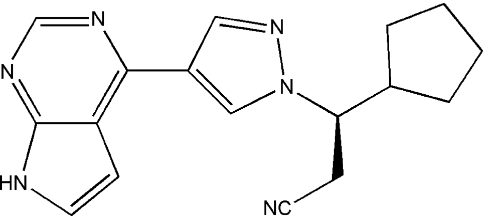
Ruxolitinib, (3R)-3-cyclopenty-3-[4-(7H-pyrrolo[2,3-d]pyrinidin-4-yl)pyrazole-1-yl]propanentrile, has the chemical formula C17H18N6 and is an oral inhibitor of JAK1 and JAK2, which is approved for the treatment of intermediate- and high-risk myelofibrosis (MF) and high-risk polycythemia vera (PV). Ruxolitinib selectively inhibits the proliferation of JAK2-driven Ba/F3 cells and these effects are correlated with decreased levels of phosphorylated JAK2 and of signal transducer and activator of transcription 5 (STAT5) (Plosker, Reference Plosker2015; Lussana et al., Reference Lussana, Cattaneo, Rambaldi and Squizzato2018). Thus, in November 2011, the U.S. FDA approved ruxolitinib for the treatment of patients with intermediate or high-risk myelofibrosis. On May 24, 2019, ruxolitinib was approved by FAD for steroid-refractory acute graft-versus-host disease (GVHD) in adult and pediatric patients 12 years and older. The chemical structure of ruxolitinib is shown in Figure 1 and its salts, such as ruxolitinib phosphate, hydrochloride, sulfate, besylate, and their polymorphism forms were reported (Albrecht and Selig, Reference Albrecht and Selig2016; Bernal-Vazquez et al., Reference Bernal-Vazquez, Lazcano-Seres, Juarez-Lagunas, Cano-Herrera, Van, Cedillo-Cruz and Mendoza2016; Tieger et al., Reference Tieger, Tozickova, Tkadlecova, Dammer and Gurgut2017). But the free form of ruxolitinib has not been reported yet. The crystal structures of ruxolitinib and its salts have also not been found yet.
Chemical structure of ruxolitinib.

We have not found this compound in the CSD database (Groom et al., Reference Groom, Bruno, Lightfoot and Ward2016) or in the PDF4+ database (Gates-Rector and Blanton, Reference Gates-Rector and Blanton2019). Therefore, we have decided to characterize this compound by X-ray powder diffraction and X-ray single-crystal diffraction techniques. In our study, we present powder data for ruxolitinib.
II. EXPERIMENTAL
A. Sample preparations
The sample was supplied by Zhejiang Ausun Pharmaceutical Co., LTD (purity >99.9%) and used without further purification. Dissolving ruxolitinib (500 mg) in the mixture of ethyl acetate and butyl acetate (10 ml 1:1 v/v) at reflux temperature and slow cooling of the solutions yielded crystals of ruxolitinib. Then, the crystals were dried, smashed, screened through 50 μm mesh size, and mounted on a flat zero-background plate.
B. Powder diffraction data collection
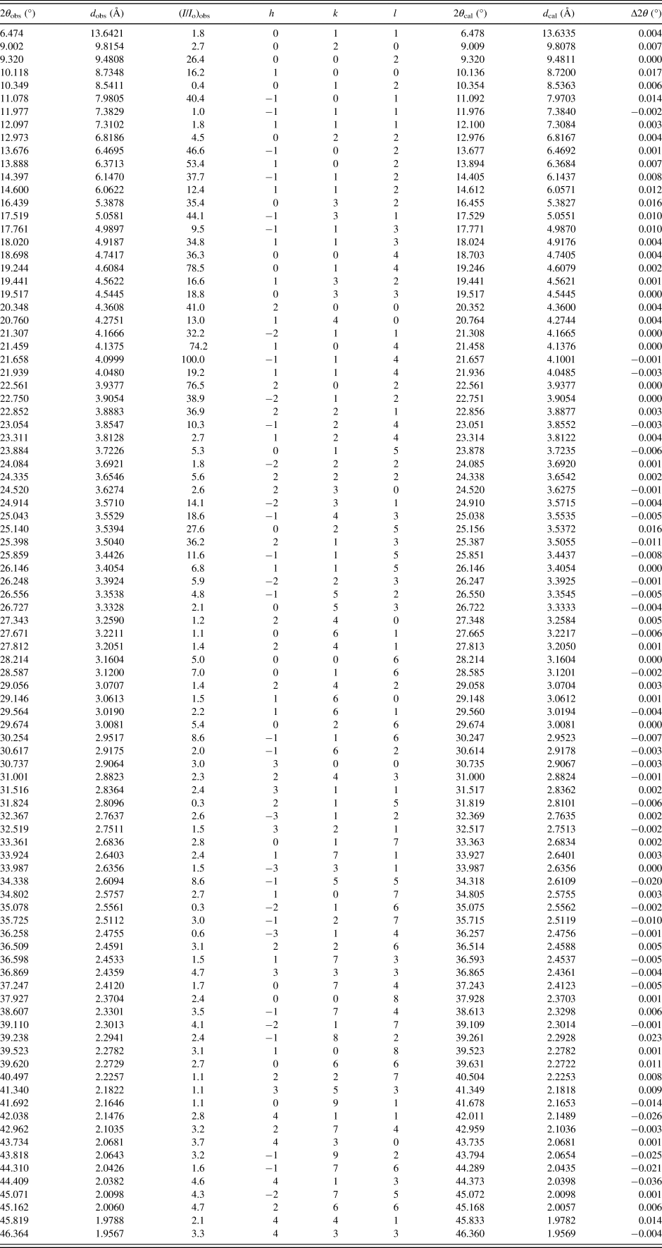
X-ray powder diffraction data was measured on a SmartLab diffractometer with fixed divergence slits and a D/tex Ultra 250 detector at room temperature. The diffractometer was configured in parafocusing Bragg-Brentano geometry. Data were collected over a 2-θ range of 5° to 50° with a step size of 0.02° and a counting time of 1.0 s/step using a CuKα radiation at a powder of 40 kV and 180 mA. CuKβ radiation was removed using a divergent beam Ni filter. The software package MDI-Jade version 7.5 (Materials Data Inc., USA) was used to smooth the data, fit the background, strip off the Kα 2 component and obtain the peak positions and intensities (Table I). The Kα 1 (λ = 1.5406 Å) wavelength was used in converting observed 2-θ to d-spacing.
X-ray powder diffraction data of ruxolitinib.

C. Single-crystal diffraction data collection
X-ray single-crystal diffraction data were collected at 296(2) K with a Bruker D8 Venture diffractometer with CuKα radiation (λ = 1.54178 Å) for cell determination and subsequent data collection. Data reduction was performed by APEX3 software and multi-scan absorption correction was applied. Using Olex2 (Dolomanov et al., Reference Dolomanov, Bourhis, Gildea, Howard and Puschmann2009), the structure was solved with the ShelXT (Sheldrick, Reference Sheldrick2015a) structure solution program using intrinsic phasing and refined with the ShelXL (Sheldrick, Reference Sheldrick2015b) refinement package using Least Squares minimization.
III. RESULTS AND DISCUSSION
Indexing of the experimental X-ray diffraction patterns and unit-cell refinements was done using MDI-Jade (Materials Data Inc., 2002). The cell refinement results which were combined with the results of single-crystal diffraction and characteristics of a chiral molecule, showed that ruxolitinib belonged to monoclinic with space group P21 and unit-cell parameters: a = 8.7211(5) Å, b = 19.6157(15) Å, c = 18.9645(10) Å, β = 90.903(6)°, unit-cell volume V = 3243.85 Å3, Z = 8, ρ = 1.2546 g/cm3. The figure of merit is F 30 = 203.9(0.0059,30) (Smith and Snyder, Reference Smith and Snyder1979). The values of 2θ obs, d obs, I obs, h, k, l, 2θ cal, d cal, and Δ2θ are listed in Table I.
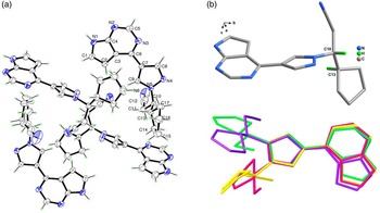
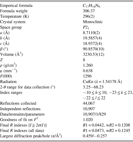
The single-crystal experiment was carried out at the temperature of 296 K and the structure solution was obtained (space group P21, a = 8.7110(2) Å, b = 19.5857(4) Å, c = 18.9372(4) Å, β = 90.8570(10)°, unit-cell volume V = 3230.53(10) Å3, Z = 8, ρ = 1.260 g/cm3). The detailed single-crystal data and the experimental data are summarized in Table II. The figures were drawn with ORTEP-3 (Oak Ridge Thermal Ellipsoid Plot) and Diamond (Brandenburg and Putz, Reference Brandenburg and Putz2005). The asymmetric units of the title compound contain four ruxolitinib molecules, which have slightly different conformations, and mainly shows the different orientation of pyrazole-1-yl]propanentrile. Meanwhile, the groups of 7H-pyrrolo[2,3-d]pyrinidin in four molecules are dislocated slightly each other, which are revealed from torsion angles of C9–C7–C6–N3, C3–C6–C7–C9 and their correspondings. But the configurations of chiral C atoms (C10, C10A, C10B, C10C) of four ruxolitinib molecules in the asymmetric unit are the same “R” configuration (Figure 2). Intermolecular hydrogen bond interactions N–H⋯N and other weak intermolecular interactions link ruxolitinib molecules, forming two-dimensional layer packing along bc-directions.
(a) Asymmetric unit of ruxolitinib shown in thermal ellipsoid model with 30% probability. (b) Overlay of configurations for four ruxolitinib molecules.

Crystal and experimental data of ruxolitinib.

The comparison of the experimental powder diffraction pattern and the simulated pattern derived from the single-crystal data is shown in Figure 3. Results showed that both single-crystal and powder diffraction methods can get the similar structure data and the deviations of the unit-cell parameters and unit-cell volume were between 0.05 and 0.41%.
X-ray powder diffraction pattern (black line) and the simulated pattern of the crystal structure (red line) of ruxolitinib.

IV. DEPOSITED DATA
The Crystallographic Information Framework (CIF) file was deposited with the ICDD. The data can be requested at pdj@icdd.com.
FUNDING
This work was financially supported from Zhejiang University Experimental Technology Research (SYBJS202204).