INTRODUCTION
Eubacteria of the genus Epulopiscium (here referred to as ‘epulos’ to distinguish them from reference to other bacteria) have been found as intestinal symbionts in numerous species of surgeonfish (Fishelson et al., Reference Fishelson, Montgomery and Myrberg1985; Clements et al., Reference Clements, Sutton and Choat1989; Bresler et al., Reference Bresler, Montgomery, Fishelson and Pollak1998). They were given the name Epulopiscium fishelsoni by Montgomery & Pollak (Reference Montgomery and Pollak1988), later suggested to be prokaryotes (Clements & Bullivant, Reference Clements and Bullivant1991), and subsequently confirmed to be Eubacteria by 16S rDNA analysis (Angert et al., Reference Angert, Clements and Pace1993). Clements et al. (Reference Clements, Sutton and Choat1989) assigned these prokaryotes to ten morphotypes (A–J) dependent upon their sizes, shapes, number of daughter cells (endospores) inside their cell walls and whether or not binary fission had been observed. The concept of endospores within large intestinal bacteria was first noted by Kunstyr et al. (Reference Kunstyr, Schiel, Kaup, Uhr and Kirchhoff1988); while examining Metabacterium polysporum from rodents. If daughter cells have formed a true spore wall (see figures 3A, B in Flint et al., Reference Flint, Drzymalski, Montgomery, Southam and Angert2005) they are considered to be endospores and are phase-bright in the light microscope. The presence of one or more daughter cells or endospores within the mother cell of Epulopiscium sp. has been documented in several publications (Fishelson et al., Reference Fishelson, Montgomery and Myrberg1985; Montogomery & Pollak, Reference Montgomery and Pollak1988; Clements et al., Reference Clements, Sutton and Choat1989; Bresler et al., Reference Bresler, Montgomery, Fishelson and Pollak1998; Flint et al., Reference Flint, Drzymalski, Montgomery, Southam and Angert2005).
Epulopiscium-like bacteria have been described from surgeonfish living in and around reefs of the Red Sea, Australia, Guam, Tuvalu, Moorea (French Polynesia), Ishigaki Island (southern Japan) and the Hawaiian Islands. This is the first report of their occurrence in surgeonfish living in the Atlantic Ocean.
For the present study, several Epulopiscium-like morphotypes were isolated from three species of surgeonfish that live in the reef systems of Flat Cay, which is about 2.75 km south-east of St Thomas, US Virgin Islands. Their forms and sizes, and certain characteristics of the daughter cells (endospores) inside mother cells are presented here. Also, they are tentatively delegated to specific ‘form types’ as described by Clements et al. (Reference Clements, Sutton and Choat1989). A sample of Epulopiscium-like bacteria was isolated by one of us (W.L.M.) from the intestines of the surgeonfish, Acanthurus tractus, from waters of southern Florida. It was studied as part of a PhD thesis (Flint, Reference Flint2006).
MATERIALS AND METHODS
Most of the host surgeonfish specimens, representing Acanthurus tractus (recently differentiated from A. bahianus by Bernal & Rocha, Reference Bernal and Rocha2011), A. coeruleus and A. chirurgus, were captured in 5 to 12 m of water, by aquarium nets in the early evening from Flat Cay (18°17.8′N 64°59.2′W), St Thomas, US Virgin Islands. These collections were made on various dates: three during October 2006, and one on 30 March 2007. Fish were held for up to 18 days after capture in a semi-natural aquarium (with running natural seawater, the tank exposed to natural sunlight, and native coralline algae, adherent to the aquarium glass, as a food source). In most samples, gut contents were removed at about mid-day, although some were removed and fixed at about 1800 hours and others at about 2300 hours. Small samples of gut contents were fixed in phosphate buffered 4.0% (w/v) formaldehyde and shipped to one of us (J.N.G.). Samples were taken from different locations in the intestines: near the anus, 3–4 cm up from the anus, and about mid-gut. Specimens of microbes to be measured and examined for morphological characters were selected as they were encountered on the slide. For a special winter study, two A. tractus were collected off the University of the Virgin Islands dock from less than 2 m of water, at 2000 hours on 12 December 2010. These fish were inactive, ‘sitting’ in reef crevices, at the time of collection; this is consistent with observed behaviour of strictly diurnal surgeonfish elsewhere (W.L.M., personal observation). They were placed in a mesh basket and hung in the water off the dock. They had no access to food. One fish was sacrificed and gut contents collected after 4.5 hours (at 0115 hours) and the second was sacrificed two days later at 1315 hours, after being held for ~41 hours. In each case, the gut was exposed and 1 cm lengths excised, slit open, and fixed. Samples were taken mid-gut, 3 cm up from the anus, and a section midway between these two.
Fixed samples were placed under a cover slip and six―full side to side―survey scans made over each slip. The bacteria were examined and measurements made with the aid of an ocular micrometer, without staining, under phase-contrast light microscopy in a Zeiss microscope. Some digital micrographs were taken using the same microscope.
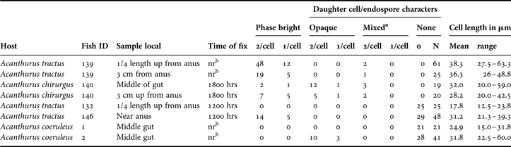
Counts were made of all cells encountered, and, in most cases, each was scored for the number of apparent daughter cells it contained. Daughter cells were classified as either opaque or phase-bright. Phase-bright cells are distinguished from opaque cells by possessing very heavy cell walls and appear to be true bacterial endospores (Clements et al., Reference Clements, Sutton and Choat1989; Bresler et al., Reference Bresler, Montgomery, Fishelson and Pollak1998; Flint et al., Reference Flint, Drzymalski, Montgomery, Southam and Angert2005). In some cases, a mother cell contained one opaque and one phase-bright daughter; these were termed ‘mixed’ in Table 1.
Mother cell sizes and daughter, or endospore, characteristics from three species of Caribbean surgeonfish. Fish identifiers were either field numbers or standard lengths.

a, mixed = with both phase bright and opaque; b, time of fixation not recorded.
The epulos from the South Florida collection were examined by a PhD student at Cornell University, and their morphotypes determined. Their forms are compared to those from Flat Cay, the US Virgin Islands.
RESULTS
The Epulopiscium-like morphs (hereafter termed epulos) from each surgeonfish varied in size and had differences in phase-contrast light refraction for the daughter cells (endospores). Daughter cells ranged from very small at the ends of the cells (Figures 1 & 7) to large enough to contact or overlap slightly at the cell's centre (Figure 4); however, in some cases no daughter cells were clearly visible (Figure 8). Epulo cells from hosts in the Pacific Ocean form phase-bright endospores, particularly at night, but daughter cells formed during the day lack the heavy endospore wall (Fishelson et al., Reference Fishelson, Montgomery and Myrberg1985; Clements et al., Reference Clements, Sutton and Choat1989; Bresler et al., Reference Bresler, Montgomery, Fishelson and Pollak1998; Robinow & Angert, Reference Robinow and Angert1998; Flint et al., Reference Flint, Drzymalski, Montgomery, Southam and Angert2005). Thus, daughter cells range from nearly invisible to ‘phase-bright’ (Clements et al., Reference Clements, Sutton and Choat1989; Flint et al., Reference Flint, Drzymalski, Montgomery, Southam and Angert2005) to opaque under phase-contrast light microscopy. Data for their size ranges, in length, are shown in Table 1.
Epulopisicum fishelsoni from surgeonfish (genus Acanthurus) from the island of St Thomas in the western Atlantic Ocean: (1) cell from host fish A. coeruleus with two small endospores at ends of the ‘mother cell’; (2) from host: A. tractus, the two endospores take up ~ two-thirds of the mother cell volume; (3) host: A. tractus, endospores nearly in contact at centre of mother cell; (4) host: A. chirurgus, endospores are overlapped at the centre; (5) host: A. tractus, only one endospore taking up about 60% of the cell length; (6) host: A. coeruleus, very faint opaque daughter cell at right (arrow head) and small, slightly bright one, on left in figure; (7) host: A. tractus, epulo with very small endospore at one end; (8) host: A. coeruleus, no daughter cells or endospores are apparent. Scale bars: 2, 3, 10 µm; 1, 4, 6, 7, 8, 20 µm.

Gut contents of two Acanthurus coeruleus were examined. In one (A. coeruleus No. 1; Table 1) 21 epulos were observed, and none were seen to have visible daughter cells. In the second A. coeruleus (No. 2), 41 epulos were examined and one daughter cell was seen in three, while two daughters were observed in ten cells; none of those was phase-bright. However, for many samples and host fish, daughter cells were phase-bright; thus, endospores (Figures 1–5 & 7; Table 1). Daughter cells varied from very small (each about 15% of the cell's length (Figure 1)) to large enough to almost fill 1/2 or slightly more of the ‘mother’ cell's length (Figures 3 & 4). When more than 1/2 the ‘mother’ cell's length, the daughter cells were bright endospores and appeared to be in contact, or nearly so, with each other (Figure 4). On occasion, epulos had only one endospore daughter—located in one half of the cell (Figures 5 & 7). In the host fish A. coeruleus, daughter cells were opaque to slightly phase-bright; often, they were difficult to see (Figure 6). In one epulo of A. tractus a single and very small phase-bright endospore could be seen at one end (Figure 7).
In A. tractus 139, the time of day for collecting and fixing were not recorded. For the ‘1/4 length up from anus’ location, 61 epulo cells were examined and of these, 48 (78.7%) contained two phase-bright endospores and 12 (19.7%) contained only one (Table 1). At the site 3 cm up from the anus, 25 epulos were observed and, of these, 19 (76%) had two phase-bright endospores and five (20%) contained only one (apparent) endospore. A single additional cell from the latter site had one phase-bright endospore, and one opaque daughter cell.
Using the full width of the ocular scale as transect, three scans were made across the cover-slip to count and characterize the epulos from A. tractus (146 mm). A total of 48 epulos were counted and of those, 19 (39.6%) contained very distinct phase-bright endospores, and 29 (60.4%) were without apparent endospores (Table 1). In marked contrast, no daughter cells were apparent in the 25 epulos examined from another A. tractus (No. 132); they were collected and fixed between 1100 and 1200 hours, in late November.
In this study, phase-bright endospores were often observed in epulos examined from the intestines of two fish species: A. tractus and A. chirurgus. In contrast, no phase-bright endospores were observed in the 61 epulos observed in two A. coeruleus specimens; however, they had many with amorphic opaque cytoplasm, and a few containing opaque daughter cells (Table 1).
In the ‘special winter study’, gut contents were surprisingly sparse. No epulos were seen in the four samples taken at 0115 hours. There were only two cells observed in the four samples taken in the afternoon, at 1315 hours; one of the two epulos was without apparent daughter cells and the second had two daughters; each of these was slightly phase-bright. These data are based on six scans of each preparation from four samples from each of two fish.
To summarize, briefly on their dimensions, the epulos described here ranged in length from 12.5 µm to 60 µm, with the average length, within the fish specimens, varying from 17.8 µm to 38.3 µm (Table 1). They were never observed to contain more than one or two daughter cells, or endospores.
DISCUSSION
Clements et al. (Reference Clements, Sutton and Choat1989) recorded ten morphotypes of large eubacteria from Pacific Ocean surgeonfish, but they have not been previously recorded in the literature from surgeonfish that inhabit the Atlantic Ocean. Bacteria related to, or in, the genus Epulopiscium have been described, thus far, as intestinal symbionts in roughly half the species of Pacific surgeonfish examined (Clements et al., Reference Clements, Sutton and Choat1989; unpublished; Montgomery & Clements, unpublished). Here, we add to that list three more host surgeonfish: Acanthurus tractus, A. coeruleus and A. chirurgus from the Caribbean; thus, the western Atlantic Ocean.
It is noteworthy that the fish from St Thomas contained epulo morphs after several hosts were held in salt water aquaria for up to 18 days. Thus, the aquarium habitat appears to have been relatively normal for food and other environmental factors. In a previous study (Flint et al., Reference Flint, Drzymalski, Montgomery, Southam and Angert2005), epulos were observed in fish that had been held in salt-water aquaria for a few hours only, and none were held in captivity longer. In the seminal paper first describing epulos (Fishelson et al., Reference Fishelson, Montgomery and Myrberg1985), when the surgeonfish A. nigrofuscus was ‘starved for 2–3 days’ they became free of these symbionts.
Two of the host species examined in this study, A. tractus and A. chirurgus, routinely feed over sandy bottoms and have gizzard-like stomachs (Beebe & Tee Van, Reference Beebe and Van-Tee1928; Randall, Reference Randall2002), while the third, A. coeruleus, feeds over shallow coral reefs and has a thin walled stomach (Longley et al., Reference Longley, Schmitt and Taylor1925; Randall, Reference Randall2002). Thus, A. coeruleus is algivorous, and the first two qualify as detritivorous. No surgeonfish studied here is planktivorous.
Clements et al. (Reference Clements, Sutton and Choat1989) presented evidence that, of the acanthurid fish they studied, those with gizzard-like stomachs, often ingesting ‘large amounts of sand’, contained smaller epulo bacteria. In the present study, all three host fish species contained epulos of approximately the same average size: 30.1, 30.9 and 28.4 µm long; thus, relatively small. Therefore, these results support the evidence of a relationship between stomach morphology and food intake, and the size of their gut symbionts for two of the host fish: A. tractus and A. chirurgus. However, the results do not support that relationship for A. coeruleus because of its thin walled stomach. Nevertheless, it is noteworthy that Clements et al. (Reference Clements, Sutton and Choat1989) stated: ‘…it is possible that the relationships between host feeding-type and protist [a term used prior to their determination as prokaryotes] type may have a locality component.’
Thus, relative to sizes, the epulos studied here best fit morphotypes F and G (Clements et al., Reference Clements, Sutton and Choat1989). The various forms of the daughter cells depicted by Clements et al. (Reference Clements, Sutton and Choat1989) ranged from ‘cigar-oval’ to ‘oval’, to ‘cigar’ to ‘elongate’. The epulos of this study were cigar-oval or cigar in shape. Of the hundreds of specimens examined, none showed binary fission characteristic of elongate forms of epulos. Consequently, the epulos described here do not fit neatly within the criteria established by Clements et al. (Reference Clements, Sutton and Choat1989), but are close to the characteristics of their types D and E.
In another study, three morphotypes of epulos were observed from the single A. tractus collected from southern Florida. These were all cigar shaped and one averaged 21.1 µm long by 3.1 µm wide (N = 35); another was 28.7 µm by 2.7 µm (N = 55); the third was 30.3 µm by 3.5 µm (N = 55). Thus, the sizes from that study compare well with the data here. These Florida based data are in an unpublished PhD thesis (Flint, Reference Flint2006).
In several studies of epulos, the cells have been described as having one or more phase-bright daughter cells (thus, endospores) within the mother cell (see Flint et al., Reference Flint, Drzymalski, Montgomery, Southam and Angert2005 for a review). As noted, this phase-bright appearance is characteristic of bacterial endospores for intestinal symbionts from at least a rodent and surgeonfish (Angert, Reference Angert2006). Phase-bright endospores have been observed most predictably at night (Flint et al., Reference Flint, Drzymalski, Montgomery, Southam and Angert2005). In the present study, 60% of the epulos from A. chirurgus No. 140 (3 cm up from the anus) collected at about 1600 hours and fixed at about 1800 hours, were phase-bright; hence, considered to be ‘mature’ (Table 1). However, at ‘mid-gut’ in the same specimen, only ~16% were phase-bright. Similarly, in two A. tractus (compare Nos. 132 and 146) (Table 1) epulos collected ‘near the anus’ had many phase-bright endospores while those upstream of the anus had no endospores. Thus, the formation of endospores may be dependent at least in part upon their location in the intestinal tract and the nutrient quality and quantity at that location. Two A. coeruleus specimens (Table 1) had no phase-bright endospores. Unfortunately, records were not kept of the time of day/night at which gut contents were fixed from these fish.
Of special interest is that phase bright endospores in this study are of quite different sizes, including a few very small ones at the poles of the mother cells (Figures 1 & 7). This seems a curious phenomenon because endospore walls appear to be thick and possibly non-flexible; hence, possibly, daughter cells develop into endospores when at different sizes. Except for the very small polar endospores, similar endospore size differences have been shown also in epulos from different acanthurid fish species from the Pacific Ocean (Flint et al., Reference Flint, Drzymalski, Montgomery, Southam and Angert2005).
During the night, endospores were deemed to be dormant (figure 6 in Flint et al., Reference Flint, Drzymalski, Montgomery, Southam and Angert2005) and usually emerged from the ‘mother cell’ at about day-break. When they emerge, the mother cell wall appears to tear or burst open centrally and one end of each endospore can be seen at that site, and often protruding slightly (Ward et al., Reference Ward, Clements, Choat and Angert2009). However, at this time, it is not known if this might be a tearing that results from fixation; thus be an artefact.
The December collection results are likely due to the probability that the fish have not fed for 3–4 hours and the gut has been largely evacuated of contents. It is well known that, at least in the Red Sea study (Montgomery et al., Reference Montgomery, Myrberg and Fishelson1989), surgeonfish feed in the winter at the same rates as during other seasons, but for fewer hours.
It is considered note-worthy that none of the fish examined contained ciliated protozoa in their gut lumen. Based on numerous studies (see: Grim et al., Reference Grim, Clements and Byfield2002 for a review), this lack of protists was not expected.
ACKNOWLEDGEMENTS
We thank Amber McCammon of the University of the Virgin Islands, St Thomas, US Virgin Islands, for help with fish collections. Also thanked are Therry The of the Imaging Facility at Northern Arizona University (NAU) who helped with photomicrography with a computer controlled con-focal microscope set for phase contrast optics, Daniel Boone of NAU for resolving a difficult problem with the plate, and Professor Esther Angert, Cornell University, New York, for help with some background information and valuable advice. Finally, we are pleased to thank Professor Kendall D. Clements (University of Auckland, New Zealand) who provided valuable critique and important new information.