INTRODUCTION
The Anaplasma species are gram negative obligate intracellular bacteria that infect diverse cell types, worldwide (Berger, Reference Berger2014). These are important tick-associated bacteria of veterinary and public health significance; cause anaplasmosis in various animal species and humans. The genus Anaplasma comprises of A. marginale, A. marginale subsp. centrale, A. ovis, A. bovis, A. platys, A. phagocytophilum and Aegyptianella pullorum. The A. bovis, A. platys and A. phagocytophilum species previously belong to genus Ehrlichia now included in genus Anaplasma after reclassification based on 16SrRNA and groEl genes in 2001 (Dumler et al. Reference Dumler, Barbet, Bekker, Dasch, Palmer, Ray, Rikihisa and Rurangirwa2001). Anaplasma marginale, A. marginale subsp. centrale, and A. bovis cause bovine anaplasmosis. Anaplasma ovis is the agent of ovine/caprine anaplasmosis. Anaplasma marginale, A. marginale subsp. centrale, and A. ovis infect erythrocytes. Anaplasma platys infects canine platelets, responsible for canine infectious cyclic anaplasmosis. Anaplasma bovis infects monocytes of ruminants (Sreekumar et al. Reference Sreekumar, Anandan, Balasundaram and Rajavelu1996; Rar and Golovljova, Reference Rar and Golovljova2011; Zobba et al. Reference Zobba, Anfossi, Pinna Parpaglia, Dore, Chessa, Spezzigu, Rocca, Visco, Pittau and Alberti2014); whereas A. phagocytophilum is a zoonotic pathogen that infect a wide range of vertebrate hosts including domestic and wild animals, birds, reptiles, rodents and humans infecting granulocytes (Foggie, Reference Foggie1951). Nevertheless, A. centrale is the least pathogenic among Anaplasma species. The distribution of A. centrale is limited to Mediterranean countries such as Turkey, Italy and Tunisia (Aktas et al. Reference Aktas, Altay and Dumanli2011; Zobba et al. Reference Zobba, Anfossi, Pinna Parpaglia, Dore, Chessa, Spezzigu, Rocca, Visco, Pittau and Alberti2014; Belkahia et al. Reference Belkahia, Ben Said, Alberti, Abdi, Issaoui, Hattab, Gharbi and Messadi2015a ). In terms of developing vaccines and diagnostic assays, it is important to know the similarities of closely related organisms. Currently, limited reviews are available on these intracellular blood-borne pathogens. Therefore, there is a need to provide updated information on ecology, epidemiology and major similarities of all Anaplasma species of family Anaplasmataceae for effective prevention and control of anaplasmosis in animals and humans.
ANAPLASMA PHAGOCYTOPHILUM
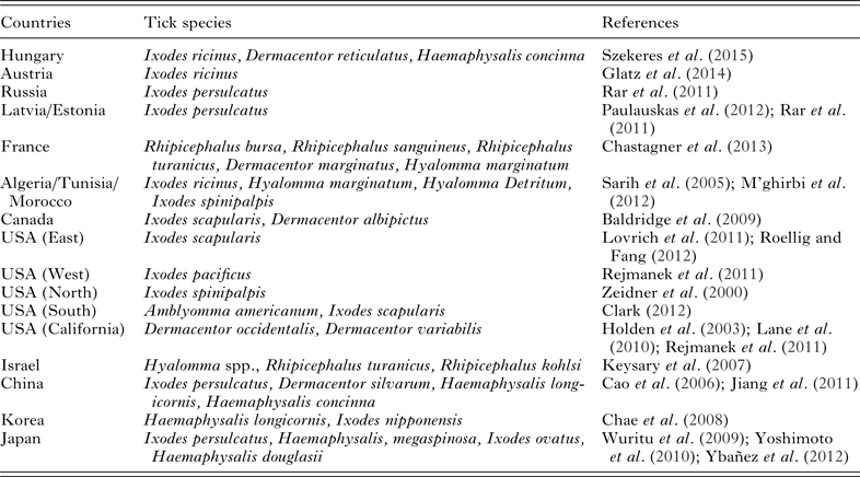
Anaplasma phagocytophilum is a hetero-genetic zoonotic tick-borne bacteria of diverse hosts, mainly transmitted by Ixodes, Dermacentor, Haemaphysalis, Amblyomma, Hyalomma, Rhipicephalus species of ticks (Jiang et al. Reference Jiang, Jiang, Yu, Zhang, Gao, Zhan, Sun, Zhang, Zhang, Liu, Wu, Xu and Cao2011; Clark, Reference Clark2012; M'ghirbi et al. Reference M'ghirbi, Yaïch, Ghorbel and Bouattour2012; Chastagner et al. Reference Chastagner, Bailly, Leblond, Pradier and Vourc'h2013; Szekeres et al. Reference Szekeres, Claudia Coipan, Rigó, Majoros, Jahfari, Sprong and Földvári2015). This has been detected in Asia, Europe, Africa and America (Kawahara et al. Reference Kawahara, Rikihisa, Lin, Isogo, Tahara, Itagaki, Hiramitsu and Tajima2006; M'ghirbi et al. Reference M'ghirbi, Yaïch, Ghorbel and Bouattour2012; Kang et al. Reference Kang, Kim, Choi, Nam, Chae, Chong, Klein, Ko and Chae2013; Djiba et al. Reference Djiba, Mediannikov, Mbengue, Thiongane, Molez, Seck, Fenollar, Raoult and Ndiaye2013; Stuen et al. Reference Stuen, Pettersen, Granquist, Bergström, Bown and Birtles2013a ; Borthakur et al. Reference Borthakur, Deka, Bhattacharjee and Sarmah2014; Razzaq et al. Reference Razzaq, Khosa, Ahmad, Hussain, Saeed, Khan, Shaikh, Ali and Iqbal2015; Table 1). Prevalence varies from region to region depending upon tick exposure, immunity and age (CDC, 2013). Infection in domestic ruminants is generally known as tick-borne fever, responsible for significant economic losses especially in Northern Europe (Stuen, Reference Stuen2007; Grøva et al. Reference Grøva, Olesen, Steinshamn and Stuen2011).
Detection of Anaplasma phagocytophilum from different vertebrate hosts a

a Detection of A. phagocytophilum from 1998 to 2015.
b Organism detected in tissue or blood specimen.
c In parenthesis target gene has been mentioned.
d Nested PCR.
e Reverse line blot hybridization.
f Real time PCR.
g Commercial kit.
h Species involved were not available.
Clinical findings
High fever, dullness, in-appetence, sudden drop in milk production, low weight gain, coughing, abortion, stillbirth and low fertility in sheep and reduced semen quality in rams are important clinical signs in cattle and sheep (CFSPH, 2013) but clinical signs differ among variants/strains involved. Less severe cases recover within few days and death is unusual outcome (Grøva et al. Reference Grøva, Olesen, Steinshamn and Stuen2011; CFSPH, 2013). The TBF may develop concurrent infections with Borrelia burgdorferi and/or A. marginale and followed by secondary infections (tick pyemia and pasteurellosis) (Hofmann-Lehmann et al. Reference Hofmann-Lehmann, Meli, Dreher, Gönczi, Deplazes, Braun, Engels, Schüpbach, Jörger, Thoma, Griot, Stärk, Willi, Schmidt, Kocan and Lutz2004; Berger, Reference Berger2014). Co-infection may alter disease pathogenesis and complicate diagnosis resulting in more severe clinical disease. These factors affect treatment and prognosis as well. Co-infected dogs with B. burgdorferi further predispose to Bartonella vinsonii subs. berkhoffii infection (Grab et al. Reference Grab, Nyarko, Barat, Nikolskaia and Dumler2007). Single A. phagocytophilum infected dogs and cats show generalized non-specific signs (CFSPH, 2013). Horses (>3 years) develop sever disease including fever, depression anorexia, icterus, ataxia, petechial hemorrhages and distal limb edema with severe myopathy (CFSPH, 2013). Fever, headache, myalgias and chills are the important clinical findings of human anaplasmosis (CFSPH, 2013). Leucopenia (marked neutropenia), thrombocytopenia, anaemia and/or elevated liver enzymes are common hematological and biochemical finding in humans and animals (especially dogs and cats) (Bakken et al. Reference Bakken, Dumler, Chen, Eckman, Van Etta and Walker1994; Aguero-rosenfeld et al. Reference Aguero-rosenfeld, Horowitz, Wormser, Mckenna, Nowakowski, Munoz and Dumler1996; CFSPH, 2013; Ravnik et al. Reference Ravnik, Tozon, Smrdel and Zupanc2011; Dondi et al. Reference Dondi, Russo, Agnoli, Mengoli, Balboni, Alberti and Battilani2014; Savidge et al. Reference Savidge, Ewing, Andrews, Aucoin, Lappin and Moroff2016; Khatat et al. Reference Khatat, Defauw, Marynissen, Van de Maele, van Dongen and Daminet2015). Bacterium show purple colour mulberry-like micro-colonies after staining with Romanowsky stain called Morulae (Rikihisa, Reference Rikihisa2011). Enlargement of spleen (4–5 times) with subscapular bleeding is the most significant post-mortem finding of tick-borne fever in sheep, reindeer and roe deer (Gordon et al. Reference Gordon, Brownlee, Wilson and Macleod1932; Øverås et al. Reference Øverås, Lund, Ulvund and Waldeland1993; Stuen, Reference Stuen2003).
Epidemiology
Infection has been noticed in cattle, sheep, goat, horse, donkey, dog, cat and wild ruminants (Table 1). Anaplasma phagocytophilum is most commonly been found in red deer, roe deer, fallow deer with highest prevalence (10–98%) in red deer and roe deer population (Hulínská et al. Reference Hulínská, Langøová, Pejèoch and Pavlásek2004; Zeman and Pecha, Reference Zeman and Pecha2008; Scharf et al. Reference Scharf, Schauer, Freyburger, Petrovec, Schaarschmidt-Kiener, Liebisch, Runge, Ganter, Kehl, Dumler, Garcia-Perez, Jensen, Fingerle, Meli, Ensser, Stuen and Von Loewenich2011; Hapunik et al. Reference Hapunik, Vichova, Karbowiak, Wita, Bogdaszewski and Pet'ko2011; Overzier et al. Reference Overzier, Pfister, Herb, Mahling, Böck and Silaghi2013). Exact reservoir hosts for A. phagocytophilum in animals and humans are not known to date. However, the pathogen has been detected in sika deer, white-tailed deer, Korean water deer, chamois, European bison, wild boar, Alpine ibex, mouflon, mule deer, elk, ilama, alpaca, Swedish moose and birds (Table 1). Small mammals: Dusky-footed wood rats, white-footed mice, vole, eastern chipmunk, squirrel, Virginia opossum and striped skunk. Insectivorous mammals: Shrew, hedgehog. Reptiles and snakes: Northern alligator lizard and Pacific gopher snake. Others: Gray fox, cotton tail rabbit, American black bear, timber wolf, raccoon and European brown bear (Table 1).
High-risk outdoor behaviour (such as gardening and hiking), immunocompromised individuals (cancer treatments, HIV infection and prior organ transplants) and blood transfusion are important risk factors for human granulocytic anaplasmosis (HGA) (Leiby and Gill, Reference Leiby and Gill2004; Zhang et al. Reference Zhang, Liu, Xu, Zhang, Jin, Li, Lu, Li, Chang, Zhang, Fan, Cao, Bao, Zhang, Guan, Cheng, Tian, Wang, Yu, Yu, Wang, Zhang, Tang, Yin, Lao, Wu, Li, Li, Xu, Shi and Huang2014; Atif, Reference Atif2015).
Higher degree of genetic, pathogenic and host tropisms exist in A. phagocytophilum isolates (Baráková et al. Reference Baráková Granquist, Derdáková, Carpi, Rosso, Collini, Tagliapietra, Ramponi and Rizzoli2014). The 16S rRNA, p44, groEL heat-shock protein, msp4 and ankA genes are important genetic markers to study genetic diversity (Granquist et al. Reference Granquist, Aleksandersen, Bergstrom, Dumler, Torsteinbo and Stuen2010a ; Silaghi et al. Reference Silaghi, Scheuerle, Friche Passos, Thiel and Pfister2011a , Reference Silaghi, Hamel, Thiel, Pfister, Passos and Rehbein b ).
Transmission: Ixodes (I.) ricinus is the major vector in Europe; I. pacificus in Western USA; Dermacentor (D.) occidentalis and D. variabilis in California (Holden et al. Reference Holden, Boothby, Anand and Massung2003; Lane et al. Reference Lane, Mun, Peribáñez and Fedorova2010; Rejmanek et al. Reference Rejmanek, Nieto, Barash and Foley2011); I. scapularis in Eastern USA (Lovrich et al. Reference Lovrich, Jobe, Kowalski, Policepatil and Callister2011; Roellig and Fang, Reference Roellig and Fang2012); Amblyomma americanum and I. scapularis in Southern USA; I. spinipalpis in North Colorado (USA) (Zeidner et al. Reference Zeidner, Burkot, Massung, Nicholson, Dolan, Rutherford, Biggerstaff and Maupin2000); I. scapularis and D. albipictus in Canada (Baldridge et al. Reference Baldridge, Scoles, Burkhardt, Schloeder, Kurtti and Munderloh2009); I. persulcatus, I. nipponensis, I. ovatus, D. silvarum, Haemaphysalis (H.) douglasii, H. megaspinosa, H. japonica, H. longicornis in Asia (China, Korea, Japan, Russia; Table 2); I. ricinus, I. spinipalpis, Hyalomma (Hy.) marginatum, Hy. Detritum in North Africa (Algeria, Tunisia and Morocco) (Sarih et al. Reference Sarih, M'ghirbi, Bouattour, Gern, Baranton and Postic2005; M'ghirbi et al. Reference M'ghirbi, Yaïch, Ghorbel and Bouattour2012); R. turanicus, Rhipicephalus kohlsi in Israel (Keysary et al. Reference Keysary, Massung, Inbar, Wallach, Shanas, Mumcuoglu and Waner2007). The pathogen has also been detected in I. persulcatus, H. concinna, D. reticulates and I. ventalloi ticks (Table 2).
Detection of Anaplasma phagocytophilum in different tick species

As mentioned above, A. phagocytophilum has been detected in various tick species, but the vector competence of I. ricinus, I. pacificus, I. scapularis and I. spinipalpis has so far been proved (Woldehiwet, Reference Woldehiwet2010). Identification of reservoir hosts and competent vectors are the important research area that needs to be explored. There are reports of mechanical transmission by deer ked (Lipoptena cervi) from Cervides (Víchová et al. Reference Víchová, Majláthová, Nováková, Majláth, Čurlík, Bona, Komjáti-Nagyová and Peťko2011). Similarly, transplacental (lambs and calves), perinatal, nosocomial and blood transfusions associated transmissions has been reported (Bakken et al. Reference Bakken, Dumler, Chen, Eckman, Van Etta and Walker1994; Horowitz et al. Reference Horowitz, Kilchevsky, Haber, Aguero-Rosenfeldt, Kranwinkel, James, Wong, Chu, Liveris and Schwartz1998; Dhand et al. Reference Dhand, Nadelman, Aguero-Rosenfeld, Haddad, Stokes and Horowitz2007; Zhang et al. Reference Zhang, Liu, Ni, Li, Yu, Yu, Wan, Li, Liang, Jiang, Jing, Run, Luan, Fu, Zhang, Yang, Wang, Dumler, Feng, Ren and Xu2008; Annen et al. Reference Annen, Friedman, Eshoa, Horowitz, Gottschall and Straus2012; Henniger et al. Reference Henniger, Henniger, Grossmann, Distl, Ganter and von Loewenich2013; Reppert et al. Reference Reppert, Galindo, Breshears, Kocan, Blouin and de la Fuente2013). The epidemiology of anaplasmosis is further complicated by these modes of transmissions.
Diagnosis
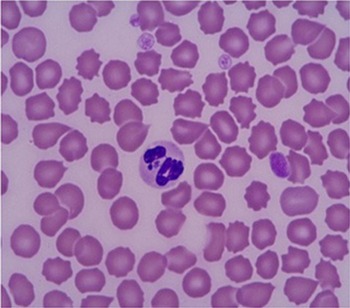
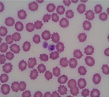
Anaplasma phagocytophilum is rarely diagnosed only on the basis of clinical signs; usually laboratory tests are required for confirmation. Intra-cytoplasmic inclusions can be observed in granulocytes in peripheral blood smear (Fig. 1). Disease can be confirmed by electron microscopy of blood or organs smears, cell culture or immune-histochemistry (Lepidi et al. Reference Lepidi, Bunnell, Martin, Madigan, Stuen and Dumler2000; Stuen et al. Reference Stuen, Granquist and Silaghi2013b ). The “SNAP®4Dx® ELISA” (dog), also detect antibodies for sheep and horse (Granquist et al. Reference Granquist, Stuen, Crosby, Lundgren, Alleman and Barbet2010b ; Hansen et al. Reference Hansen, Christoffersen, Thuesen, Petersen and Bojesen2010); “MegaFLUO® ANAPLASMA phagocytophilum” (dog, horse) and “ImmunoRun Canine Anaplasma Antibody Detection kit” (dog) are commercially available diagnostic kits for in-house detection of A. phagocytophilum antibodies in plasma or serum; as per specifications of manufacturer. The indirect immunofluorescence assay is the gold standard for HGA (Center for Disease Control and Prevention) (CDC, 2013). Conventional, real-time and nested PCR techniques has been developed for blood and tissue samples targeting 16S rRNA, groEL, msp4, ankA and p44 genes of A. phagocytophilum (Chen et al. Reference Chen, Dumler, Bakken and Walker1994; Courtney et al. Reference Courtney, Kostelnik, Zeidner and Massung2004; Alberti et al. Reference Alberti, Zobba, Chessa, Addis, Sparagano, Pinna Parpaglia, Cubeddu, Pintori and Pittau2005). There is no such test available that can differentiate persistent and chronic Anaplasma infections.
Anaplasma phagocytophilum in dog's granulocyte (Sainz et al. Reference Sainz, Roura, Miró, Estrada-Peña, Kohn, Harrus and Solano-Gallego2015); magnification 100×.

Treatment
Ovine granulocytic anaplasmosis can be treated and possibly eliminated by long acting oxytetracycline at 10–20 mg kg−1 (Stuen and Bergstrom, Reference Stuen and Bergstrom2001). Similarly, oxytetracycline is also effective against horses and other ruminants (Atif et al. Reference Atif, Khan, Khan, Ashraf and Avais2012b ; Siska et al. Reference Siska, Tuttle, Messick, Bisby, Toth and Kritchevsky2013; The Merck Veterinary Manual, 2014); whereas doxycycline showed efficacy against feline and canine granulocytic anaplasmosis (CFSPH, 2013). Oxytetracycline, rifampin, doxycycline hyclate and levofloxacin are effective against HGA (Goodman et al. Reference Goodman, Nelson, Vitale, Madigan, Dumler, Kurtti and Munderloh1996; Wormser et al. Reference Wormser, Dattwyler, Shapiro, Halperin, Steere, Klempner, Krause, Bakken, Strle, Stanek, Bockenstedt, Fish, Dumler and Nadelman2006; Zeidner et al. Reference Zeidner, Massung, Dolan, Dadey, Gabitzsch, Dietrich and Levin2008; CDC, 2013; Bakken and Dumler Reference Bakken and Dumler2015).
ANAPLASMA BOVIS
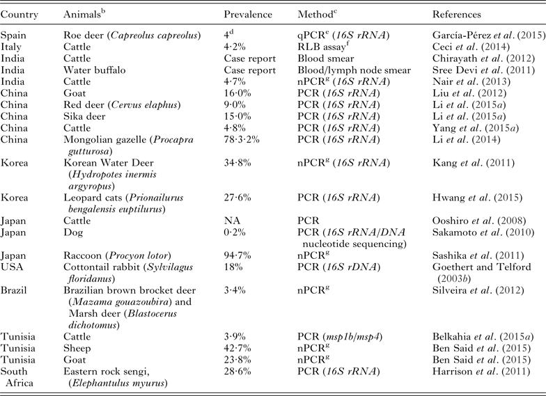
Anaplasma bovis infects monocytes of diverse hosts. This has been described for the first time in cattle in 1936 (Donatien and Lestoquard, Reference Donatien and Lestoquard1936). Since it has been reported from Spain, Italy, India, China, Korea, Japan, America, Brazil, Tunisia and South Africa from domestic and wild animals (Sakamoto et al. Reference Sakamoto, Ichikawa, Sakata, Matsumoto and Inokuma2010; Harrison et al. Reference Harrison, Bown and Horak2011; Sashika et al. Reference Sashika, Abe, Matsumoto and Inokuma2011; Chirayath et al. Reference Chirayath, Lakshmanan, Pillai, Alex and Rejitha2012; Liu et al. Reference Liu, MA, Wang, Wang, Peng and Li2012; Ceci et al. Reference Ceci, Iarussi, Greco, Lacinio, Fornelli and Carelli2014; Li et al. Reference Li, Chen, Liu, Liu, Yang, Li, Li, Luo and Yin2015a ; Yang et al. Reference Yang, Li, Liu, Liu, Niu, Ren, Chen, Guan, Luo and Yin2015a ; Hwang et al. Reference Hwang, Oh, Lee and Chun2015; Belkahia et al. Reference Belkahia, Ben Said, Alberti, Abdi, Issaoui, Hattab, Gharbi and Messadi2015a ; García-Pérez et al. Reference García-Pérez, Oporto, Espí, Del Cerro, Barral, Povedano, Barandika and Hurtado2015; Ben Said et al. Reference Ben Said, Belkahia, Karaoud, Bousrih, Yahiaoui, Daaloul-Jedidi and Messadi2015; Table 3).
Detection of Anaplasma bovis among vertebrate hosts from different countries a

NA = not available.
a Detection of A. bovis from 2003–2015.
b Organism detected in tissue or blood specimen.
c In parenthesis target gene has been mentioned.
d Number of animals found positive.
e Real time PCR.
f Reverse line blot hybridization.
g Nested PCR.
Clinical findings
Anaplasma bovis infection is characterized by fever, drop in milk production, weakness, obvious weight loss, pale mucous membranes, enlargement of prescapular lymph nodes and death in stressed or naïve cattle (Losos, Reference Losos1986; Sreekumar et al. Reference Sreekumar, Anandan, Balasundaram and Rajavelu1996).
Epidemiology
Cattle and goats are the major reservoirs of this pathogen (Goethert and Telford, Reference Goethert and Telford2003 Reference Goethert and Telford b ). Infection has also been detected in other animals including buffalo, dog, roe deer, red deer, sika deer, Korean water deer, Brazilian brown brocket deer, marsh deer, Mongolian gazelle, leopard cat, raccoon, cotton tail rabbit and eastern rock sengi (Table 3). The first molecular phylogenetic study of A. bovis was performed in Japan using groEL and gltA genes (Ybañez et al. Reference Ybañez, Sashika and Inokuma2014). Prevalence of A. bovis varies from region to region depending upon the test method and specie type. Prevalence in domestic ruminants range from 3·94·8 to 39·9% and 9 to 15% in wild Cervids (Red deer and Sika deer) (Ceci et al. Reference Ceci, Iarussi, Greco, Lacinio, Fornelli and Carelli2014; Belkahia et al. Reference Belkahia, Ben Said, Alberti, Abdi, Issaoui, Hattab, Gharbi and Messadi2015a ; Li et al. Reference Li, Chen, Liu, Liu, Yang, Li, Li, Luo and Yin2015a ; Yang et al. Reference Yang, Li, Liu, Liu, Niu, Ren, Chen, Guan, Luo and Yin2015a ; Njiiri et al. Reference Njiiri, Bronsvoort, Collins, Steyn, Troskie, Vorster, Thumbi, Sibeko, Jennings, Van Wyk, Mbole-Kariuki, Kiara, Poole, Hanotte, Coetzer, Oosthuizen, Woolhouse and Toye2015; Table 3).
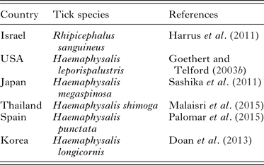
Transmission: Haemaphysalis megaspinosa, H. shimoga, H. punctata, H. longicornis, H. leporispalustris and Rhipicephalus sanguineus species of ticks are important vector of A. bovis (Table 4) but the vector competence of ticks needs to be further evaluated.
Molecular detection of Anaplasma bovis in different tick species

Diagnosis
Examination of Leishman's stained peripheral blood smear of cow show acidophilic A. bovis elementary bodies in monocytes or lymphocytes with eosinophilia (Sreekumar et al. Reference Sreekumar, Anandan, Balasundaram and Rajavelu1996; Chirayath et al. Reference Chirayath, Lakshmanan, Pillai, Alex and Rejitha2012). Nested PCR targeting 16S rRNA gene has been used for the detection of bacterial DNA in ticks, domestic and wild ruminants (Kawahara et al. Reference Kawahara, Rikihisa, Lin, Isogo, Tahara, Itagaki, Hiramitsu and Tajima2006; Doan et al. Reference Doan, Noh, Choe, Yoo, Kim, Reddy, Quyen, Nguyen, Nguyen, Kweon, Jung, Chang and Kang2013; Li et al. Reference Li, Chen, Liu, Liu, Yang, Li, Li, Luo and Yin2015a ; Yang et al. Reference Yang, Li, Liu, Liu, Niu, Ren, Chen, Guan, Luo and Yin2015a ).
Treatment
So far, there are no controlled treatment studies available for A. bovis. In a case study, infection has been treated with oxytetracycline at the dose rate of 20 mg kg−1 intravenous along with 500 ml normal saline. This showed marked improvement in clinical signs (Chirayath et al. Reference Chirayath, Lakshmanan, Pillai, Alex and Rejitha2012).
ANAPLASMA OVIS
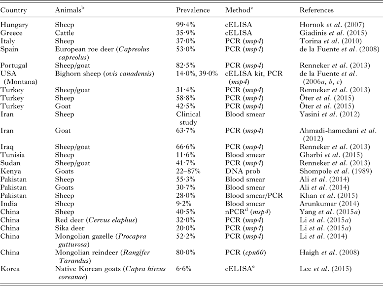
Anaplasma ovis is an important intraerythrocytic tick-borne pathogen of goats, sheep and wild small ruminants worldwide (Shompole et al. Reference Shompole, Waghela, Rurangirwa and Mcguire1989; Zaugg et al. Reference Zaugg, Goff, Foreyt and Hunter1996; Yabsley et al. Reference Yabsley, Davidson, Stallknecht, Varela, Swift, Devos and Dubay2005; de la Fuente et al. Reference de la Fuente, Atkinson, Naranjo, Fernandez, Mera, Mangold, Keating and Kocan2007; Yasini et al. Reference Yasini, Khaki, Rahbari, Kazemi, Amoli, Gharabaghi and Jalali2012; Ybañez et al. Reference Ybañez, Sashika and Inokuma2014; Gharbi et al. Reference Gharbi, Omri, Jedidi, Zorii and Darghouth2015; Khan et al. Reference Khan, Khan, Tufail, Saleem and Masood2015; Li et al. Reference Li, Chen, Liu, Liu, Yang, Li, Li, Luo and Yin2015a ; Lee et al. Reference Lee, Jung and Kwak2015; Yang et al. Reference Yang, Li, Liu, Liu, Niu, Ren, Chen, Guan, Luo and Yin2015a ; Table 5). Ovine anaplasmosis has been considered a problem for poor countries but scientists have also detected this bacterium in developed countries such as USA, Hungary, Greece, Italy, Spain, Portugal and Turkey (de la Fuente et al. Reference de la Fuente, Atkinson, Hogg, Miller, Naranjo, Almazán, Anderson and Kocan2006a ; Hornok et al. Reference Hornok, Elek, de la Fuente, Naranjo, Farkas, Majoros and Földvári2007; de la Fuente et al. Reference de la Fuente, Ruiz-Fons, Naranjo, Torina, Rodríguez and Gortázar2008; Torina et al. Reference Torina, Galindo, Vicente, Di Marco, Russo, Aronica, Fiasconaro, Scimeca, Alongi, Caracappa, Kocan, Gortazar and de la Fuente2010; Renneker et al. Reference Renneker, Abdo, Salih, Karagenç, Bilgiç, Torina, Oliva, Campos, Kullmann, Ahmed and Seitzer2013; Giadinis et al. Reference Giadinis, Katsoulos, Chochlakis, Tselentis, Ntais, Lafi, Karatzias and Psaroulaki2015; Öter et al. Reference Öter, Çetinkaya, Vurusaner, Toparlak and Ergünay2015). The occurrence of A. ovis is not widespread in these countries yet, but the climate change and traditional farming (pasturage on meadows) may lead to the spread of this pathogen.
Detection of Anaplasma ovis among vertebrate hosts from different countries a

a Detection of A. ovis from 1989 to 2015 including a clinical study.
b Organism detected in tissue or blood specimen.
c In parenthesis target gene has been mentioned.
d Nested PCR.
e Detected Anaplasma species.
Clinical findings
Acute infection is characterized by fever, weakness, anaemia, pallor mucous membrane, lower milk production, weight loss, jaundice, abortion and often death (Shompole et al. Reference Shompole, Waghela, Rurangirwa and Mcguire1989; Kocan et al. Reference Kocan, de la Fuente, Guglielmone and Melendez2003). Anaplasma ovis does not cause clinical disease in cattle. Clinical disease cause great economic losses in resource limited countries where sheep and goat are the major component of livestock. The clinical signs are variable based on age, breed and health status of animal (Splitter et al. Reference Splitter, Twiehaus and Castro1955; Shompole et al. Reference Shompole, Waghela, Rurangirwa and Mcguire1989). Animals recovered from clinical disease become life-long carriers. Persistent infections may cause low weight gain (>14%) in lambs (Klabi, Reference Klabi2011). Splenectomized animals, inter-current infections, malnutrition and pregnancy render animals more susceptible to anaplasmosis (Splitter et al. Reference Splitter, Anthony and Twiehaus1956).
Epidemiology
In recent years, its occurrence have been reported from all over the world such as Hungary, Greece, Italy, Spain, Portugal, USA, Turkey, Iran, Iraq, Tunisia, Sudan, Kenya, Pakistan, India, China and Korea (Table 5).
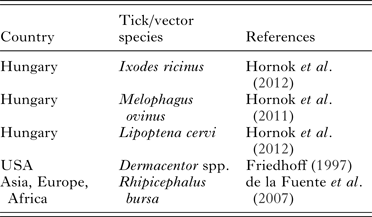
Transmission: Anaplasma ovis is biologically transmitted by soft and hard tick species including R. bursa, Dermacentor andersoni, R. turanicus, Ornithodoros lahorensis and mechanically by biting Diptera. Nonetheless, sheep ked (Melophagus ovinus) has also been suspected to transmit A. ovis (Table 6).
Detection of Anaplasma ovis in different tick/vector species

Diagnosis
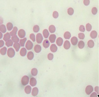
Acute disease is usually diagnosed on the basis of clinical signs, detection of organism in blood smear and hematology (Rassouli et al. Reference Rassouli, Kafshdouzan, Zow and Ghodrati2015). This bacterium is deeply stained and appears dense, round bodies located at the centre or margins of the erythrocytes (Fig. 2).
Anaplasma ovis in sheep's erythrocyte (Jalali et al. Reference Jalali, Khaki, Kazemi, Bandehpour, Rahbari, Jalali and Yasini2013); magnification 100×.

Treatment
Oxytetracycline is the most effective drug followed by imidocarb dipropionate and diaminazene in sheep and goats (Ali et al. Reference Ali, Ijaz, Durrani, Ali, Mehmood and Sabir2014).
ANAPLASMA MARGINALE
Anaplasma marginale has been described by Sir Arnold Theiler in erythrocytes of cattle as “marginal points” (Theiler, Reference Theiler1910). Bovine anaplasmosis (BA) is caused by A. marginale, characterized by fever, severe anaemia, pale mucous membranes, jaundice, brownish urine, decreased milk production, abortion, hyper excitability, weight loss and mortality during acute phase of the infection (Richey and Palmer, Reference Richey and Palmer1990; Wanduragala and Ristic, Reference Wanduragala, Ristic, Woldehiwet and Ristic1993; Ooshiro et al. Reference Ooshiro, Zakimi, Matsukawa, Yafuso, Katagiri and Inokuma2009; Jaswal et al. Reference Jaswal, Bal, Singla, Gupta and Brar2013). The BA is responsible for economic losses of 48·13 million USD in Tanzania and $26 million Australian dollars due to mortality in Australia (Kivaria, Reference Kivaria2006; Sackett and Holmes, Reference Sackett and Holmes2006). Hemolytic anaemia is the major clinical manifestation of A. marginale infection in cattle (Atif et al. Reference Atif, Khan, Iqbal and Roheen2012a ). Animals recovered from clinical disease become life-long carriers (Palmer et al. Reference Palmer, Futse, Knowles and Brayton2006). Numerous strains of A. marginale exist. They differ in morphology, antigenic characteristics, protein sequence and biological transmission (Smith et al. Reference Smith, Levy, Kuhlenschmidt, Adams, Rzechula, Hardt and Kocan1986; Barbet et al. Reference Barbet, Palmer, Myler and Mcguire1987; Kocan et al. Reference Kocan, de la Fuente, Guglielmone and Melendez2003, Reference Kocan, de la Fuente, Blouin and Garcia-Garcia2004).
Epidemiology
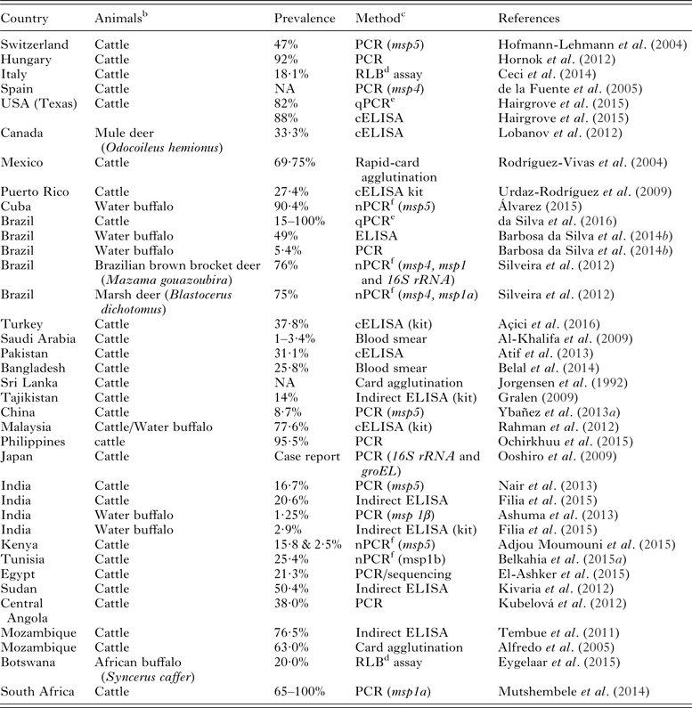
The BA is widely distributed throughout the world especially in tropical and subtropical (400N–320S) regions, including Asia, Far East Asia, Central Asia, Southern Europe, America and Africa (Table 7). Increase in prevalence and distribution of anaplasmosis is due to increase in movement and transportation of cattle from endemic to non-endemic regions (Saetiew et al. Reference Saetiew, Simking, Inpankaew, Wongpanit, Kamyingkird, Wongnakphet, Stich and Jittapalapong2015).
Detection of Anaplasma marginale from different countries a

NA = not available.
a Detection of A. marginale from 1992 to 2015.
b Organism detected in tissue or blood specimen.
c In parenthesis target gene has been mentioned.
d Reverse line blot hybridization.
e Real-time PCR.
f Nested PCR.
Clinical anaplasmosis mostly occurs in cattle but other ruminants usually develop persistent infection. However, natural or experimental infection has been reported in buffaloes (Bubalus bubalis), African buffalo (Syncerus caffer), black-tailed deer (Odocoileus hemionus columbianus), white-tailed deer (Odocoileus virginianus), marsh deer (Blastocerus dichotomus), Brazilian brown brocket deer (Mazama gouazoubira), mule deer (Odocoileus hemionus), bighorn sheep (Ovis canadensis), pronghorn antelope (Antelocapra americana), elk (Cervus elaphus nannodes), rocky mountain elk (Cervus elaphus nelson), giraffes (Giraffa camelopardalis) and bison (Bison bison), (Stiller et al. Reference Stiller, Leatch and Kuttler1981; Kuttler, Reference Kuttler1984; Davidson and Goff, Reference Davidson, Goff, Williams and Barker2001; Lobanov et al. Reference Lobanov, Gajadhar, Al-Adhami and Schwantje2012; Silveira et al. Reference Silveira, Rabelo and Ribeiro2012; Eygelaar et al. Reference Eygelaar, Jori, Mokopasetso, Sibeko, Collins, Vorster, Troskie and Oosthuizen2015; Table 7). Most of the earlier reports on wildlife were based on serology. There is a need for advanced studies using molecular techniques.
Risk factors vary from region to region and various epidemiological and managemental factors contribute for occurrence of the disease in an area. During a risk factors study, it was revealed that heavy tick-infested crossbred cattle of more than 4 year of age was significantly at higher risk as compared to low tick infested indigenous cattle. On the contrary, sex and health status of the animals had no effect on the occurrence of A. marginale infection (Atif et al. Reference Atif, Khan, Muhammad and Ahmad2013). Other farm management factors associated with BA include stall feeding, moderate acaricide frequency, use of ivermectin and unhygienic needles. Animals receiving avermectin (ivermectin) were found at higher risk compared to pyrethroid and organophosphate class of acaricide. Farm type (beef, dairy, mix), herd size, livestock species at farm, sources of animals and farm instruments were not significantly involved. Moderate climatic regions are at higher risk as compared with arid and cooler regions for the occurrence of A. marginale infection in cattle (Tembue et al. Reference Tembue, da Silva, da Silva, Pires, Baldani, Soares, Massard and da Fonseca2011; Ashraf et al. Reference Ashraf, Khan, Khattak, Ali, Shaikh, Ali and Iqbal2013; Atif et al. Reference Atif, Khan, Muhammad and Ahmad2013). Da Silva and da Fonseca (Reference da Silva and Da Fonseca2014) reported Bos indicus, primiparous tick infested, high milk producing female cattle with high-density grazing are at higher risk for A. marginale infection (da Silva and da Fonseca, Reference da Silva and Da Fonseca2014).
There are six major surface proteins (MSPs), while MSP2 and MSP3 play a vital role in the aetiology of disease because they undergo antigenic variation (Palmer et al. Reference Palmer, Futse, Knowles and Brayton2006). The MSP1a, MSP4 and MSP5 are antigenically similar within strains because they are encoded by single gene while MSP1b, MSP2 and MSP3 belong to multigene families and hence antigenically different (Kocan et al. Reference Kocan, de la Fuente, Guglielmone and Melendez2003). The msp1a and msp4 genes have been used as constant genetic marker for the differentiation of A. marginale strains (de la Fuente et al. Reference de la Fuente, Bussche and Kocan2001; Kocan et al. Reference Kocan, de la Fuente, Blouin, Coetzee and Ewing2010b ). The Msp1a genes do not provide phylogenetic information, whereas msp4 provide information (Kocan et al. Reference Kocan, de la Fuente, Step, Blouin, Coetzee, Simpson, Genova and Boileau2010a ; Cabezas-Cruz and de la Fuente, Reference Cabezas-Cruz and de la Fuente2015). Major surface protein 5 is highly conserved in Anaplasma species and is being effectively used as diagnostic antigen for serodiagnosis in competitive ELISA (cELISA; Torioni de Echaide et al. Reference Torioni de Echaide, Knowles, Mcguire, Palmer, Suarez and McElwain1998).
The lifecycle starts with the ingestion of blood by vector ticks. The initial replication of A. marginale takes place in gut cells; bacterium infects the salivary glands of ticks and transmits disease to cattle during blood meal (Kocan et al. Reference Kocan, de la Fuente, Guglielmone and Melendez2003). After gaining entry in the host cells A. marginale multiply in erythrocytes; they divide up to eight initial bodies within its outer membrane. Along with rupture of infected erythrocytes, the parasite membrane also ruptures releasing the initial bodies into blood stream to invade other erythrocytes (Stewart et al. Reference Stewart, Dalgliesh and Tratt1981). Later the infected erythrocytes are phagocytized by bovine spleen and reticuloendothelial cells leading to hemolytic anaemia and icterus (Kocan et al. Reference Kocan, de la Fuente, Guglielmone and Melendez2003; Fig. 3).
Life cycle of Anaplasma marginale in erythrocytes and tick gut cells (Marcelino et al. Reference Marcelino, de Almeida, Ventosa, Pruneau, Meyer, Martinez, Lefrançois, Vachiéry and Coelho2012).

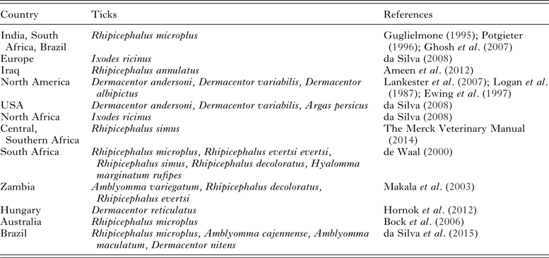
Transmission: Rhipicephalus, Ixodes, Dermacentor and Amblyomma genera of ticks are the important vectors of Anaplasma species (Table 8). A total of 17 species of ticks have been experimentally proved to transmit A. marginale (The Merck Veterinary Manual, 2014). Rhipicephalus microplus is the major vector of A. marginale in various countries including India, South Africa, Brazil, Malaysia and Australia (Guglielmone et al. Reference Guglielmone1995; Potgieter, Reference Potgieter1996; Mirwan, Reference Mirwan2003; Ghosh et al. Reference Ghosh, Azhahianambia and Yadav2007; Table 8). Ixodes ricinus is the main vector in Europe; Dermacentor (D.) andesrsoni, D. variabilis and D. albipictus in North America (Table 8). Some tick species or genotypes are selective for the transmission of specific A. marginale strains (Scoles et al. Reference Scoles, Broce, Lysyk and Palmer2005). Intrastadial and interstadial (one stage to another stage) transmission is reported while transovarial transmission does not occur (Kocan et al. Reference Kocan, Goff, Stiller, Claypool, Edwards, Ewing, Hair and Barron1992a , Reference Kocan, Stiller, Goff, Claypool, Edwards, Ewing, Mcguire, Hair and Barron b ). In endemic region, animal-to-animal transmission usually results from infested mother to suckling calves (Kocan, Reference Kocan1994). Experimental and clinical studies have proved that infected erythrocytes move from infected cows to their offspring through placenta (Zaugg, Reference Zaugg1985; Potgieter and Van Rensburg, Reference Potgieter and Van Rensburg1987; Salabarria and Pino, Reference Salabarria and Pino1988; Costa et al. Reference Costa, de Magalhães, de Oliveira, Carvalho, de Almeida, Machado and Munhoz2015; Silvestre et al. Reference Silvestre, Silveira, Meneses, Facury-Filho, Carvalho and Ribeiro2016). Barbosa Da Silva reported transplacental transmission using indirect enzyme-linked immunosorbent assay (ELISA) and nested PCR in calves (Barbosa da Silva et al. Reference Barbosa da Silva, de Santana Castro and Fonseca2014a ). Blood-contaminated needles, ear tagging devices, castration, dehorning equipments and nose tongs contribute in mechanical transmission within the herd (Kocan et al. Reference Kocan, de la Fuente, Guglielmone and Melendez2003; The Merck Veterinary Manual, 2014). Biting flies were not able to experimentally transmit infection (Scoles et al. Reference Scoles, Broce, Lysyk and Palmer2005, Reference Scoles, Miller and Foil2008). Recently Haematopinus tuberculatus, sucking lice have been reported as the potential vector of A. marginale in buffaloes (da Silva et al. Reference da Silva, Lopes, Diaz, Tonin, Stefani and Araújo2013). The transplacental and mechanical routes do not contribute much in the transmission of A. marginale infection.
Detection of Anaplasma marginale in different tick species

Diagnosis
Anaemia, pale to yellow mucous membranes, brown lymph nodes, jaundice, enlarged spleen and distended gall bladder are important clinical findings of BA (The Merck Veterinary Manual, 2014). Diagnosis is based on giemsa stained blood smear (Fig. 4), serology, nucleic acid detection and sub-inoculation of blood into splenectomized calf. Giemsa stained blood smear examination is inexpensive, rapid and most widely used method for the diagnosis of tick-borne diseases (Salih et al. Reference Salih, Hassan and El Hussein2007), but this technique is not suitable in persistently infected carrier animals with low levels of parasitaemia (Kocan et al. Reference Kocan, de la Fuente, Blouin, Coetzee and Ewing2010b ).
Anaplasma marginale in erythrocytes of cattle (The Merck Veterinary Manual, 1998); magnification 100×.

There are problems of sensitivity, reproducibility, interpretation and non-specific reactions associated with these serological tests (OIE, 2015). The results of serological assays should be interpreted with caution because of possible cross-reactivity implications. Sub-inoculation of blood from suspected into splenectomized calf is a gold standard for the demonstration of Anaplasma-free status (Coetzee et al. Reference Coetzee, Apley and Kocan2006). But this method is expensive, time consuming and may not be good for validation of assays.
World animal health organization recommended cELISA for serodiagnosis of anaplasmosis in cattle. The cELISA is cost effective test and also commercially available as diagnostic kit “Anaplasma Antibody Test Kit” (cELISA); VMRD Inc., Pullman, WA, USA for the detection of early and persistent Anaplasma infection. Recently improved cELISA, uses monoclonal antibody with 100% sensitivity and 99·7% specificity at 30% cutoff (Chung et al. Reference Chung, Wilson, Bandaranayaka-Mudiyanselage, Kang, Adams, Kappmeyer, Knowles, McElwain, Evermann, Ueti, Scoles, Lee and McGuire2014).
Card agglutination test is sensitive, may be performed in laboratory as well as under field conditions and results can be obtained within few minutes. There are problems of non-specific reactions, reproducibility and interpretation of the test results as well as preparation of the antigen which is the suspension of A. marginale which differs from batch to batch and laboratory to laboratory. Card agglutination and complement fixation tests have been replaced with ELISA for regional and international movement of cattle (OIE, 2008). Newly improved, competitive ELISA has obvious advantage because of greater sensitivity 100% and specificity 99·7% (Chung et al. Reference Chung, Wilson, Bandaranayaka-Mudiyanselage, Kang, Adams, Kappmeyer, Knowles, McElwain, Evermann, Ueti, Scoles, Lee and McGuire2014).
Recently developed nested PCRs based on groEL, 16SrRNA (longer sequence), msp1α, msp5, groEL and gltA genes to characterize A. marginale isolates. Ybañez and associates developed nPCR which is highly specific and sensitive with a detection limit of two copies per PCR (Ybañez et al. Reference Ybañez, Sivakumar, Battsetseg, Battur, Altangerel, Matsumoto, Yokoyama and Inokuma2013a ). Furthermore, (Bilgiç et al. Reference Bilgiç, Karagenç, Simuunza, Shiels, Tait, Eren and Weir2013) developed multiplex and single PCR assays for simultaneous detection of A. marginale (166 bp), Theileria annulata (312 bp) and Babesia bovis (265 bp) in cattle. The test is effective for single or simultaneous detection of anaplasmosis, tropical theileriosis and bovine babesiosis in field samples.
Previous molecular studies targeted 16S rRNA, msp1α, msp1β, msp4, msp5, gltA and heat-shock protein (groEL) genes for the detection and characterization of A. marginale isolates (Hornok et al. Reference Hornok, Elek, de la Fuente, Naranjo, Farkas, Majoros and Földvári2007; Bilgiç et al. Reference Bilgiç, Karagenç, Simuunza, Shiels, Tait, Eren and Weir2013; Ybañez et al. Reference Ybañez, Sivakumar, Battsetseg, Battur, Altangerel, Matsumoto, Yokoyama and Inokuma2013a , Reference Ybañez, Sivakumar, Ybañez, Ratilla, Perez, Gabotero, Hakimi, Kawazu, Matsumoto, Yokoyama and Inokuma b ; Belkahia et al. Reference Belkahia, Ben Said, Alberti, Abdi, Issaoui, Hattab, Gharbi and Messadi2015a ).
Anaplasmosis is the most imperative cause of hemolytic anaemia in cattle (Hofmann-Lehmann et al. Reference Hofmann-Lehmann, Meli, Dreher, Gönczi, Deplazes, Braun, Engels, Schüpbach, Jörger, Thoma, Griot, Stärk, Willi, Schmidt, Kocan and Lutz2004). The important feature of anaplasmosis is extra vascular hemolytic anaemia, results due to phagocytosis of parasitized erythrocytes in spleen and bone marrow (Jain, Reference Jain1993). The disease is divided into three stages i.e. prepatent phase, acute phase [41–47 days post infection (dpi)] and late phase (50–70 dpi). No change occurs in blood parameters during prepatent phase in A. marginale-infected cattle (Haider, Reference Haider1991) except transitory decrease in lymphocytes (Alfonso et al. Reference Alfonso, Medina, Fazzino and Caballero1996). Anaplasmosis significantly affect packed cell volume (Marufu et al. Reference Marufu, Chimonyo, Dzama and Mapiye2010). Increase in concentration of monocytes and neutrophils occur in later stages of the disease (Alfonso et al. Reference Alfonso, Medina, Fazzino and Caballero1996).
Hypochromic and macrocytic anaemia, increase in total white cell counts, reduced platelet counts, blood aspartate aminotransferase, serum bilirubin, glutamic dehydrogenase, gamma glutamyltransferase and blood urea nitrogen have been reported by Riond et al. (Reference Riond, Meli, Braun, Deplazes, Joerger, Thoma, Lutz and Hofmann-Lehmann2007) in Swiss cattle infected with A. marginale along with co-infection of other vector-borne pathogens. There is a positive correlation with the increase of parasitaemia and mean corpuscular fragility (MCF) and lactate dehydrogenase (LDH), while superoxide dismutase (SOD) had negative correlation with parasitaemia level (Nazifi et al. Reference Nazifi, Razavi, Mansourian, Nikahval and Moghaddam2008). Furthermore, increase of total protein, total bilirubin, alanine amino transferase has been reported during BA (Ashuma et al. Reference Ashuma, Singla, Kaur, Bal, Batth and Juyalm2013).
Treatment
Oxytetracycline has been approved for parenteral use for treatment and control. In addition, chlortetracycline is approved for use in continuous feed for the control of bovine anaplasmosis in the USA (FDA, 2009). Antimicrobial therapy is mainly focused to treat clinical disease and to minimize disease effects during vector season. Splenectomized calves experimentally infected with A. marginale were successfully treated with 1–3 intramuscular injections of oxytetracycline administered at 10–20 mg kg−1 body weight and doxycycline at the dose rate of 100 mg kg−1 intramuscular (Kuttler and Simpson, Reference Kuttler and Simpson1978). Chloramphenicol and rolitetracycline had been proved effective in the treatment of clinical and carrier states of anaplasmosis (Sharma et al. Reference Sharma, Benerjee and Gautam1977).
Keeping Anaplasma free herds is one the important strategy for controlling bovine anaplasmosis. The carrier free status is required as per free trade policy for the movement of cattle between endemic and non-endemic regions of the world (Reinbold, Reference Reinbold2009).
There are conflicting reports on the chemosterilization of persistent infection. A recent study on carrier elimination, oxytetracycline (22 mg kg−1 IV once in a day for five days) and imidocarb dipropionate (5 mg kg−1 IM twice, 7 days apart) eliminated 86·67 and 73·33% of A. marginale infections, respectively; in adult naturally infected carrier cattle at 56th post-treatment day. The carrier clearance was confirmed by cELISA followed by sub-inoculation of blood in sero-negative splenectomized calves (Atif et al. Reference Atif, Khan, Khan, Ashraf and Avais2012b ). Chemosterilization of A. marginale carriers has been attained by oral chlortetracycline hydrochloride and injectable formulations of oxytetracycline antibiotics (Magonigle et al. Reference Magonigle, Renshaw, Vaughn, Stauber and Frank1975; Roby et al. Reference Roby, Simpson and Amerault1978; Kuttler, Reference Kuttler1983; Rogers and Dunster, Reference Rogers and Dunster1984; Kocan et al. Reference Kocan, de la Fuente, Blouin, Coetzee and Ewing2010b ; Reinbold et al. Reference Reinbold, Coetzee, Hollis, Nickell, Riegel, Olson and Ganta2010; Turse et al. Reference Turse, Scoles, Deringer, Fry and Brown2014).
ANAPLASMA PLATYS
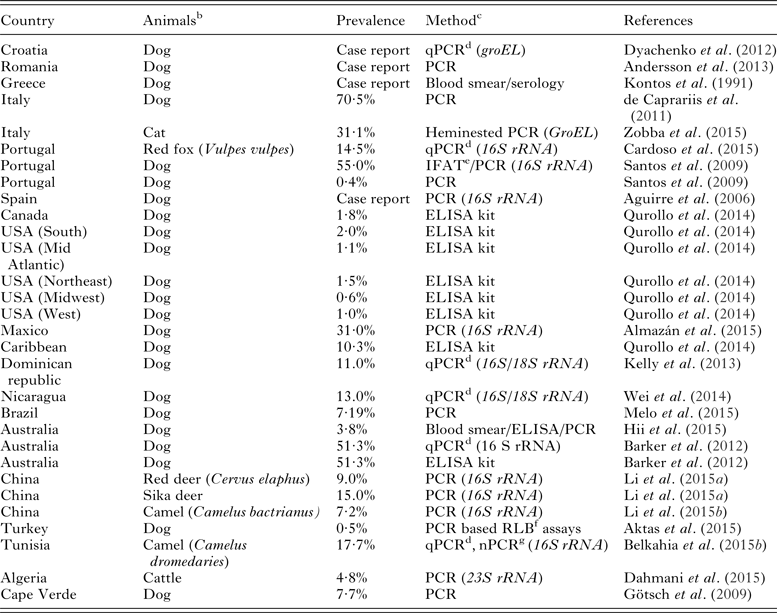
Anaplasma platys, a zoonotic obligate intracellular alphaproteobacteria replicates in platelets (Harvey et al. Reference Harvey, Simpson and Gaskin1978). In 1978, a clinical syndrome was first reported in a dog in Florida (USA) called as canine infectious cyclic thrombocytopenia (CICT) caused by Ehrlichia platys (now known as A. platys) (Harvey et al. Reference Harvey, Simpson and Gaskin1978; Dumler et al. Reference Dumler, Barbet, Bekker, Dasch, Palmer, Ray, Rikihisa and Rurangirwa2001). Anaplasma platys has been reported worldwide (Table 9). Prevalence in dogs from different European countries ranges from 0·4 to 70·5% using molecular methods (de la Fuente et al. Reference de la Fuente, Torina, Naranjo, Nicosia, Alongi, LaMantia and Kocan2006b ; Santos et al. Reference Santos, Alexandre, Sousa, Nuncio, Bacellar and Dumler2009; de Caprariis et al. Reference de Caprariis, Dantas-Torres, Capelli, Mencke, Stanneck, Breitschwerdt and Otranto2011; Table 9).
Detection of Anaplasma platys in vertebrate hosts from different countries a

a Detection of Anaplasma platys from 1991 to 2015.
b Organism detected in tissue or blood specimen.
c In parenthesis target gene has been mentioned.
d Real-time PCR.
e Immunofluorescence Antibody Test.
f Reverse line blot hybridization.
g Nested PCR.
Clinical findings
Incubation period in dog ranges from 7 to 14 days. After an experimental infection bacterium can be demonstrated in the peripheral blood after 8–15 days and develop severe thrombocytopenia. This recurs at 10 days interval. Later the cyclic pattern diminishes and thrombocytopenia level declines and completely resolves slowly. This cyclic infection includes low cyclic bacteremia and thrombocytopenia (Harvey, Reference Harvey and Greene2006).
Usually dogs remain asymptomatic. Fever, lethargy, anorexia, weight loss, anaemia, icterus, petechiae, nasal discharge, lymphadenopathy and lymphadenomegaly are important clinical signs of CICT. However, highly pathogenic strains of A. platys cause serious uveitis and pancytopenia after direct or autoimmune damage to platelets (Harvey et al. Reference Harvey, Simpson and Gaskin1978; Glaze et al. Reference Glaze, Gaunt and Babin1986; Huang et al. Reference Huang, Unver, Perez, Orellana and Rikihisa2005; Aguirre et al. Reference Aguirre, Tesouro, Ruiz, Amusategui and Sainz2006). Single-case clinical study showed bilateral uveitis and epistaxis in a dog. The clinical findings mentioned in earlier studies did not rule out concurrent infections using molecular tools (Harrus et al. Reference Harrus, Aroch, Lavy and Bark1997; Sainz et al. Reference Sainz, Amusategui and Tesouro1999; Alleman and Heather, Reference Alleman and Heather2008; de Caprariis et al. Reference de Caprariis, Dantas-Torres, Capelli, Mencke, Stanneck, Breitschwerdt and Otranto2011; Dyachenko et al. Reference Dyachenko, Pantchev, Balzer, Meyersen and Straubinger2012). Concurrent infections with E. canis have been reported because R. sanguineus ticks transmit both the pathogens (The Merck Veterinary Manual, 2014). Mostly A. platys and Babesia vogeli concurrent infections have been reported (Otranto et al. Reference Otranto, Testini, Dantas-Torres, Latrofa, Diniz, Caprariis, LIA, Mencke, Stanneck, Capelli and Breitschwerdt2010).
Epidemiology
Anaplasma platys has been detected in dog, cat, cattle, camel and wild ruminants (Lima et al. Reference Lima, Soares, Ramos, Araujo, Ramos, Souza, Faustino and Alves2010; Qurollo et al. Reference Qurollo, Balakrishnan, Cannon, Maggi and Breitschwerdt2014; Dahmani et al. Reference Dahmani, Davoust, Benterki, Fenollar, Raoult and Mediannikov2015; Table 9). The organism has not been detected in wild carnivores i.e. foxes, jackals and wolves (Sainz et al. Reference Sainz, Roura, Miró, Estrada-Peña, Kohn, Harrus and Solano-Gallego2015). Age, sex or breed is not the important determinants for CICT (Sainz et al. Reference Sainz, Roura, Miró, Estrada-Peña, Kohn, Harrus and Solano-Gallego2015). Anaplasma platys infection has been reported in two human families in the USA and two individuals in Venezuela (Maggi et al. Reference Maggi, Mascarelli, Havenga, Naidoo and Breitschwerdt2013; Arraga-Alvarado et al. Reference Arraga-Alvarado, Qurollo, Parra, Berrueta, Hegarty and Breitschwerdt2014).
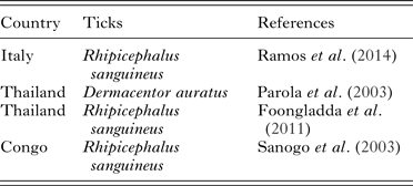
Transmission: The R. sanguineus is the principal vector in Europe and all around the world (Sainz et al. Reference Sainz, Roura, Miró, Estrada-Peña, Kohn, Harrus and Solano-Gallego2015; Table 10). However, the experimental transmission has not yet been proven (Simpson et al. Reference Simpson, Gaunt, Hair, Kocan, Henk and Casey1991). Other likely route of transmission is blood transfusion (Gaunt et al. Reference Gaunt, Beall, Stillman, Lorentzen, Diniz and Chandrashekar2010; Table 11). Screening of donor blood before transfusion is warranted because of associated zoonotic potential.
Molecular detection of Anaplasma platys in different tick species

The characteristic of Anaplasma species (Rickettsiales, Anaplasmataceae)

Diagnosis
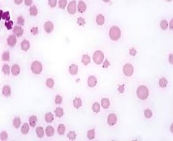
Anaplasma platys morulae in platelets can be detected in stained blood smear but this method is less sensitive at persistent or low level of infection (Shaw et al. Reference Shaw, Binns, Birtles, Day, Smithson and Kenny2005; Fig. 5). This bacterium can also be found in megakaryocytes (de Tommasi et al. Reference de Tommasi, Baneth, Breitschwerdt, Stanneck, Dantas-Torres, Otranto and de Caprariis2014), but the usefulness of bone marrow cytology needs to be evaluated. The serological test should be interpreted with caution. Positive serological tests do not mean active infection but there might be the case in which infection has been resolved. Usually serological test followed by molecular diagnosis (PCR/DNA sequencing) is recommended that indicate active infection. The PCR is not a definite diagnosis for persistently infected dogs with low level of bacteremia because of the cyclic peaks and lowering of the organism (Alleman and Heather, Reference Alleman and Heather2008). Spleen or peripheral blood with EDTA is required for PCR.
Anaplasma platys in dog's platelet (Sainz et al. Reference Sainz, Roura, Miró, Estrada-Peña, Kohn, Harrus and Solano-Gallego2015); magnification 100×.

Moreover, concurrent infections with other vector-borne infectious agents such as E. canis, Rickettsia conorii or B. vogeli may aggravate the clinical disease along with abnormal laboratory findings (Santos et al. Reference Santos, Alexandre, Sousa, Nuncio, Bacellar and Dumler2009; Gaunt et al. Reference Gaunt, Beall, Stillman, Lorentzen, Diniz and Chandrashekar2010; de Caprariis et al. Reference de Caprariis, Dantas-Torres, Capelli, Mencke, Stanneck, Breitschwerdt and Otranto2011). Consequently, when an infection of one specific pathogen is diagnosed, a complete work-up is required to rule out concurrent infections with other vector-transmitted pathogens (Sainz, Reference Sainz, Miró and Solano-Gallego2011). Fifty percent of Ehrlichia canis infected dogs show a titre of A. platys infection during concurrent infection but no cross-reactivity exist between these pathogens (Stillman et al. Reference Stillman, Monn, Liu, Thatcher, Foster, Andrews, Little, Eberts, Breitschwerdt, Beall and Chandrashekar2014; The Merck Veterinary Manual, 2014). However, a cross-reactivity has been reported between A. phagocytophilum and A. platys (Solano-Gallego et al. Reference Solano-Gallego, Trotta, Razia, Furlanello and Caldin2006; Chandrashekar et al. Reference Chandrashekar, Mainville, Beall, O'connor, Eberts, Alleman, Gaunt and Breitschwerdt2010; Zobba et al. Reference Zobba, Anfossi, Pinna Parpaglia, Dore, Chessa, Spezzigu, Rocca, Visco, Pittau and Alberti2014, Reference Zobba, Anfossi, Visco, Sotgiu, Dedola, Pinna Parpaglia, Battilani, Pittau and Alberti2015).
Treatment
Anaplasma Platys infections can be treated with doxycycline at the dose rate of 5–10 mg kg−1 q for 8–10 days (Sainz et al. Reference Sainz, Roura, Miró, Estrada-Peña, Kohn, Harrus and Solano-Gallego2015). Steroids are only indicated particularly when immune-mediated complications encounter. In these conditions, prednisolone/prednisolone should be given at the dose rate of 0·5–2 mg kg−1 day−1. Usually, most of dogs recover after acute infection with effective treatment (Wen et al. Reference Wen, Rikihisa, Mott, Greene, Kim, Zhi, Couto, Unver and Bartsch1997; Harrus et al. Reference Harrus, Waner, Aizenberg and Bark1998; Breitschwerdt et al. Reference Breitschwerdt, Hegarty and Hancock1998). Thrombocytopenia characteristically disappears about 1 week after the initiation of therapy (Chang et al. Reference Chang, Su and Pan1997).
Control
Currently, no commercial vaccines are available for A. platys. Ticks control plays a key role in infection prevention. The R. sanguineus ticks are mainly located indoors in cooler regions and found outdoor in warmer areas. Outdoor populations of R. sanguineus are active at 10–12 °C. Collar, pour-on, or spot-on acaricidal products can be selected based upon preference and therapeutic needs. Immediate response is the elimination of attached ticks responsible for pathogen transmission. To maximize the effectiveness, the acaricide must be applied at the recommended dosage according to manufacturer's instructions (Sainz et al. Reference Sainz, Roura, Miró, Estrada-Peña, Kohn, Harrus and Solano-Gallego2015). Tick control methods should be implemented according to local and environmental conditions. For appropriately control knowledge of tick seasonality, ecology, stages and status of multiple tick infestations is necessary.
PREVENTION AND CONTROL OF ANAPLASMOSIS
Acaricides are mostly used for the control of ticks. Drug resistance and residues are the problems associated with the use of acaricides. Housing should not favourable the growth and multiplication of vector ticks. Establishment of buffer zone between domestic animal housings and vegetations can help to control vectors (Jonsson et al. Reference Jonsson, Mayer, Matschoss, Green and Ansell1998). Anti-tick vaccines would be a good alternative to control infection. These vaccines are environment friendly; decrease the occurrence of tick-borne diseases by reducing tick infestation, and limit acaricide use (Graf et al. Reference Graf, Gogolewski, Leach-BING, Sabatini, Molento, Bordin and Arantes2004; de la Fuente et al. Reference de la Fuente, Almazán, Blouin, Naranjo and Kocan2006c , Reference de la Fuente, Moreno-C.I.D., Canales, Villar, de la Lastra, Kocan, Galindo, Almazán and Blouin2011). Understanding vector pathogen interactions can play an important role for the control of vector-borne diseases. Tick vaccines based on tick proteins Q38, SUB, SILK, TROSPA, BM86/BM95, 64P interfere with tick vector competence resulting in lower tick infection and tick-borne diseases (Labuda et al. Reference Labuda, Trimnell, Lľcková, Kazimírová, Davies, Lissina, Hails and Nuttall2006; Willadsen, Reference Willadsen2006; Zivkovic et al. Reference Zivkovic, Esteves, Almazán, Daffre, Nijhof, Kocan, Jongejan and de la Fuente2010; Antunes et al. Reference Antunes, Galindo, Almazán, Rudenko, Golovchenko, Grubhoffer, Shkap, do Rosário, de la Fuente and Domingos2012; Hajdǔsek et al. Reference Hajdǔsek, Síma, Ayllón, Jalovecká, Perner, de la Fuente and Kopáček2013; Merino et al. Reference Merino, Antunes, Mosqueda, Moreno-Cid, de la Lastra, Rosario-Cruz, Rodríguez, Domingos and de la Fuente2013). Similarly, Akirin and Subolesin proteins have the potential candidate for universal anti-tick vaccine against major vector species and vector-borne diseases (de la Fuente et al. Reference de la Fuente, Moreno-CID, Galindo, Almazan, Kocan, Merino, Perez de la Lastra, Estrada-Peña and Blouin2013). Recently, vector-pathogen vaccine comprising A. marginale MSP1a and tick Subolesin proteins showed efficacy in lowering tick infestation, lower weight of female ticks (Boophilus microplus) and reduced A. marginale infection in cattle (Torina et al. Reference Torina, Moreno-CID, Blanda, Fernández de Mera, de la Lastra, Scimeca, Blanda, Scariano, Briganò, Disclafani, Piazza, Vicente, Gortázar, Caracappa, Lelli and de la Fuente2014). Immunization of high-risk human population and animal reservoirs would help in the control of anaplasmosis.
The universal vaccine for any Anaplasma specie is not yet available which is effective against diverse strains. Blood based A. marginale subsp. centrale has been used as live vaccine in South Africa, Australia, Israel and several other South American countries for more than three decades against bovine anaplasmosis (Kocan et al. Reference Kocan, de la Fuente, Step, Blouin, Coetzee, Simpson, Genova and Boileau2010a , Reference Kocan, de la Fuente, Blouin, Coetzee and Ewing b ). Anaplasma marginale subsp. centrale vaccine has proved effective in preventing clinical disease against field strains. This blood-based vaccine had the potential to transmit other blood-borne pathogens during inoculation (Rogers et al. Reference Rogers, Dimmock, de Vos and Rodwell1988). Because of inherent danger this vaccine has not been permitted by the European Union and USA (Rogers et al. Reference Rogers, Dimmock, de Vos and Rodwell1988). Studies have shown that vaccination caused mild clinical signs after vaccination and protect severe disease, whereas some studies have suggested that A. centrale strain provided slight to no protection (Abdala et al. Reference Abdala, Pipano, Aguirre, Gaido, Zurbriggen, Mangold and Guglielmone1990). Recently, A. centrale has been cultured in vitro in tick cell lines with the possibility of safer vaccine for bovine anaplasmosis (Bell-Sakyi et al. Reference Bell-Sakyi, Palomar, Bradford and Shkap2015). Various proteins have been recommended as vaccine candidates but the main issue associated with development of successful vaccine is the selection of suitable conserved antigen, existence of different variants, antigenic variations and lack of cross protection studies (Stuen et al. Reference Stuen, Granquist and Silaghi2013b ).
Control measures vary from region to region. Prevention and control mainly focus on vector control, use of antibiotics, breeding Anaplasma-free herds, prevention of mechanical transmission, use of live and killed vaccines. Detection, elimination and chemosterilization policy may be launched (Reinbold et al. Reference Reinbold, Coetzee, Hollis, Nickell, Riegel, Olson and Ganta2010) where no endemic stability exist. However, this approach is costly and may not work for the poor livestock farmers. Testing of animals at 3 weeks interval, vector control and purchase of animals from Anaplasma-free areas is a good option to prevent the entry of infected animals.
COMMON FEATURES OF ANAPLASMA SPECIES
-
(1) Persistent infection is the common sequel after acute disease.
-
(2) Serologic cross reactivity; because MSP5 proteins is conserved among all Anaplasma species.
-
(3) Breed resistance, biological, mechanical and transplacental transmission potentials.
-
(4) The 16SrRNA gene based classification and similarity in family Anaplasmataceae.
-
(5) A. phagocytophilum and A. platys have blood associated zoonotic potential.
-
(6) Major common prevention and control strategies [References are mentioned in the text above. Comparative features of Anaplasma species are mentioned in Table 11].
CONCLUSION
All age groups are susceptible but prevalence increases with age. Movement of young animals from tick-free area to tick-infested areas is the major risk for anaplasmosis in animals. High-risk outdoor tick exposure activities and blood transfusion are important risk factor for HGA. These organisms attribute a reasonable degree of similarity. No successful vaccine is available for any Anaplasma specie, effective against ecologically diverse strains. Identification of novel antigenic parts had been the major effort for the development of a universal Anaplasma vaccine. Major problem associated with development of a successful vaccine is the selection of suitable conserved antigen, existence of different variants, antigenic variations and lack of cross-protection studies (Stuen et al. Reference Stuen, Granquist and Silaghi2013b ). The advances in epidemiology, biotechnology, genetic engineering, vector ecology, immunology, cell culture, clinical, experimental and longitudinal studies should be utilized to reveal ecology and infection biology for effective prevention and control of anaplasmosis in humans, domestic and wild animals.
ACKNOWLEDGEMENTS
I am grateful to Dr Georg Gerhard Duscher from Institute of Parasitology, Department of Pathobiology, University of Veterinary Medicine, Vienna, Austria for valuable comments on anaplasmosis.
FINANCIAL SUPPORT
This review paper received no financial support from any funding agency.