Introduction
The micropyle is the entrance through which spermatozoa without acrosomes pass for fertilization. Although the numbers vary slightly depending on the taxa, teleost fishes generally have one micropyle in the region of the animal pole (Coward et al., Reference Coward, Bromage, Hibbitt and Parrington2002). According to previous studies and despite few differences between closely related species, morphological characteristics of the micropyle can be used as an important indicator for taxonomic purposes (Riehl, Reference Riehl1980; Hirai and Yamamoto, Reference Hirai and Yamamoto1986; Chen et al., Reference Chen, Shao and Yang1999) because they differ according to taxa. This fundamental difference can be described as the most basic of barriers to prevent cross-breeding. Nevertheless, there is a phenomenon in which this barrier collapses in the natural world, leading to hybridization.
Hybrids are individuals resulting from hybridization between different species, meaning by the combination of different genes. In some cases hybrids play an important role in the sympatric speciation process (Chevassus, Reference Chevassus1983; Barton and Hewitt, Reference Barton and Hewitt1985; Abbott et al., Reference Abbott, Albach, Ansell, Arntzen, Baird, Bierne, Boughman, Brelsford, Buerkle, Buggs, Butlin, Dieckmann, Eroukhmanoff, Grill, Cahan, Hermansen, Hewitt, Hudson, Jiggins, Jones, Keller, Marczewski, Mallet, Martinez-Rodriguez, Möst, Mullen, Nichols, Nolte, Parisod, Pfennig, Rice, Ritchie, Seifert, Smadja, Stelkens, Szymura, Väinölä, Wolf and Zinner2013). Natural hybrids are often found to exist in limited populations in their native environments and generally are not fertile; hybrids alone cannot maintain a population and their traits cannot be passed on to the next generation. One of the results of common reproductive isolation, especially among species presenting with post-zygotic isolation, is hybrid sterility (Scopece et al., Reference Scopece, Widmer and Cozzolino2008). Obviously, there is also a pre-zygotic mechanism that prevents hybrid formation.
The genus of Cobitis hybrids distributed in Korea is derived from two parental groups of Cobitis fishes, Cobitis hankugensis and Iksookimia longicorpa (first reported as C. longicorpus by Kim et al. (Reference Kim, Choi and Nalbant1976)). They maintain their lineage of almost all females with the extremely rare males (Park et al., Reference Park, Kim and Ko2011). Three types of the hybrid are identified through microsatellite loci analysis [i.e. one diploid hybrid: one C. hankugensis locus with one I. longicorpa locus (HL), two triploid hybrids: one C. hankugensis locus with two I. longicorpa locus (HLL) and two C. hankugensis locus with one I. longicorpa locus (HHL)]. In the above unisexual fishes, the rare males that do occur have been considered sterile with low counts of sperm cells. Such an abnormal reproductive mode has attracted attention. According to previous research, the C. hankugensis–I. longicorpa hybrid forms through the normal hybridogenesis process and then combines with a haploid male through a premeiotic endomitosis mechanism to form a triploid. These triploids generate haploid eggs after heterogeneous allele removal and are fertilized with the male genome. For homozygous, intact C. hankugensis or I. longicorpa, when heterozygous, they form the HL-type diploid C. hankugensis–I. longicorpa complex again (Lee, Reference Lee1992, Reference Lee1995; Ko, Reference Ko2009; Yun, Reference Yun2017). To date, research on these unisexual fishes in the wild has tended to focus only on this process, while the structure of the micropyle remains poorly understood. Therefore, this study for Korean unisexual fishes sought to understand and compare the fine structure of the micropyle, given that it may be useful for identifying species and hybrids as a taxonomic characteristic.
Materials and methods
Ethics, sample collection and identification
All of the fish in this study were treated based on the ‘Ethical review process by Chonbuk National University Institutional Animal Care and Use Committee (JBNUIACUC)’.
Specimens of I. longicorpa, C. hankugensis and their hybrids were collected during their spawning period in Ram Stream, Nakdong River basin, Namwon, Jeollabuk-do, April to June 2019 (35°27′26′′N, 127°36′26′′E) with a cast net (6 × 6 mm mesh). In total, 25 individuals were collected: I. longicorpa (n = 5), C. hankugensis (n = 5), HL type (n = 5), HLL (n = 5) and HHL type (n = 5). The identification of the hybrid type followed the allele dosage effects using microsatellite loci (Yun, Reference Yun2017).
Egg collection and ultrastructural analysis
Under MS-222 (Sigma Aldrich Co., St. Louis, USA) anaesthesia conditions, the oocyte of each fish was collected under gentle pressure through the abdomen and water, blood and urine were carefully removed to avoid contamination. For scanning electron microscopy observation, the collected oocytes (30 eggs from five females of each species) were fixed in 2.5% glutaraldehyde solution with 0.1 M phosphate buffer at pH 7.4. Then the eggs were fixed in 1% osmium tetroxide solution with 0.1 M phosphate buffer for 6 h and dehydrated using a graded series of ethanol and tert-butyl alcohol and freeze dried using evaporation under vacuum (VFD-21S, Vacuum Device Co., Ltd, Ibaragi, Japan). The dried samples were coated with osmium tetroxide using ion sputtering (HPC-1SW, Vacuum Device Inc., Tokyo, Japan) and then observed under a field emission scanning electron microscope (Carl Zeiss, SUPRA40VP, Germany). The evaluation of the number of attachments over the egg surface was measured 10 times for each egg in the range 10 × 10 µm.
Statistical analysis
Egg diameter and morphometric values of micropylar components were analyzed using a one-way analysis of variance (ANOVA) and Tukey’s post hoc test in SPSS v.22. The level of significance was set at 5% (P < 0.05). Data are presented as mean and standard deviation.
Results
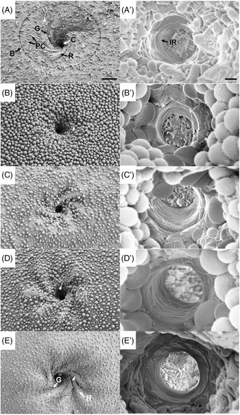
In a scanning electron microscopic observation, all unfertilized eggs had only a single micropyle complex composed of a funnel-shaped micropylar region and a manhole-shaped micropylar canal on its innermost. Repetitive rotational grooves and ridges were observed in the other four species of micropylar regions except for C. hankugensis. In all species, the micropylar canal has grooves and ridges down to the egg surface. The direction of the spirals in the micropylar region and the canal was counterclockwise from the outer boundary of the micropyle towards the centre (Figure 1).
Schematic diagram of micropyle complex of Iksookimia longicorpa, Cobitis hankugensis (exceptionally no spiral pattern in the micropylar region), and their hybrids. C, micropyle canal; G, groove; IR, inner ridge; R, outer ridge.

However, their detailed morphological characteristics differed slightly by type. The unfertilized eggs of I. longicorpa were an average of 1.36 mm in diameter, similar in size to those of the HLL hybrid (1.35 mm). C. hankugensis with 1.11 mm was the smallest. The diameters of HL type and HHL type were 1.43 mm and 1.21 mm, respectively (Table 1).
Comparison of microstructural characters of the unfertilized eggs of in Iksookimia longicorpa, Cobitis hankugensis and their natural hybrids. Values within each column with different letters are significantly different (P < 0.05)

The micropylar region was covered with numerous pore canals being surrounded by attachments that showed villous and granular forms (Figure 2 and Table 1). I. longicorpa and the HLL hybrid had a villus type and the number of villi was in the ranges 6–9 and 15–19, respectively. In addition, the other three species, C. hankugensis, HL hybrid and HHL hybrid, had a granular type and were 20–27, 13–19 and 20–27 in number, respectively. Unlike the other two types, in particular, the pore canals of C. hankugensis were barely exposed due to compacted arranged granules (Figure 2B).
Scanning electron microscopy of micropylar region (A–E) and pore (A′–E′) in Iksookimia longicorpa (A, A′), Cobitis hankugensis (B, B′) and their natural hybrids, HL (C, C′), HHL (D, D′) and HLL (E, E′). The white triangle represents surface attachment, where (A) and (E) are villus type, and (B), (C) and (D) are granule type. B, micropylar region boundary; C, micropyle canal; G, groove; IR, inner ridge; PC, micropyle pore canal; R, outer ridge. Bars indicates 10 µm (A–E) and 1 µm (A′–E′).

Inside the micropylar region, counterclockwise spirals formed at least four to eight grooves in four species: I. longicorpa, HL type, HHL type and HLL type. Interestingly, however, for C. hankugensis, these spiral structures were not observed. Their absence may be due the many granules covering the entire egg surface as well as the micropylar region, but the surface that was covered with the granules was slightly curved and traces of grooves could be presumed. In addition, the multilayered grooves of the inner canal that were not fully connected were observed equally in all species (Figure 2A′–E′ and Table 1).
Micropylar regions were easily distinguished by the boundary where the chorion sank inward abruptly (Figure 2A). The HLL hybrid had the largest diameter of 49 µm and the C. hankugensis was the smallest at 35.36 µm. I. longicorpa and the HL hybrid were similar, 38.46 µm and 38.72 µm, respectively, while the diameter of the HHL hybrid was 44.93 µm (Table 1).
The circular opening of the canal was placed in the centre of the funnel. In contrast with the egg diameter, the micropylar canal diameter was the smallest in I. longicorpa (3.38 µm) and the C. hankugensis (3.56 µm) was larger than that of I. longicorpa. The largest species were the HHL hybrid (4.37 µm), followed by HL and HLL at 3.98 µm and 4.01 µm, respectively (Table 1).
Upon analysis of the ratio of the size of the micropylar canal to the egg diameter, I. longicorpa, HL hybrid and HLL hybrid were less than 0.3% and both C. hankugensis and HHL hybrid were greater than 0.3%, indicating that the size of the micropylar canal was relatively larger than that of the other three species (Table 1).
Discussion
In the evolution of vertebrates, fish have developed distinctive reproductive strategies such as the micropyle for optimization of mass spawning and oviparity. These processes can be found by backtracking from Teleostei to Agnatha. The hagfish, Eptatretus burgeri, has been reported to have a micropylar-like structure and spermatozoa containing acrosomes, which are predominantly observed in mammals (Morisawa, Reference Morisawa1999). Acrosomes are also observed in sturgeon, a ray-finned fish with micropyles varying in number from 2 to 52 (Ginsburg, Reference Ginsburg1968; Debus et al., Reference Debus, Winkler and Billard2008; Psenicka et al., Reference Psenicka, Rodina and Linhart2010). However, among teleosts, the largest group of fish comprising 96% of fish species (Nelson, Reference Nelson2006), a single spermatozoon without an acrosome passes through a single micropyle and fuses with the egg. This is supported by a series of excellent reviews published continuously since the 1950s (Yamamoto, Reference Yamamoto1952; Riehl and Schulte, Reference Riehl and Schulte1977; Hart, Reference Hart1990; Coward et al., Reference Coward, Bromage, Hibbitt and Parrington2002). In scanning electron microscope observation, it was confirmed that not only do the relatively recently differentiated cobitid fish have one micropyle [30–35 million years ago (MYA); Slechtová et al., Reference Slechtová, Bohlen and Perdices2008], but that C. hankugensis and I. longicorpa and their hybrids do as well, a characteristic typical of the teleost.
Studies on the morphological characteristics of micropyles have distinguished them into several types. Riehl and Kock (Reference Riehl and Kock1989) focused on the size of the micropylar pit and canal: deep pit and a short canal (type I), flat pit and a longer canal (type II), only canal without a pit (type III) and two pits and a short canal (type IV). In a more recent study, Yanagimachi et al. (Reference Yanagimachi, Harumi, Matsubara, Yan, Yuan, Hirohashi, Iida, Yamaha, Arai, Matsubara, Andoh, Vines and Cherr2017) classified these into three types based on the form in which the micropylar region is depressed: flat or slightly depressed micropylar region with manhole-like canal (type I), flat or slightly depressed micropylar region with funnel-like canal (type II) and a deeply depressed micropylar region such as a sinkhole or one with distinct grooves with a short canal (type III). In this study, we demonstrated the micropyle structure of all fishes with the same appearance with a shallow funnel-type micropylar region and a short manhole-type canal at the centre. Although our results did not correctly belong to the previous classification scheme, they were most similar to the type III suggested by Yanagimachi et al. (Reference Yanagimachi, Harumi, Matsubara, Yan, Yuan, Hirohashi, Iida, Yamaha, Arai, Matsubara, Andoh, Vines and Cherr2017).
The grooves situated on the surface of the micropylar region guide sperm into the micropylar canal, this was mainly observed in species corresponding to the type III proposed by Yanagimachi et al. (Reference Yanagimachi, Harumi, Matsubara, Yan, Yuan, Hirohashi, Iida, Yamaha, Arai, Matsubara, Andoh, Vines and Cherr2017) and their effectiveness has already been proven (Amanze and Iyengar, Reference Amanze and Iyengar1990). Although this varies depending on the species, I. longicorpa and three hybrids that received its genes had similar counterclockwise spiral structures, while in C. hankugensis spirals were absent. These spiral grooves are very distinctive even in the sister groups: Misgurnus anguillicaudatus has irregular grooves with sinkhole-shaped micropylar regions (Yanagimachi et al., Reference Yanagimachi, Harumi, Matsubara, Yan, Yuan, Hirohashi, Iida, Yamaha, Arai, Matsubara, Andoh, Vines and Cherr2017) and Kichulchoia multifasciata has only funnel-shaped micropylar regions without grooves and ridges (Kim et al., Reference Kim, Kim and Park2011). Only toadfish (Thalassophryne amazonica) and anabantoid fishes showed a spiral pattern around the micropyle, but the extent of the pattern was not limited to the micropylar region and appeared throughout the chorion (Riehl and Kokoscha, Reference Riehl and Kokoscha1993; Britz et al., Reference Britz, Kokoscha and Riehl1995; Britz and Toledo-Piza, Reference Britz and Toledo-Piza2012). Consequently, the morphological characteristics of the micropylar region, such as the spiral of I. longicorpa or the non-spiral of C. hankugensis observed in this study seem to be species specific. It is unclear what advantage is offered by the linear structures appearing in the majority of grooves and equally unclear what the presence of the spiral structures or the absence of the grooves identified in this study means. More considerable research on the topic is required. In addition, the existence of the grooves was dependent on the L gene, which can be seen for the grooves of I. longicorpa; for HLL with two L gene this was clearer than on HL and HHL hybrids. The structure of the micropyle canal is a comparatively well defined component among the micropylar complex. Funnel-type structures observed in some fish such as Oryzias (Iwamatsu et al., Reference Iwamatsu, Onitake, Matsuyama, Satoh and Yukawa1997) and Sparidae (Chen et al., Reference Chen, Shao and Yang1999) are intended to guide sperm to the centre of the canal. A similar manhole-type structure was also seen in this study.
On morphological analysis, the egg diameter of I. longicorpa was significantly similar to that of the HLL hybrid. There was no significant relationship, but the C. hankugensis and HHL hybrid egg diameters were smaller than in other examples, and the HL hybrid was the largest among them. This seems to be related to the reproductive mode of the C. hankugensis and I. longicorpa hybrids, in which the HL hybrid produces diploid eggs (HL) and HHL and HLL hybrids produce haploid eggs, H and L, respectively (Yun, Reference Yun2017). As genome size affects egg diameter (Hardie and Hebert, Reference Hardie and Hebert2004), the diploid egg, in the HL hybrid, had the largest diameter. The HLL hybrid with the haploid L gene had a size similar to that of the L and the HHL hybrids; the haploid H gene produced eggs of small size, as found for C. hankugensis. However, other metrics did not follow the ‘genome-size relationship’. Namely, diploid eggs showed smaller diameters in micropylar region and canal than triploids, HHL and HLL hybrids, which are believed to be the result of ‘cell size coordination by ploidy level’ were identified in many organisms (Mendell et al., Reference Mendell, Clements, Choat and Angert2008; Matsunaga et al., Reference Matsunaga, Katagiri, Nagashima, Sugiyama, Hasegawa, Hayashi and Sakamoto2013). Nevertheless, for canal diameter, the difference was not significant between species. As there has been no research on this so far, a new approach will therefore be needed to understand the relevance of the ploidy level and micropylar components in hybrids.
The attachment architecture of the egg membrane may play a role in adhesive or non-adhesive properties in demersal eggs (Mito, Reference Mito1979). Oryzias latipes are among well reported examples of this type of adhesive envelope with long filaments on its entire egg surface, and which attach its fertilized eggs to substrates such as seaweed. Moreover, the number and length of the adhesive envelope have been used to identify between O. latipes and O. sinensis (Hart et al., Reference Hart, Pietri and Donovan1984, Kim and Park, Reference Kim and Park2021). In addition, villus or granular types have been already known for the family Cobitidae, as well as teleost fishes, as a tool for species identification (Mazzini et al., Reference Mazzini, Callaini and Mencarelli1984; Park, Reference Park1996; Park and Kim, Reference Park and Kim1997). We confirmed in this study that there have been two types, a villus and a granular form. Interestingly, the shape and number of the attachment structures in Cobitis hybrids were dependent on their parental species, not being fixed in a regular form. In case the L gene was dominant, the villus type with a small number was shown, whereas, in the dominant H gene, the granule type with a large number appeared. Exceptionally, for the HL hybrid, the attachment appeared to be affected by the H gene in shape and by the L gene in number.
The micropyle canal is the part through which the head of spermatozoa directly passes and has a very important role in taxonomic terms (Ginsburg, Reference Ginsburg1968; Kobayashi and Yamamoto, Reference Kobayashi and Yamamoto1981). Interestingly, there are reviews about the relationship between the size of the micropylar canal and the head of spermatozoa. In Pleuronectes obscurus, Yanagimachi et al. (Reference Yanagimachi, Cherr, Matsubara, Andoh, Harumi, Vines, Pillai, Griffin, Matsubara, Weatherby and Kaneshiro2013) reported that the size of the sperm head was ∼87% of the micropyle canal. According to Solis Murgas et al. (Reference Solis Murgas, Paulino, Palhares, Miliorini, Alves and Oliveira Felizardo2017), the corresponding ratios of the four neotropical fishes, Prochilodus lineatus, Salminus brasiliensis, Piaractus mesopotamicus and Brycon orbignyanus, were reported to be 31%, 19%, 23% and 43%, respectively. As such, the ratio varied depending on the species, but the important thing was, of course, that the size of the micropyle canal was larger than the head of the sperm and is a fundamental feature of teleost fishes (Psenicka et al., Reference Psenicka, Rodina and Linhart2010). In previous research, we could obtain only limited information about the morphology of C. hankugensis and I. longicorpa hybrid sperm (Park et al., Reference Park, Kim and Ko2011). In that study, the size of the sperm head was 1.7 µm for I. longicorpa, 2.0 µm for C. hankugensis and 3.9 µm for the hybrid and when calculated in relation to the canal size of all species in this study, sperm of I. longicorpa was 39–50% and sperm of C. hankugensis was 46–59% and hybrid sperm were identified as 89–115%, respectively. Based on this, it was possible to carefully infer the answer as to why this hybrid population consisted mostly of females. Although that value was measured without the identification of three hybrids, in this population, male hybrids are basically unable to participate in the breeding competition as a result of their abnormally large diameter spermatozoa head.
Consequently, given that research on unisexual fishes showing polyploidy has focused mainly on genetic aspects, results based on micropyle may be used as another key for identifying and estimating hybrid populations from generation to generation.
In conclusion, the main finding of this study is the basic morphological characteristics on the micropyle of the I. longicorpa, C. hankugensis and their natural hybrids. They all have in common a single micropyle composed of a funnel-shaped micropylar region that can guide sperm to the centre of the micropyle with a manhole-type micropylar canal. The groove patterns with their counterclockwise spirals in the micropylar region were species specific and, moreover, C. hankugensis lacked the groove. These differences suggested a strong possibility for the participation of the L gene in the manifestation of the groove: I. longicorpa and HLL hybrid with two sets of L genes showed more distinct grooves than did HL and HHL hybrids with one set of L genes. The egg diameter, which is directly affected by the genome size, was the largest in the HL hybrid, which forms diploid eggs and in other somatic characteristics of the micropyle; the values were relatively high in triploid hybrids. Consequently, I. longicorpa had a large egg diameter with a small micropyle canal, whereas C. hankugensis had a small egg diameter with a relatively large micropyle canal. In addition, their hybrids showed the intermediate values of the parent species, being particularly dependent on the ploidy of the egg. Now, in addition to the many studies that have been performed on unisexual fishes, this type of approach may be helpful for obtaining insight into determining polyploidy or complicated taxa.
Data availability
Research data are not shared.
Competing interest
The authors declare no conflict of interest.