Introduction
Rhino-orbital-cerebral mucormycosis was a rare disease in the pre-coronavirus disease 2019 (Covid-19) era. It has been known to cause disease in immunosuppressed individuals. However, with the onset of the recent Covid-19 pandemic, and the rampant use of steroids with subsequent hyperglycaemia in diabetic patients, our country has witnessed a significant rise in the numbers of patients with rhino-orbital-cerebral mucormycosis. Studies have reported that the orbit is the most common site for extrasinus involvement of mucormycosis.Reference Sreshta, Dave, Varma, Nair, Bothra and Naik1,Reference Therakathu, Prabhu, Irodi, Sudhakar, Yadav and Rupa2
The imaging findings in such patients are often helpful in diagnosing early disease, which can be lifesaving, as the disease has high morbidity and mortality owing to the rapid progression and angio-invasive nature of the causative fungi. Often, subtle signs of orbital invasion through natural pathways can be seen on imaging scans, long before the advent of frank bone destruction or onset of ocular symptoms. It is imperative for us to actively examine these particular sites of involvement, in order to diagnose the condition early and delineate its extension, which can greatly aid surgical management. This pictorial review illustrates the various pathways of spread of mucormycosis to the orbit and the relevant imaging anatomy.
Clinical features
Early non-ocular signs and symptoms include facial pain, headache, fever, epistaxis, nasal discharge, nasal mucosa ulceration and crusting.Reference Mukherjee, Raichura and Alam3,Reference Yohai, Bullock, Aziz and Markert4 Common ophthalmic manifestations include decreased vision, proptosis, ophthalmoplegia, and other conditions such as chemosis, ptosis and corneal anaesthesia.Reference Mukherjee, Raichura and Alam3,Reference Johnson5 Hemiparesis, hemiplegia, disorientation, coma or death may occur in cases of brain involvement. Often, subtle signs of orbital spread can be detected on imaging scans, even before the ocular symptoms manifest.
Pathways of disease spread to orbit
After inhalation of mucormycosis spores, these proliferate in the nasal mucosa, and then spread to the paranasal sinus with subsequent involvement of the orbit and brain. However, in a few patients, orbital involvement may be seen without significant involvement of the paranasal sinus, via the route of the nasolacrimal duct or pterygopalatine fossa.Reference Sreshta, Dave, Varma, Nair, Bothra and Naik1
The disease can understandably spread to the orbit through congenital dehiscence or destruction of the lamina papyracea. However, more often it is seen to creep along perivascular channels, and along the medial wall of the orbit (anterior and posterior ethmoidal foramina) and the inferior wall of the orbit (infra-orbital foramen), with natural pathways along the inferolateral wall (inferior orbital fissure) and inferomedial wall of the orbit (nasolacrimal duct).Reference Sreshta, Dave, Varma, Nair, Bothra and Naik1,Reference Herrera, Dublin, Ormsby, Aminpour and Howell6–Reference Rangel-Guerra, Martínez, Sáenz, Bosques-Padilla and Estrada-Bellmann8
Anterior ethmoidal foramen
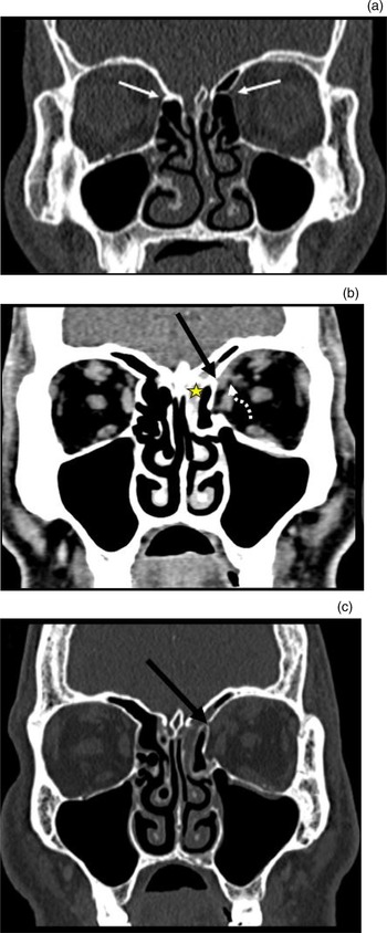
The anterior ethmoidal foramen is a small opening present in the ethmoid bone lateral to the olfactory groove on either side. It is situated about the middle of the lateral margin of the olfactory groove. It can be best seen on the coronal plane (Figure 1a). It transmits the anterior ethmoid artery, vein and nerve.
(a) Coronal reformatted computed tomography (CT) image at bone window settings, showing normal anterior ethmoidal foramina (white arrows). (b & c) Contrast-enhanced coronal reformatted CT images at soft tissue and bone window settings in a patient with coronavirus disease 2019 associated mucormycosis, depicting thickening in left anterior ethmoidal air cells (yellow star), extending into the superomedial extraconal compartment of the orbit via the anterior ethmoidal foramen (black arrows), with involvement of the superior oblique muscle (curved white arrow).

This represents one of the earliest pathways of disease spread from anterior ethmoid air cells to the orbit across its medial wall. From here, the disease involves the adjacent superior oblique muscle (Figure 1b and 1c).
Posterior ethmoidal foramen
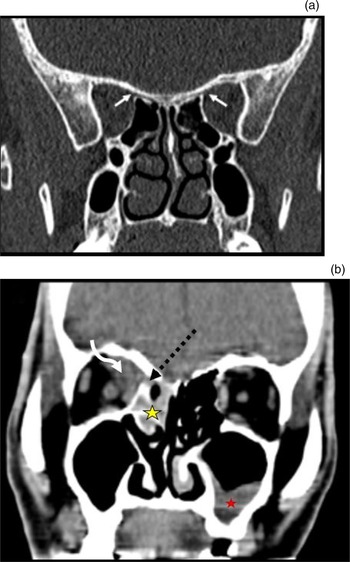
The posterior ethmoidal foramen is an opening in the ethmoid bone, approximately 12–15 mm posterior to the anterior ethmoidal foramen (Figure 2a). It transmits the posterior ethmoidal artery, vein and nerve.
(a) Coronal reformatted computed tomography (CT) image at bone window settings, showing normal posterior ethmoidal foramina (solid white arrows). (b) Contrast-enhanced coronal reformatted CT image at soft tissue window settings in a patient with coronavirus disease 2019 associated mucormycosis, revealing soft tissue thickening involving the right posterior ethmoidal air cells (yellow star), with extension into the superomedial extraconal and intraconal compartment of the orbit (curved arrow) via the posterior ethmoidal foramen (dashed black arrow). Mucosal thickening is also seen in the left maxillary sinus (red star).

This serves as a conduit for the spread of infection from posterior ethmoidal air cells and the sphenoid sinus to the orbit (Figure 2b).
Nasolacrimal duct
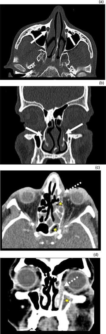
The nasolacrimal duct is a channel to drain the tears from the eyes to the nasal cavity. It forms the terminal part of nasolacrimal apparatus and spans approximately 17 mm in length. It consists of an intraosseous part (15 mm), which runs within the nasolacrimal canal of the maxilla, and a membranous part (2 mm), which runs in the nasal mucosa, terminating as a small opening in the inferior meatus (Figure 3a and 3b).
(a) Axial and (b) coronal reformatted computed tomography (CT) images at bone window settings, showing a normal nasolacrimal duct (solid white arrows). (c) Axial and (d) coronal contrast-enhanced images at soft tissue window settings in a patient with coronavirus disease 2019 associated mucormycosis, depicting mucosal thickening in the left nasal cavity with contiguous soft tissue within the left nasolacrimal duct extending into the lacrimal sac (dashed white arrows). Mucosal thickening is also noted in the left ethmoid, sphenoid and maxillary sinuses (yellow stars).

This is another common channel that leads to the spread of infection from the inferior meatus of the nasal cavity to the orbit, reaching the lacrimal sac (Figure 3c and 3d).
Infra-orbital foramen or canal
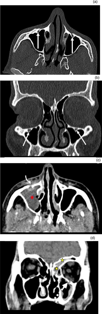
Along the floor of the orbit in its posterior aspect lies the infra-orbital groove, which continues as the infra-orbital canal, and terminates as an opening in the maxilla known as the infra-orbital foramen (Figure 4a and 4b). The infra-orbital nerve, artery and vein traverse through this foramen (Figure 4c and 4d).
(a) Axial and (b) coronal reformatted computed tomography (CT) images at bone window settings, showing normal infra-orbital foramina (solid white arrows). (c) Contrast-enhanced axial image at soft tissue window settings in a patient with coronavirus disease 2019 (Covid-19) associated mucormycosis, depicting soft tissue thickening in the right maxillary sinus (red star), with extrasinus extension via the infra-orbital foramen which is widened (curved white arrow). (d) Contrast-enhanced coronal image at soft tissue window settings in another patient with Covid-19 associated mucormycosis, revealing mucosal thickening in the left maxillary sinus, with extension into the inferior compartment of the orbit via the infra-orbital foramen (white dashed arrow). Mucosal thickening is also seen in the left frontal and ethmoid sinuses (yellow stars).

The disease spreads across this canal to involve the inferior extraconal fat and then goes on to involve the inferior rectus muscle. Through the foraminal opening, the infection can also spread into the preantral fat.
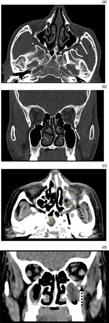
Inferior orbital fissure
The inferior orbital fissure is an opening between the lateral wall and orbital floor. Its boundaries consist of the greater wing of the sphenoid bone superiorly, the maxilla and orbital process of the palatine bone inferiorly, and the zygomatic bone laterally. It opens into the posterolateral aspect of the orbital floor and joins medially with the pterygomaxillary fissure at a right angle (Figure 5a and 5b). It transmits the infra-orbital nerve and zygomatic nerve (from the maxillary division of trigeminal nerve), infra-orbital artery, infra-orbital vein, branch of the inferior ophthalmic vein, and orbital branches from the pterygopalatine ganglion.
(a) Axial and (b) coronal reformatted computed tomography (CT) images at bone window settings, showing normal anatomy of the pterygopalatine fossa (thick arrow), pterygomaxillary fissure (curved arrow) and inferior orbital fissure (thin straight arrows). (c) Axial and (d) coronal contrast-enhanced CT images at soft tissue window settings in a patient with coronavirus disease 2019 associated mucormycosis, revealing soft tissue in the left pterygopalatine fossa (thick black arrow) extending into the inferolateral extraconal compartment of the orbit via the inferior orbital fissure (black dashed arrows). Mucosal thickening is also seen in the bilateral maxillary and sphenoid sinuses (yellow stars).

The disease process can spread from the nasal cavity to the pterygopalatine fossa through the sphenopalatine foramen, and from the pterygopalatine fossa to the inferior orbital fissure via the pterygomaxillary fissure. Thus, it is an important route of spread of mucormycosis from the pterygopalatine fossa, which acts as a reservoir of disease in these cases and is a major neurovascular crossroad in the head and neck region (Figure 5c and 5d).
Conclusion
Familiarity with the pathways of spread of mucormycosis to the orbit is essential so that these sites can be actively examined for early signs of invasion.
While disease within the nasal cavity and paranasal sinuses may be minimal, extrasinus extension through these foramina can provide a clue to the diagnosis of rhino-orbital-cerebral mucormycosis.
Knowledge of the routes of spread is equally important for the surgeons as it can help them to appropriately manoeuvre their surgical approach for the management or prognostication of such cases.
Competing interests
None declared