INTRODUCTION
Malaria is one of the major causes of morbidity and mortality in developing countries. Human malaria may be caused by any one of four Plasmodium species: P. falciparum, P. vivax, P. malariae, and P. ovale. P. falciparum is the parasite that causes the most severe form of malaria.
In natural infections, P. falciparum infected erythrocytes (IEs) circulate in the peripheral blood for a period corresponding to roughly 18 hours after invasion (ring stage) (Silamut et al. Reference Silamut, Phu, Whitty, Turner, Louwrier, Mai, Simpson, Hien and White1999). Later, in blood-stage development, parasite-encoded adhesion molecules are inserted into the erythrocyte membrane and adhesion of IEs to host endothelium receptors is observed. The adhesion to the host endothelium prevents the circulation of IEs and may assist the parasite in evading clearance of mature forms in the spleen (Pasloske and Howard, Reference Pasloske and Howard1994). Severe pathology is observed when IEs sequester themselves deep within the vascular beds of tissues or in high numbers in different vital organs. The accumulation results in capillary blockage and the shortage of oxygen and nutrients. In addition, the production of toxic metabolites and other factors by the parasite induces a host pro-inflammatory response that can lead to vascular damage and organ dysfunction (Miller, Good and Milon, Reference Miller, Good and Milon1994; Warrell, Reference Warrell1987). The adhesion to host cell endothelium receptors is essential for the survival of the parasite.
The principal molecule mediating IE adhesion is P. falciparum erythrocyte membrane protein 1 (PfEMP1) – a large, highly variant parasite antigen protein, encoded by the polymorphic var multigene family (Baruch et al. Reference Baruch, Pasloske, Singh, Bi, Ma, Feldman, Taraschi and Howard1995; Smith et al. Reference Smith, Chitnis, Craig, Roberts, Hudson-Taylor, Peterson, Pinches, Newbold and Miller1995; Su et al. Reference Su, Heatwole, Wertheimer, Guinet, Herrfeldt, Peterson, Ravetch and Wellems1995). PfEMP1 determines the host adhesion receptor preference of the IEs. Examples of these are CD36, intercellular adhesion molecule 1 (ICAM-1), vascular cell adhesion molecule 1 (VCAM-1), E-selectin and chondroitin sulfate A (CSA) (Craig and Scherf, Reference Craig and Scherf2001). Exposure of PfEMP1 on the IE surface stimulates a variant specific antibody response by the host (Miller et al. Reference Miller, Baruch, Marsh and Doumbo2002). There are approximately 60 var genes per haploid genome, which are predominantly situated in the subtelomeric regions of the parasite genome (Gardner et al. Reference Gardner, Hall, Fung, White, Berriman, Hyman, Carlton, Pain, Nelson and Bowman2002). Only a single variant of the var gene family is expressed at the surface of an IE at any given time. P. falciparum switches expression from one var gene to another, successively (Scherf et al. Reference Scherf, Hernandez-Rivas, Buffet, Bottius, Benatar, Pouvelle, Gysin and Lanzer1998). The switching in expression between the different var genes allows the parasite to undergo clonal antigenic variation, thus eluding the host's immune response and maintaining a persistent infection. In addition to its role in immune evasion, switching in PfEMP1 expression is associated with fundamental changes in parasite adhesion phenotype leading to sequestration in various tissues and ultimately results in variable disease outcomes (for review see Kyes, Horrocks and Newbold, Reference Kyes, Horrocks and Newbold2001).
EMERGENCE OF UNIQUE PARASITE VARIANTS IN PREGNANT WOMEN
In malaria-holoendemic areas, clinically protective immunity is acquired during the first 5–10 years of life. In these areas adults had experienced several malaria episodes from the time they were born throughout their lives. Consequently, many have developed a repertoire of anti-PfEMP1 antibodies against the most common variants of the membrane protein. This form of adaptive immunity enables them to curb P. falciparum infection, thus rarely experiencing severe malaria. Pregnant women in such endemic areas, especially during their first pregnancy, are an exception. P. falciparum infection during first pregnancy poses serious health risks for the mother and her baby, and can lead to maternal anaemia, foetal growth retardation, low birth weight, premature delivery and even death of both mother and child (Menendez, Reference Menendez1995; Rogerson and Brown, Reference Rogerson and Brown1997). This foeto-maternal syndrome is best described by the term ‘pregnancy associated malaria’ (PAM). Principally during first pregnancy, IEs massively sequester to the maternal side of the syncytiotrophoblast (a multinucleated cell layer derived from the foetus that lines the intervillous space that is in contact with maternal blood) that lines the placenta. IEs mainly accumulate in the intervillous space and only a sub-population appears to be directly attached to the syncytiotrophoblast surface (Muthusamy et al. Reference Muthusamy, Achur, Bhavanandan, Fouda, Taylor and Gowda2004). Placental IE sequestration is a virulence phenomenon exhibited by P. falciparum and is apparently not shared by other malarial parasites.
PAM is associated with the emergence of a subpopulation of IEs that are expressing PfEMP1 proteins that are structurally, antigenically and functionally distinct from those expressed by parasites that infect children and non-pregnant women. The placenta supports the clonal expansion of a unique subset of parasites that do not adhere to the usual endothelial receptors used by IEs like CD36 and ICAM-1, but instead bind to CSA and possibly other receptors (see below) (Fried and Duffy, Reference Fried and Duffy1996). Since the occurrence of a PfEMP1 variant with CSA-binding ability is low in non-pregnant patients, during first pregnancy women are deficient in antibodies against this PfEMP1 form. However, after multiple (more than one or two) pregnancies women (in malaria endemic areas) become immune to CSA-binding infected erythrocytes (IECSA) by producing specific antibodies associated with resistance to infection and to improved pregnancy outcomes (Duffy and Fried, Reference Duffy and Fried2003; Fried et al. Reference Fried, Nosten, Brockman, Brabin and Duffy1998b). Sera of multigravida women possess antibodies that recognize IECSA and inhibit their sequestration, whereas such antibodies are detected at very low levels in the sera of children, men or women before the third trimester of their first pregnancy (Fried et al. Reference Fried, Nosten, Brockman, Brabin and Duffy1998b; Ricke et al. Reference Ricke, Staalsoe, Koram, Akanmori, Riley, Theander and Hviid2000). Primigravida women, who do not yet have effective titers of antibodies against the IECSA, offer a new niche for the sequestration and proliferation of these parasites.
A switch from CD36 binding to CSA binding may change the tissue tropism of IEs leading to extensive sequestration of IEs in placental intervillous space. This is probably a key event in malaria pathogenesis during pregnancy. Nonetheless, despite all the accomplishments achieved in PAM research, the adhesive tropism of placental parasites is still enigmatic. Available evidence from different laboratories suggests that a particular var gene, var2CSA, is selectively expressed during placental infection and in IEs selected to adhere to CSA in vitro (Gamain et al. Reference Gamain, Trimnell, Scheidig, Scherf, Miller and Smith2005; Salanti et al. Reference Salanti, Staalsoe, Lavstsen, Jensen, Sowa, Arnot, Hviid and Theander2003; Viebig et al. Reference Viebig, Gamain, Scheidig, Lepolard, Przyborski, Lanzer, Gysin and Scherf2005). It is believed that var2CSA PfEMP1 plays a major role in PAM (Salanti et al. Reference Salanti, Dahlback, Turner, Nielsen, Barfod, Magistrado, Jensen, Lavstsen, Ofori and Marsh2004). In IEs with non-CSA binding abilities, var2CSA is silent and different members of the var gene family are transcribed during chronic infection. Switching in transcription from one var gene to another is associated at the molecular level with epigenetic changes in the gene locus (Duraisingh et al. Reference Duraisingh, Voss, Marty, Duffy, Good, Thompson, Freitas-Junior, Scherf, Crabb and Cowman2005; Freitas-Junior et al. Reference Freitas-Junior, Hernandez-Rivas, Ralph, Montiel-Condado, Ruvalcaba-Salazar, Rojas-Meza, Mancio-Silva, Leal-Silvestre, Gontijo and Shorte2005; Ralph and Scherf, Reference Ralph and Scherf2005). The rate at which parasites switch expression from one var gene to another is difficult to measure in patients (Peters et al. Reference Peters, Fowler, Gatton, Chen, Saul and Cheng2002) and only some studies from in vitro cultured parasites are available (Horrocks et al. Reference Horrocks, Pinches, Christodoulou, Kyes and Newbold2004). Therefore, an intriguing question is how a var gene that is apparently not expressed (or only rarely) in patients comes to be the predominant var gene expressed during pregnancy. We discuss in this review how the IECSA phenotype may arise in vivo by two different pathways which may act independently or in concert.
SELECTION MECHANISM
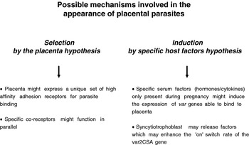
During pregnancy a new highly vascularized organ, the placenta, emerges in the woman's body. The unusual vascular bed of the placenta could, in theory, select for IEs expressing rare variant binding phenotypes (var2CSA) that circulate in very small numbers in the peripheral circulation of the primigravida. This would be possible if the placental structure was able to support the binding of IEs that do not bind to host receptors which are expressed in men and women. These parasites, when in contact with placental receptors such as glycosaminoglycans, could adhere to them and expand as a clonal population.
The placental intervillous space contains an extracellular proteoglycan matrix, with the main glycosaminoglycan being CSA (Achur et al. Reference Achur, Valiyaveettil, Alkhalil, Ockenhouse and Gowda2000; Alkhalil et al. Reference Alkhalil, Achur, Valiyaveettil, Ockenhouse and Gowda2000). As previously mentioned, epidemiological and molecular data strongly suggest that CSA is the principal receptor for IE adhesion in the placenta. However, it is important to note that the glycosaminoglycan CSA is expressed in non-placental tissues. Therefore, it is a striking observation that IECSA are only present at high levels during pregnancy and that anti-IECSA antibodies are largely restricted to pregnant women. Indeed, CSA is expressed throughout the surface of endothelial cells in the microvasculature of other target organs, such as the brain and the lung (Dittman and Majerus, Reference Dittman and Majerus1990; Wong et al. Reference Wong, Hofman, Ishii and Fisher1991). Furthermore, it has been demonstrated that IECSA cytoadhere to monkey brain microvascular endothelial cells and human lung epithelial cells, apparently expressing chondroitin sulfate (Robert et al. Reference Robert, Pouvelle, Meyer, Muanza, Fujioka, Aikawa, Scherf and Gysin1995; Traore et al. Reference Traore, Muanza, Looareesuwan, Supavej, Khusmith, Danis, Viriyavejakul and Gay2000).
One explanation for placental tropism could be that the placental receptors are somehow different from the vascular receptors in other organs. CSA is a glycosaminoglycan composed of repeating disaccharide units of D-glucuronic acid linked to N-acetyl-D-galactosamine with a sulfate group at position C4 of the galactosamine. CSA molecules are very heterogeneous in their sulfate content, and this property influences their ability to support IE binding (Achur et al. Reference Achur, Valiyaveettil and Gowda2003). An unusually low-sulfated form of CSA is expressed in the placenta, and efficiently supports placental IE binding (Achur et al. Reference Achur, Valiyaveettil, Alkhalil, Ockenhouse and Gowda2000). These low-sulfated glycosaminoglycans are most likely the main receptor for the opportunistic cytoadhesion of IEs in the placenta (Fried et al. Reference Fried, Domingo, Gowda, Mutabingwa and Duffy2006).
IECSA-specific sequestration has also been observed in male Saimiri sciureus monkeys (Pouvelle et al. Reference Pouvelle, Meyer, Robert, Bardel and Gysin1997) and in vitro studies mimicking physiological conditions have also shown that IEsCSA cytoadhere strongly enough to resist the wall shear stresses observed in the microvasculature (Pouvelle et al. Reference Pouvelle, Traore, Nogueira, Pradines, LePolard and Gysin2003). Therefore, it is perplexing that a well-adapted phenotype for microvessel sequestration is limited to placental cytoadhesion.
The presence of other co-receptors in the placenta that bind IEs efficiently or that favour CSA adhesion could help to explain the exclusivity of placental binding. Placental and laboratory isolates with differences in adhesion patterns to CSA receptors and with the ability to bind to several receptors in vivo have been reported (Avril et al. Reference Avril, Traore, Costa, Lepolard and Gysin2004; Beeson et al. Reference Beeson, Rogerson, Cooke, Reeder, Chai, Lawson, Molyneux and Brown2000; Rasti et al. Reference Rasti, Namusoke, Chene, Chen, Staalsoe, Klinkert, Mirembe, Kironde and Wahlgren2006; Tuikue Ndam et al. Reference Tuikue Ndam, Fievet, Bertin, Cottrell, Gaye and Deloron2004). In addition to CSA, another member of the glycosaminoglycan family, hyaluronic acid (HA), and nonimmune immunoglobulins (IgG and IgM) have been proposed to mediate IE adhesion and sequestration in the placenta (Beeson et al. Reference Beeson, Rogerson, Cooke, Reeder, Chai, Lawson, Molyneux and Brown2000; Creasey et al. Reference Creasey, Staalsoe, Raza, Arnot and Rowe2003; Flick et al. Reference Flick, Scholander, Chen, Fernandez, Pouvelle, Gysin and Wahlgren2001; Rasti et al. Reference Rasti, Namusoke, Chene, Chen, Staalsoe, Klinkert, Mirembe, Kironde and Wahlgren2006), but evidence for their roles is still unclear (Fried et al. Reference Fried, Domingo, Gowda, Mutabingwa and Duffy2006; Rasti et al. Reference Rasti, Namusoke, Chene, Chen, Staalsoe, Klinkert, Mirembe, Kironde and Wahlgren2006). Placental isolates were shown to bind HA in vitro (Beeson et al. Reference Beeson, Rogerson, Cooke, Reeder, Chai, Lawson, Molyneux and Brown2000), however, this result might have been influenced by a CSA contamination in the HA preparations used (Fried et al. Reference Fried, Domingo, Gowda, Mutabingwa and Duffy2006; Rasti et al. Reference Rasti, Namusoke, Chene, Chen, Staalsoe, Klinkert, Mirembe, Kironde and Wahlgren2006). A recent study (Rasti et al. Reference Rasti, Namusoke, Chene, Chen, Staalsoe, Klinkert, Mirembe, Kironde and Wahlgren2006) suggested that nonimmune Igs bind to the surface of IECSA allowing IE to bind to Ig receptors (Fc receptors) in the placenta. Nevertheless, the presence of HA and Fc receptors either in the intervillous space or in the syncytiotrophoblast lining of placentas is still obscure (Fried et al. Reference Fried, Domingo, Gowda, Mutabingwa and Duffy2006; Lyden et al. Reference Lyden, Robinson, Tridandapani, Teillaud, Garber, Osborne, Frey, Budde and Anderson2001; Valiyaveettil et al. Reference Valiyaveettil, Achur, Alkhalil, Ockenhouse and Gowda2001). Some unidentified co-receptors exclusively from placental tissue could be responsible for this particular tropism.
INDUCTION MECHANISM
An alternative way by which var2CSA expressing parasites may emerge during pregnancy is through its var2CSA activation by locally-released placental factors or serum-specific factors present only during pregnancy. These may act directly or indirectly to promote switching to the expression of var2CSA. The modulation of antigenic expression by host factors would require that the parasite be able to ‘sense’ extracellular changes in its surroundings and could activate specifically one particular var gene. The fact that the promoter region of the var2CSA gene has a unique promoter sequence not shared with other var genes – which have common 5′UTR sequence types, either upsA-, upsB- or upsC-type (Gardner et al. Reference Gardner, Hall, Fung, White, Berriman, Hyman, Carlton, Pain, Nelson and Bowman2002; Lavstsen et al. Reference Lavstsen, Salanti, Jensen, Arnot and Theander2003) – provides circumstantial evidence for a possible alternative epigenetic control which may result from the parasite sensing a new extracellular environment.
Active signaling between the host and parasite has been suggested in the P. knowlesi/rhesus monkey model. In vivo experiments have suggested that antigenic variation occurs only in the presence of the host spleen, and that it may be induced by antibodies reactive with the surface variant antigens of the parasite (Barnwell et al. Reference Barnwell, Howard, Coon and Miller1983; Brown, Reference Brown1973; David et al. Reference David, Hommel, Miller, Udeinya and Oligino1983). Furthermore, experimental evidence supported the belief that the new parasite population may not appear due to the selection of variant subpopulations, but as a result of an induced phenotypic switch (Barnwell et al. Reference Barnwell, Howard, Coon and Miller1983; Galinski and Corredor, Reference Galinski and Corredor2004). These observations reinforce the hypothesis that the host environment may provide regulatory cues that influence the expression and switching of surface variant antigens.
Several physiological changes occur in pregnant women and these include dramatic increases in the levels of several hormones (Roberts, Walker and Alexander, Reference Roberts, Walker and Alexander2001). Furthermore, the mother's immune system needs to alter its normal composition to prevent rejection of the foetal allograft. This immunomodulation often increases the susceptibility to certain parasitic infections (Luft and Remington, Reference Luft and Remington1982; Shirahata et al. Reference Shirahata, Muroya, Ohta, Goto and Nakane1992). It is possible that hormones and/or cytokines or other factors released into the blood during pregnancy facilitate the activation of var2CSA.
The influence of different hormones during malaria infections has been documented. Two prominent examples are: host circadian changes in the levels of the hormone melatonin have been shown to control synchronous maturation of the parasite in vivo (Hotta et al. Reference Hotta, Gazarini, Beraldo, Varotti, Lopes, Markus, Pozzan and Garcia2000) and sex hormones have been shown to influence the length and severity of the infection in P. chabaudi (Benten et al. Reference Benten, Wunderlich and Mossmann1992a, Reference Benten, Wunderlich and Mossmannb) most likely by modifying the immune response through specific interactions (Roberts, Satoskar and Alexander, Reference Roberts, Satoskar and Alexander1996).
The role of pregnancy hormones in malaria has not been studied in great depth. A few studies have shown that the hormones cortisol and prolactin, whose levels are elevated during pregnancy, were detected at different levels among primigravida and multigravida women. Cortisol concentrations were significantly higher in primigravida as opposed to multigravida women and prolactin concentrations were significantly lower (Bouyou-Akotet et al. Reference Bouyou-Akotet, Issifou, Meye, Kombila, Ngou-Milama, Luty, Kremsner and Mavoungou2004, Reference Bouyou-Akotet, Adegnika, Agnandji, Ngou-Milama, Kombila, Kremsner and Mavoungou2005). A relationship between these hormonal changes and malaria was drawn, due to the antagonist effect these two hormones have on the immune system. This effect could explain the incidence of infection in primigravidae since the highest cortisol levels were in P. falciparum infected primigravida women (Vleugels et al. Reference Vleugels, Brabin, Eling and de Graaf1989; Bouyou-Akotet et al. Reference Bouyou-Akotet, Issifou, Meye, Kombila, Ngou-Milama, Luty, Kremsner and Mavoungou2004). Several other hormones have altered levels during pregnancy and it will be interesting to study their role in the parasite's asexual blood-stage gene expression.
Cross-talk between the endocrine and the immune system is high during pregnancy. Cytokines play an important role in placental development. Different cytokines have been localized at the placenta-uterine interface where they probably play an important role in regulation of placental development. The human placenta secretes different cytokines and inflammatory molecules both locally and to the maternal systemic circulation (Bowen et al. Reference Bowen, Chamley, Keelan and Mitchell2002). In a successful pregnancy there is a predominance of T helper type 2 cytokines at the maternal-foetal interface (Wegmann et al. Reference Wegmann, Lin, Guilbert and Mosmann1993). During malaria infection, the production of pro-inflammatory factors increases and the type 2 helper T cells response weakens (Fried et al. Reference Fried, Muga, Misore and Duffy1998a). We speculate that the changes in cytokines levels not only alter the immune balance of the pregnant woman but might be able to induce gene switching in circulating parasites. Local increases in cytokine concentration at the placenta or in the peripheral circulation may be sensed by the parasite leading to var2CSA activation.
The production of hormones and other pregnancy regulated factors in pregnant women may directly influence the parasite or modify host cellular functions enabling parasitic proliferation.
CONCLUSION
In this review we have explored two possible models that could lead to var2CSA activation during PAM (Fig. 1). The selection mechanism is generally used in the literature to explain the appearance of this new phenotype. However, it does not make clear why IEs expressing var2CSA are not detected in non-pregnancy malaria infections. The fact that the parasite adhesion receptors described so far in the placenta exist in other organs (Pouvelle et al. Reference Pouvelle, Traore, Nogueira, Pradines, LePolard and Gysin2003) and that ICAM1 is present on the surface of syncytiotrophoblasts but is not involved in placental parasite sequestration is puzzling (Beeson et al. Reference Beeson, Brown, Molyneux, Mhango, Dzinjalamala and Rogerson1999) and does not convincingly explain the placental tissue tropism by ‘selection’ alone.
Proposed mechanisms involved in placental adhesion tropism. Factors that may control the occurrence of a P. falciparum population expressing var2CSA during pregnancy.

There are examples of unknown host factors able to alter parasitic gene expression, like the P. knowlesi/rhesus monkey model described above. We favour an alternative mechanism that would imply the sensing by the parasite of specific maternal factors.
The ‘induced’ switch model is an attractive model to explain var2CSA activation given that this var gene has a 5′ region unique among the var-gene 5′ sequences. The work of several groups regarding the intrinsic switching of var variants in the absence of external immune response suggests that the immune response may not directly cause var gene switching (Horrocks et al. Reference Horrocks, Pinches, Christodoulou, Kyes and Newbold2004; Roberts et al. Reference Roberts, Craig, Berendt, Pinches, Nash, Marsh and Newbold1992). The immune responses and cytoadhesion/selection in vivo may provide a higher level of control over pre-programmed/stochastic switching events. In addition, several host factors are released into the blood circulation only during pregnancy. During the intraerythrocytic life cycle, P. falciparum exports a number of proteins to the surface of the IEs and it is possible that some of these proteins can function as receptors for hormones or cytokines. The identification of possible P. falciparum hormone or cytokine receptors may open new avenues in our understanding of the underlying molecular processes that lead to placental malaria.
ACKNOWLEDGEMENTS
The authors would like to thank, Benoit Gamain for helpful discussion and Nicola Viebig for comments on the manuscript. The authors are supported by the European Commission (BioMalPar) contract No. LSHP-CT-2004-503578 and the ‘Fonds dédié: Combattre les Maladies Parasitaires’ Sanofi Aventis – Ministère de la Recherche. MCN has financial support from the Human Frontier Science Program.