INTRODUCTION
Polyclads are flattened acoelomate animals that lack both circulatory and respiratory systems, but have a highly branched intestine and a complex reproductive system. Within the cotylean polyclads, besides the distribution of the eye groups and the presence and type of tentacles, the coloration pattern is considered a useful character for the species identification (Newman & Cannon, Reference Newman and Cannon2003), being supported by molecular data in the largest and most diverse family Pseudocerotidae (Litvaitis & Newman, Reference Litvaitis and Newman2001). Polyclads are common in shallow tropical waters and were also recorded in temperate, polar and deep-sea environments (Quiroga et al., Reference Quiroga, Bolaños and Litvaitis2006). There are about 900 species described worldwide of which 129 occur in the tropical western Atlantic (Quiroga et al., Reference Quiroga, Bolaños and Litvaitis2004a; Bolaños et al., Reference Bolaños, Quiroga and Litvaitis2007).
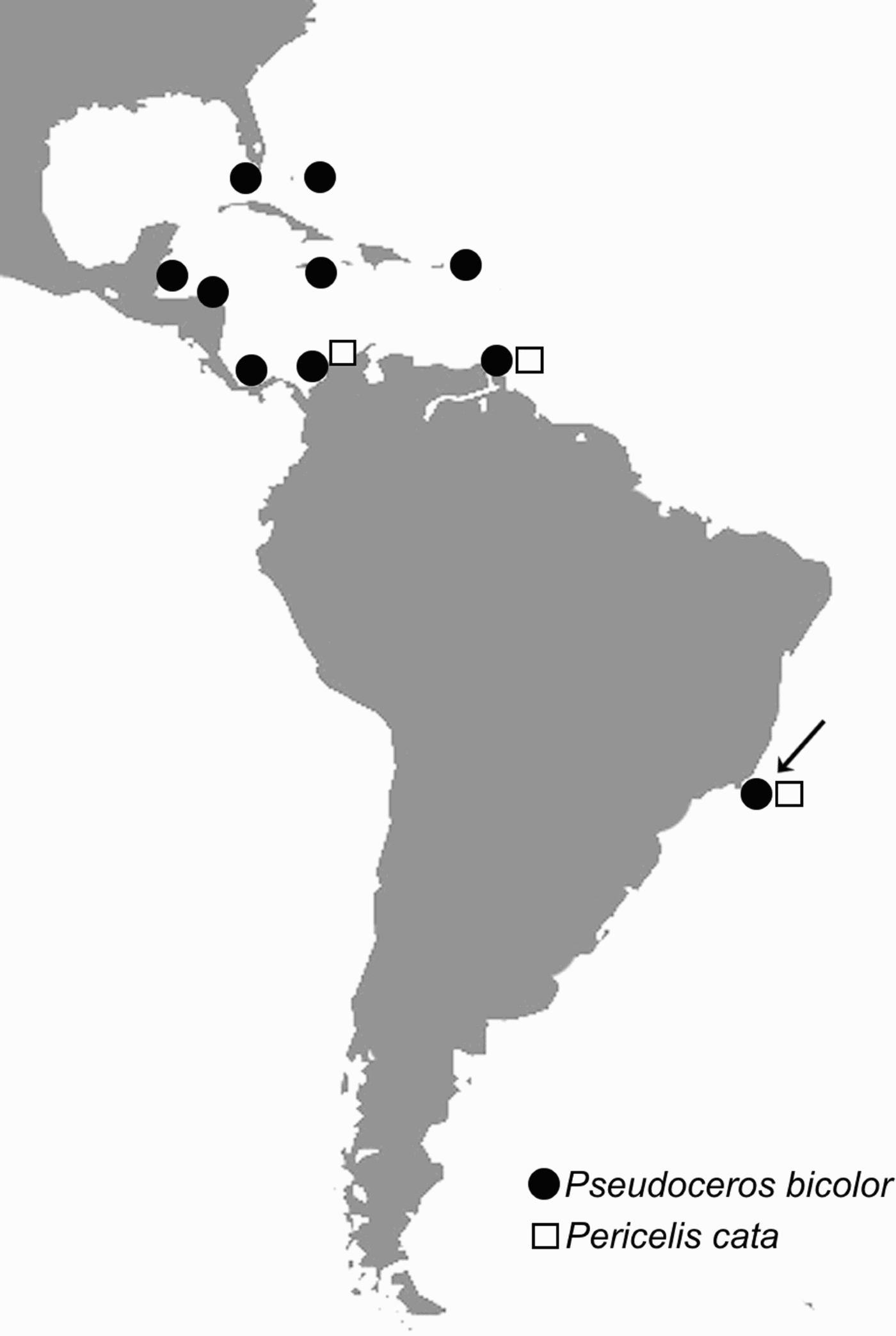
In Brazil, the works of Ernst and Eveline Marcus were valuable in describing and reporting lists of polyclad species. Most of the existing data results from collections in a relatively small area, the north coast of the State of São Paulo (Marcus, Reference Marcus1947, Reference Marcus1948, Reference Marcus1949, Reference Marcus1950, Reference Marcus1952, Reference Marcus1954). About 60 species were described from this area, 42 of these remain endemic from Brazil (Marcus & Marcus, Reference Marcus and Marcus1968; Quiroga et al., Reference Quiroga, Bolaños and Litvaitis2004a). This paper presents the first report of two polyclad species, already known from the Caribbean Sea (Figure 1), at the Brazilian coast.
Geographical distribution of Pseudoceros bicolor and Pericelis cata. The arrow indicates their records from the Brazilian coast.

MATERIALS AND METHODS
Specimens were collected manually by the authors at sites of Cabo Frio, State of Rio de Janeiro, on the south-eastern Brazilian coast. They were photographed alive, fixed on frozen 10% formalin, measured and preserved on 70% ethanol (for details see Newman & Cannon, Reference Newman and Cannon2003). Observations on informative taxonomic characters were made under stereoscopic microscope. Eyes and genital apertures were photographed with a digital camera coupled to the microscope. The identification was based on the original descriptions and the systematic classification was based on Faubel (Reference Faubel1984) and Prudhoe (Reference Prudhoe1985). All material is deposited at the Platyhelminthes Collection of the Departamento de Invertebrados, Museu Nacional/Universidade Federal do Rio de Janeiro (MNRJ-PLAT).
MATERIAL EXAMINED
Two specimens (9 × 6 mm and 8 × 6 mm, preserved), under a rock in the intertidal zone of Praia das Conchas, Cabo Frio, Brazil (22°52′15.40″S 41°58′51.86″W), 20 April 2008, V. Padula col. (MNRJ-PLAT 01).
One specimen (15 × 11 mm, preserved), under a rock in the intertidal zone of Praia das Conchas, Cabo Frio, Brazil (22°52′15.40″S 41°58′51.86″W), 7 February 2009, V. Padula col. (MNRJ-PLAT 04).
DESCRIPTION
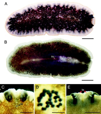
Flattened body, elliptical with undulated edge. Dorsal colour is dark, brownish black, scattered with tiny white dots; border translucent white, interrupted by greyish black bands; margin with a thin yellowish line (Figure 2A). Short and rounded pseudotentacles with a deep sinus in between their front edges. Pseudotentacles black with a narrow white border. Tentacular eyes present, also visible from the ventral side (Figure 2E); small cerebral eyes situated in the central line of the body, near the bases of the pseudotentacles and organized in two rounded groups very close together (Figure 2C, D). Short pharynx, with few folds located anteriorly, 3 mm from the fore end. Mouth located in the centre of this pharynx. Male and female gonopores and the sucker lie 1 mm from each other and are located approximately 4–5 mm from the anterior margin (Figure 2B). Uteri are visible ventrally and are purplish pigmented. Distances cited obtained from MNRJ-PLAT 04.
Pseudoceros bicolor. (A) Dorsal view of a living specimen; (B) ventral view of a living specimen and the main structures; (C) anterior region of a preserved specimen; (D) cerebral eyes; (E) ventral view of the anterior region of a preserved specimen (A–B: MNRJ-PLAT 01; C–E: MNRJ-PLAT 04). Scale bars: A, B, 1.2 mm; C, 1 mm; D, 80 µm; E, 1 mm. ce, cerebral eyes; fp, female pore; in, intestine; mp, male pore; ph, pharynx; su, sucker; te, tentacular eyes.

REMARKS
This species was previously reported from the Bahamas (type locality; Verrill, Reference Verrill1901); Curaçao (Marcus & Marcus, Reference Marcus and Marcus1968) and the Caribbean coast of Colombia (Quiroga et al., Reference Quiroga, Bolaños and Litvaitis2004b). Recently, it was listed for Florida Keys, Virgin Islands, Jamaica, Belize, Honduras and the Caribbean coast of Panama (Rawlinson, Reference Rawlinson2008). This species presents some variation concerning the intensity of the dark background colour (Newman & Canon, Reference Newman and Cannon2003: 72). The specimens herein studied fit well Verrill's original description and because of this were identified as representatives of Pseudoceros bicolor.
MATERIAL EXAMINED
One specimen (35 × 32 mm, preserved), under a rock 6 m depth at Saco do Mimi, Ilha do Papagaio, Cabo Frio, Brazil (22°53′33.26″S 41°59′7.60″W), 3 August 2008, V. Padula col. (MNRJ-PLAT 02).
One specimen (45 × 34 mm, preserved), under a rock 10 m depth at Saco da Hípica, Ilha do Papagaio, Cabo Frio, Brazil (22°53′52.44″S 41°58′43.80″W) 13 December 2008, V. Padula & E. Lanna col. (MNRJ-PLAT 03).
DESCRIPTION
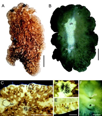
Flattened body; soft and very delicate in living specimens; crumpled marginal folds that sometimes stick together. Dorsally there is a pattern of dark pigmentation interrupted by round areas where the white parenchyma shines through. Towards the margin the white patches become smaller and the space between them decreases; dark patches contain scattered black spots of melanophores (Figure 3A). Tentacles are marginal subtle folds of the margin, 4 mm apart from each other, and are one of the darkest areas of the body, making the observation of the densely grouped tentacular eyes difficult. Cerebral eyes, situated in a light area 3.5 mm from the anterior end and organized in two rounded clusters irregularly separated (Figure 3C, D); marginal eyes, arranged in an irregular row, surround the entire margin of the body and can be seen from the ventral side at some parts (Figure 3E). Between the tentacle region and the cerebral region there are several scattered ocelli (precerebral eyes) located in two broad and loose tracts (Figure 3C). Narrow pharynx (11 × 6 mm) ruffled with numerous fine folds (Figure 3B). Male and female gonopores are closely placed and 22 mm distant from the anterior margin; sucker situated 1 mm behind the gonopores. Penial papillae directed obliquely backward (Figure 3F). Distances cited obtained from MNRJ-PLAT 02.
Pericelis cata. (A) Dorsal view of a living specimen; (B) ventral view of a living specimen; (C) anterior region of a preserved specimen; (D) cerebral eyes; (E) marginal eyes at the posterior border of the body in a preserved specimen; (F) genital pores and the sucker in a preserved specimen (A,B,F: MNRJ-PLAT 02; C–E: MNRJ-PLAT 03). Scales: A, B, 5.7 mm; C, 3 mm; D, 300 µm; E, 1 mm; F, 1 mm. ce, cerebral eyes; fp, female pore; go, gonopores; me, marginal eyes; pe, pre-cerebral eyes ph, pharynx; pp, penial papilae; su, sucker.

REMARKS
This species was previously recorded from Curaçao (type locality; Marcus & Marcus, Reference Marcus and Marcus1968) and the Caribbean coast of Colombia (Quiroga et al., Reference Quiroga, Bolaños and Litvaitis2004b). At Cabo Frio, Pericelis cata appears to be more common on deeper zones as no specimen was observed at intertidal areas. Both specimens fit well the original description, the large body and its fragility when alive appears to be very characteristic of this species. In the field both specimens examined look darker when compared to the picture herein presented.
DISCUSSION
The new records presented herein increase the number of Brazilian polyclads to 63 species. They are also important records for the State of Rio de Janeiro, where a single species was previously recorded (Corrêa, Reference Corrêa1957). This is also the first record of the genus Pericelis from Brazil. Concerning the species distribution, the present records expand their known geographical range to a transition zone between tropical and subtropical environments. Palacio (Reference Palacio1982) considered that the State of Rio de Janeiro is situated in the beginning of this transition area. Recently, in a comprehensive work concerning global marine ecoregions, Spalding et al. (Reference Spalding, Fox, Allen, Davidson, Ferdaña, Finlayson, Halpern, Jorge, Lombana, Lourie, Martin, McManus, Molnar, Recchia and Robertson2007) reaffirmed the importance of the Cabo Frio region as a limit between the tropical south-western Atlantic and the warm temperate south-western Atlantic provinces.
The occurrence of Pseudoceros bicolor and Pericelis cata in Brazil, with other 15 co-occurring polyclad species, reinforces the resemblance between the Caribbean and Brazilian marine fauna. This pattern was observed for other invertebrate groups such as molluscs (Rios, Reference Rios1994), crustaceans (Humann & Deloach, Reference Humann and Deloach2002) and also for some vertebrates such as reef fish (Rocha, Reference Rocha2003). In the case of polyclads the different oceanographic conditions and the distance apparently do not act as a barrier for their distribution on the western Atlantic. Pseudoceros bicolor and Pericelis cata, for example, are able to tolerate low seawater temperatures (under 15°C) and variations up to 10°C within a few metres depth (Valentin, Reference Valentin, Seeliger and Kjerfve2001) at Cabo Frio, resulting from seasonal local upwelling events.
The wide distribution of some species on the western Atlantic and their tolerance to different oceanographic conditions has not been questioned in previous studies on polyclads and, together with the knowledge on these species biology and ecology, are interesting subjects for future studies. As the samples in Brazil are almost restricted to intertidal areas of the State of São Paulo (Marcus, Reference Marcus1949, Reference Marcus1950, Reference Marcus1952) the collection of specimens in different bathymetric ranges in other Brazilian localities, and between northern Brazil and the Caribbean Sea, are necessary to understand the biogeography of the western Atlantic polyclads. At the same time, these new surveys will allow the increase of the knowledge on Brazilian polyclad biodiversity.
ACKNOWLEDGEMENTS
We thank CNPq–Brazil for the financial support to this work. We also thank Emilio Lanna (UFRJ) for helping in the collection of specimens and the Litoral Sub staff for dive operations at Cabo Frio. J. Maceira and S. Bahia provided useful revision of the English text.