INTRODUCTION
Although the Iberian bryozoan fauna is one of the best known in Europe, at least as regards the number of species, knowledge of the Portuguese bryozoans is still relatively scarce and fragmented. Around one hundred species are known to exist in the Portuguese circalittoral waters. Many of these species are cited in studies by Nobre (Reference Nobre1903a,Reference NobrebReference Nobre1904, Reference Nobre1937, Reference Nobre1942) and by Nobre & Braga (Reference Nobre and Braga1942), and included in lists compiled and amplified by Rosas (Reference Rosas1944) who also compiled other previous works, and in studies by Saldanha (Reference Saldanha1974, Reference Saldanha1980), who can be considered as the principal researcher dedicated to the Portuguese bryozoan fauna. On the other hand, some extensive, deep water oceanographic surveys in the 19th and 20th Centuries were carried out close to the Portuguese coasts, and the results reported by Wyville Thomson (Reference Wyville Thomson1877) (possibly the oldest reference), Jullien (Reference Jullien1882), Busk (Reference Busk1884), Calvet (Reference Calvet1906, Reference Calvet1931), Harmer (Reference Harmer1915), d'Hondt (Reference d'Hondt1974), Hayward (Reference Hayward1979) and d'Hondt & Hayward (Reference d'Hondt and Hayward1981). Other authors who have also contributed to knowledge of the Portuguese Bryozoa include Pérès (Reference Pérès1959), Gautier (Reference Gautier1961), Prenant & Bobin (Reference Prenant and Bobin1966), Marques et al. (Reference Marques, Reis, Calvário, Marques, Melo and Santos1982), Reverter-Gil & Fernández-Pulpeiro (Reference Reverter-Gil and Fernández-Pulpeiro1999a, Reference Reverter-Gil and Fernández-Pulpeiro2005, Reference Reverter-Gil and Fernández-Pulpeiro2007), Boury-Esnault et al. (Reference Boury-Esnault, Harmelin, Leloir, Saldanha and Zibrowius2001), Harmelin (Reference Harmelin2001) and Marchini et al. (Reference Marchini, Cunha and Occhipinti-Ambrogi2007). As far as we know, no other studies have included reports of bryozoan fauna from Portuguese waters; therefore we believe that to state that the literature is rather scarce is an understatement, especially as some of these studies only concern a single species, or a few nominal citations.
Approximately 140 gymnolaemate Bryozoa are known from Portuguese waters, whereas more than 400 are known in the whole of the Iberian Peninsula (300 in Atlantic waters and 260 in Mediterranean waters).
We are currently undertaking the first stage of a project entitled ‘Iberian Fauna: Bryozoa’, a long-term research project in which we aim to combine and update all previous citations for Bryozoa in the Iberian Peninsula and Balearics, and to provide new data from the collection of new material. In the present study, we report the results of the study of samples collected at three points in the Algarve (southern Portugal), where we have identified a total of 57 species of gymnolaemate Bryozoa, eight of which are new to science, as well as 16 previously unknown from the Portuguese coast.
MATERIALS AND METHODS
The samples were collected by SCUBA divers at three points located in southern Portugal, in the Armaçao de Pêra Bay. The samples were collected on 11 and 12 December, 2007.
Site 1 (37°01646′N 008°19038′W, 21 m) has a maërl bottom with abundant sand and shells as well as stones completely covered with calcareous algae. At site 2 (37°02079′N 008°18655′W, 19 m) the bottom is also maërl, but with abundant stone. Finally, site 3 (37°02423N 008°19576′W, 20 m) is located on a stone reef that runs almost the whole length of the bay and in which there are large holes.
The material was fixed with 4% formaldehyde, and once separated in the laboratory, the ctenostomes were maintained in 4% formaldehyde, and the cheilostomes were preserved in 70% alcohol or were dried. Some of the material was treated with bleach for observation by SEM.
The samples were observed and measured with a Wild MZ16 stereomicroscope and uncoated material was photographed in a LEO 435 VP scanning electron microscope.
We also revised diverse reference material stored in different institutions: Manchester Museum (MM), Museo Nacional de Ciencias Naturales, Madrid (MNCN), Natural History Museum, London (NHM) and the Muséum National d'Histoire Naturelle, Paris (MNHN).
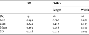
Measurements are included in the Tables. In material of the genera Rhynchozoon and Dentiporella the distance between midpoints of adjacent orifices (DO) was measured, as in these species the autozooids grow vertically in various shapes, and are therefore very difficult to measure.
RESULTS
A list of the species collected is provided. All fit the most recent descriptions. The specimens of Alcyonidium and Penetrantia were only able to be identified to genus level because of the scarce material available.
The numbers in parentheses indicate the sampling stations. Citations new to the Portuguese coast are indicated with an asterisk.
Ctenostomata
Alcyonidium sp. (Station 3)
Nolella dilatata (Hincks, 1860) (Stations 1 & 2)
*Penetrantia sp. (Station 2)
Cheilostomata
Aetea anguina (Linnaeus, 1758). (Stations 1, 2 & 3)
Aetea sica (Couch, 1844). (Station 2)
Aetea truncata (Landborough, 1852). (Station 2)
Callopora dumerilii (Audouin, 1826). (Stations 1 & 2)
*Copidozoum tenuirostre (Hincks, Reference Hincks1880). (Station 2)
Membraniporella nitida (Johnston, 1847). (Stations 1, 2 & 3)
Bugula fulva Ryland, 1960. (Station 2)
Bugula turbinata Alder, 1857. (Stations 2 & 3)
Beania mirabilis (Johnston, 1840). (Stations 1 & 2)
Beania hirtissima (Heller, 1867). (Station 2)
Caberea boryi (Audouin, 1826). (Stations 2 & 3)
Scrupocellaria scrupea Busk, 1852. (Station 2)
Mollia cristinaesp. nov. (Station 2)
*Collarina balzaci (Audouin, 1826). (Station 1)
*Puellina innominata (Couch, 1844). (Stations 1 & 2)
*Puellina arrecta Bishop & Househam, 1987. (Station 1)
Hippothoa divaricata Lamouroux, 1821. (Stations 1 & 2)
Chorizopora brongniartii (Audouin, 1826). (Stations 1 & 2)
*Trypostega venusta (Norman, 1864). (Station 2)
Escharoides coccinea (Abildgaard, 1806). (Station 2)
Smittina landsborovii (Johnston, 1847). (Stations 2 & 3)
Smittoidea reticulata (MacGillivray, 1842). (Station 1)
*Prenantia cheilostoma (Manzoni, 1869). (Station 3)
Hippoporina teresaesp. nov. (Stations 1 & 2)
Pentapora fascialis (Pallas, 1766). (Station 1)
*Schizomavella auriculata (Hassall, 1842). (Station 3)
Schizomavella cornuta (Heller, 1867). (Station 3)
Schizomavella sarniensis Hayward & Thorpe, 1995. (Stations 1, 2 & 3)
Schizomavella hastata (Hincks, Reference Hincks1862). (Stations 2 & 3)
Schizomavella linearis (Hassall, 1841). (Stations 1, 2 & 3)
Schizomavella mamillata (Hincks, Reference Hincks1880). (Stations 1 & 3)
Schizoporella cornualis Hayward & Ryland, Reference Hayward and Ryland1995. (Station 2)
Schizoporella spinosasp. nov. (Station 2)
Schizobrachiella sanguinea (Norman, Reference Norman1868). (Stations 2 & 3)
Myriapora truncata (Pallas, 1766). (Station 3)
*Hagiosynodos latus (Busk, 1856). (Station 3)
Microporella ciliata (Pallas, 1766). (Stations 1, 2 & 3)
*Microporella appendiculata (Heller, 1867). (Station 2)
Fenestrulina inesaesp. nov. (Stations 2 & 3)
*Arthropoma cecilii (Audouin, 1826). (Station 1)
Escharina vulgaris (Moll, 1803). (Station 2)
Cellepora pumicosa (Pallas, 1766). (Stations 1, 2 & 3)
Celleporina caliciformis (Lamouroux, 1816). (Stations 1, 2 & 3)
*Celleporina decipiens Hayward, 1976. (Station 2)
Celleporina derungsisp. nov. (Stations 2 & 3)
*Turbicellepora incrassata (Lamarck, 1816). (Station 2)
*Omalosecosa ramulosa (Linnaeus, 1767). (Station 1)
Dentiporella saldanhaisp. nov. (Stations 2 & 3)
Rhynchozoon bispinosum (Johnston, 1847). (Stations 2 & 3)
Rhynchozoon celestinoisp. nov. (Station 3)
Rhynchozoon rosaesp. nov. (Station 2)
*Schizotheca fissa (Busk, 1856). (Stations 1 & 2)
Stephanollona armata (Hincks, Reference Hincks1862). (Stations 2 & 3)
*Stephanollona contracta (Waters, Reference Waters1899). (Station 1)
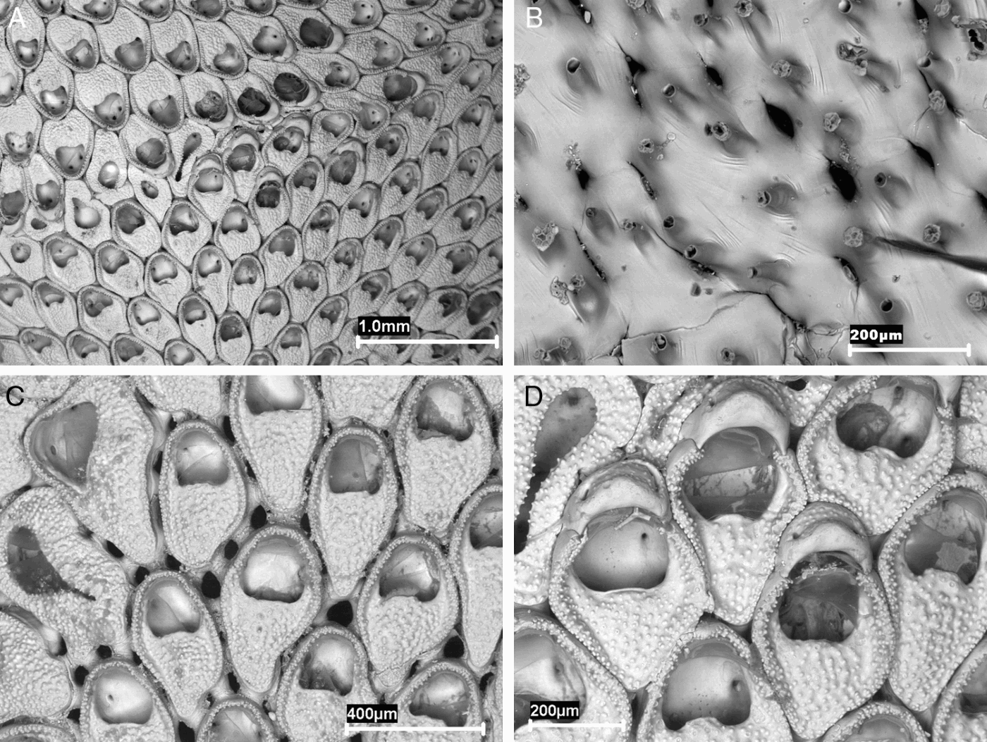
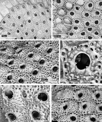
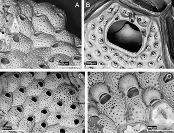
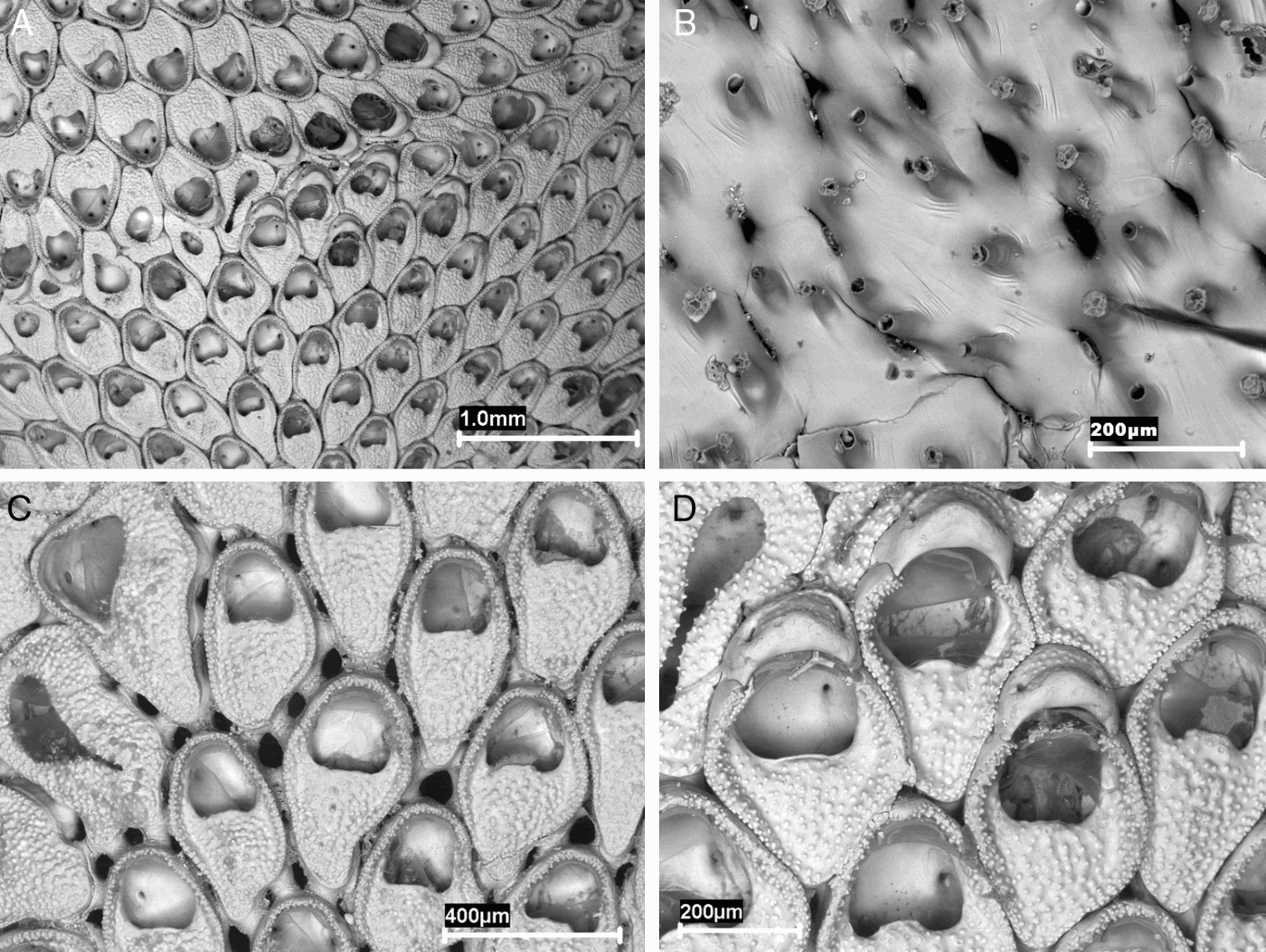
Mollia cristinae sp. nov. (A) (holotype) general view with two colony margins; (B) (paratype MNCN-25.03/3776) basal view of the autozooids with rhizoids; (C) (holotype) group of autozooids; (D) (holotype) ovicellate zooids.
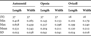
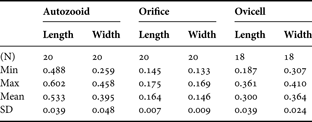
Measurements (in mm) of Mollia cristinae sp. nov. (holotype).
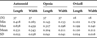
Min, minimum; Max, maximum; SD, standard deviation.
Mollia rosselii (Audouin): López de la Cuadra, Reference López de la Cuadra1991: 140, pl. 14 A–C.
TYPE MATERIAL
Holotype: MNCN-25.03/3775: Station 2. 37°02079′N 008°18655′W, 19 m, December 2007.
Paratypes: MNCN-25.03/3776, 3777: Station 2. 37°02079′N 008°18655′W, 19 m, December 2007. Two colonies.
COMPARATIVE MATERIAL EXAMINED
Lectotype of Mollia multijuncta (designated here): MM-H.1186.314, Naples 1875, Waters Coll.
Paralectotypes of Mollia multijuncta (designated here): MM-H.1186.313, MM-H.1186.315, Naples 1875, Waters Coll.
Other material: Mollia multijuncta: MM-H.1186.312, Taranto Bay, 1893, Waters Coll. MM-H.1186.310, Madeira, Waters Coll. MM-H.1186.311, Madeira, Waters Coll. Mollia patellaria: MM-H.1186.302. Naples, 1875, Waters Coll. MM-H.1186.303. Naples, 1875, Waters Coll. MM-H.1186.307. Naples, Waters Coll. MM-H.1186.308. Naples, Waters Coll. MM-H.1186.309. Naples, Waters Coll. MM-H.1186.316. Capri, Waters Coll. MM-H.1186.317. Capri, 1880, Waters Coll. MM-H.1186.318. Capri, 1880, Waters Coll. MM-H.1186.319. Capri, Waters Coll. MM-H.1186.320. Capri, Waters Coll. MM-H.1186.321. Naples or Capri. Waters Coll. MNCN-25.03/1065: SW Isla del Aire, Menorca, 15–20 m; MNCN-25.03/1511: northern Isla Dragonera, Mallorca, 31 m.
DIAGNOSIS
Autozooids disjunct, each linked to a neighbour by 1–3 tubules, frequently two. Cryptocyst occupying the proximal half of the frontal wall. Opesia semielliptical, as long as wide, with the proximal rim with a median lip. Ovicell ectoecium forming a smooth rim well developed medio-proximally; endoecium forming a central granular area distally directed, with a tiny pore in each lateral corner.
ETYMOLOGY
This species is dedicated to Mrs Cristina Taboada Montero, wife of E.F.-P.
DESCRIPTION
Colony encrusting, unilaminar, beige in colour. Zooids oval, in alternate series, fixed to the substratum by 8–10 short rhizoids placed in the periphery of the zoecial base. Autozooids disjunct, each linked to its neighbours by 9–14 (12 on average) tubular communication organs, so each zooid is linked to a neighbour by two (sometimes one or three) tubules more or less long. Frontal wall membranous, surrounded by a granular gymnocyst, raised in the distal portion and decreasing to the proximal third. Cryptocyst granular, occupying the proximal half of the frontal wall. Opesia semielliptical, as long as wide, with the proximal rim slightly convex or, more frequently, with a median lip with two lateral notches. Operculum semicircular, subterminal. Ovicell independent, closed by the operculum; ectoecium smooth, forming a calcified rim well developed medio-proximally; endoecium difficult to see in frontal view, forming a central granular area distally directed, with a tiny pore in each lateral corner. Ancestrula not known.
REMARKS
A review of the relevant literature shows that there is great confusion as regards the distinctive characters of the different species of the genus Mollia, as well as how it differs from other morphologically similar genera such as Rosseliana, Steraechmella, Gargantua, etc. Such confusion has given rise to erroneous citations, and unjustified synonyms, which can only be resolved by study of the original material and, in very few occasions, of the original references.
The material collected in Portugal is similar to existing descriptions of Mollia multijuncta, a species originally described by Waters (Reference Waters1879) as a variety of Diachoris patellaria (Moll, 1803) from material collected at Naples. However, the validity of the species has been the subject of discussion, even since the original description; Waters (Reference Waters1879) himself considered that it may be a synonym of Membranipora circumcincta Heller, 1867, an opinion not shared by the latter author. Later other authors such as Prenant & Bobin (Reference Prenant and Bobin1966) considered that both multijuncta and circumcincta are merely varieties of Mollia patellaria and did not treat them separately. Zabala (Reference Zabala1986) considered that multijuncta is synonymous with the variety circumcincta, whereas Zabala & Maluquer (Reference Zabala and Maluquer1988) considered it a variety of M. patellaria, but M. circumcincta as a different species. In summary, neither the phylogenetic position of the species nor its morphological characters were clearly established.
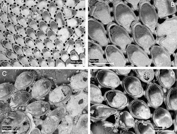
We have studied the material originally examined by Waters (Reference Waters1879) to describe the species. The material consists of three samples deposited in the Manchester Museum, one of which (MM-H.1186.315) is marked with a red dot. Waters is said to have used a red dot to mark a type (Henry McGhie, personal communication, June 2009), but it is not clear what this author interpreted as ‘Type’. Moreover, one of the samples of Mollia patellaria (MM-H.1186.302; Figure 2C) from the same locality was also marked with a red dot. None of the specimens appears to correspond to the original drawings (Waters, Reference Waters1879, pl. 10, figure 6, pl. 13, figure 4) and therefore, at least in this case, the meaning of the red dot is not clear. We have decided to designate the sample MM-H.1186.314 as the lectotype of M. multijuncta (Figure 2A,B; Table 2). This sample belongs to the type series and contains a colony that is very similar to that drawn by Waters and that also presents ovicells, which appear to have been overlooked by the author.
(A, B) Mollia multijuncta (lectotype). (A) View of the colony; (B) ovicellate zooids; (C, D) Mollia patellaria; (C) (MM-H.1186.302) margin of the colony; (D) (MNCN-25.03/1511) group of autozooids.

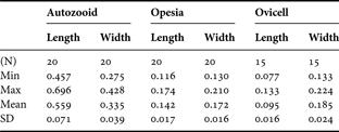
Measurements (in mm) of Mollia multijuncta (Waters) (lectotype).
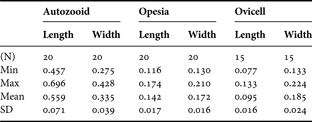
Min, minimum; Max, maximum; SD, standard deviation.
Mollia multijuncta is differentiated from M. patellaria, as described by Prenant & Bobin (Reference Prenant and Bobin1966), by the following characters, some already indicated by Waters (Reference Waters1879): the zooids are larger and more elongate; the opesia is not trifoliate; the contiguous autozooids are linked by at least two tubules, rather than one; the separation between the autozooids is very variable, and although the figure by Waters presents the zooids as highly separated, they are usually closer than in M. patellaria. Finally, the ovicell is very different in both species, as it is small in M. multijuncta, scarcely prominent, with a smooth ectoecium that forms a narrow rim clearly separated from the zoecial frame and a granular endoecium that is not clearly visible in frontal view, whereas in M. patellaria the ovicell is globose, prominent, and finely granular.
Mollia multijuncta is also differentiated from M. circumcincta, a species for which Hayward & McKinney (Reference Hayward and McKinney2002) have selected a neotype, as it has larger autozooids, with a larger opesia. However, the main difference is in the ovicell, which is smaller in M. circumcincta and appears to lack an ecotecium, whereas the endoecium is granular and the surface is continuous with the zoecial framework.
Mollia multijuncta appears to have been found on very few occasions, although the different synonyms considered by the authors, as well as the possible confusions with other species make compilation from the literature difficult. In fact, this species was cited several years later from Madeira, by the same author (Waters, Reference Waters1925). After revising the preserved material we consider that it probably belongs to a different species; nevertheless, the colonies lack ovicells, which are structures of great importance in differentiating species in Mollia and other similar genera.
Mollia cristinae sp. nov. is basically differentiated from M. multijuncta by a larger opesia, almost as long as it is wide, whereas in M. multijuncta it is clearly wider than long. In addition, the ovicell of the former is larger, and the rim of the ectoecium is more developed, especially at the mid-frontal level.
Finally, we consider that the material figured and described by López de la Cuadra (Reference López de la Cuadra1991) as Mollia rosselii (Audouin, 1826), which originates from the Isla de Tarifa and from Ceuta, corresponds to the material that we found on the coast of Portugal. It is therefore not impossible that our species had previously been cited under other different names.
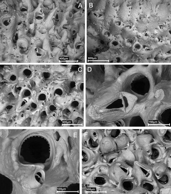
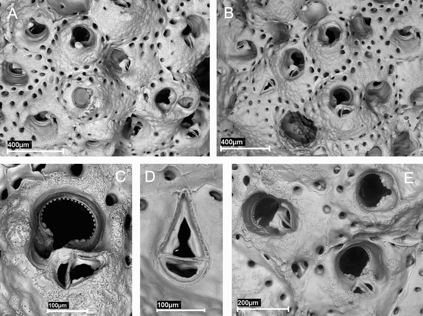
Hippoporina teresae sp. nov. (paratype MNCN-25.03/3779). (A) Margin of the colony; (B) secondary layer, with a partially covered ovicell (left); (C) autozooids with prickly peristome; (D) primary orifice; (E) avicularium; (F) ovicellate zooids.

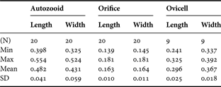
Measurements (in mm) of Hippoporina teresae sp. nov. (holotype): first layer.
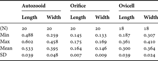
Min, minimum; Max, maximum; SD, standard deviation.
Measurements (in mm) of Hippoporina teresae sp. nov. (holotype): successive layers.
Min, minimum; Max, maximum; SD, standard deviation.
Hippoporina pertusa (Esper): López de la Cuadra, Reference López de la Cuadra1991: 189 (part), pl. 20, figure F, pl. 21, figure A.
TYPE MATERIAL
Holotype: MNCN-25.03/3778: Station 1. 37°01646′N 008°19038′W, 21 m, December 2007.
Paratypes: MNCN-25.03/3779: Station 1. 37°01646′N 008°19038′W, 21 m, December 2007. MNCN-25.03/3780: Station 2. 37°02079′N 008°18655′W, 19 m, December 2007.
OTHER MATERIAL
Dr López de la Cuadra personal Coll.: La Atunara, Mediterranean Andalusia. Figured in López de la Cuadra, Reference López de la Cuadra1991: Pl. 20, figure F, Pl. 21, figure A.
COMPARATIVE MATERIAL EXAMINED
Hippoporina acuta Cook Reference Cook1964: NHM-1963.4.16.5: Holotype, Figured.
Hippoporina lacrimosa Cook Reference Cook1964: NHM-1963.4.16.4: Holotype and paratypes, Figured.
Hippoporina pertusa (Esper, 1796): NHM-1847.9.16.46 (part): Figured by Busk (Reference Busk1854). NHM-1899.5.1.819: South Devon. Hincks Coll. NHM-1899.5.1.820: Antrim. Hincks Coll. NHM-1899.5.1.821 (part): South Devon. Hincks Coll. NHM-1911.10.1.1128: Off Antrim Coast. Norman Coll. NHM-1911.10.1.1129: Guernsey. Norman Coll. NHM-1911.10.1.1130: Brit. Barlee. Norman Coll. NHM-1911.10.1.1131: Shetland. Norman Coll. NHM-1911.10.1.1132 (part): British (Barlee). Norman Coll. NHM-1912.12.21.1043: Guernsey. Norman Coll. NHM-1930.1.4.6 (part): Millport. NHM-1936.12.30.362: Liverpool Bay, Thornely Coll. NHM-1960.4.6.20: Skye. Ryland Coll. NHM-1994.5.20.1 (part): Sound of Mull. Ryland Coll. Figured in the Synopsis.
Dr López de la Cuadra personal Coll.: La Atunara, V-1985, with ovicells.
Personal Coll.: some colonies from Ría de Vigo.
DIAGNOSIS
Colony multilaminar; autozooids rectangular, coarse nodular, perforated by areolar pores; primary orifice orbicular, with a wide shallow sinus, surrounded by a nodular peristome more developed proximally forming a thorny umbo. Ovicell scarcely prominent, covered with spiky nodules, perforated by small pores. Lateral avicularia rare.
ETYMOLOGY
This species is dedicated to Mrs Ma Teresa González García, wife of O.R.-G.
DESCRIPTION
Colony encrusting, multilaminar, beige coloured, forming large nodular masses, up to 25 cm2 in the holotype. Autozooids in the basal layer rectangular, with distal orifice, arranged in linear series, and separated by clear sutures. Autozooids in successive layers tend to be more quadrate, separated by prominent sutures and lose orientation; orifice placed more proximally, even centrally. Multiporous septula present in vertical walls. Frontal wall flat, coarsely granular, becoming prickly, evenly perforated by rounded pores deeply immersed with increasing calcification. Primary orifice orbicular, wider than long in basal zooids, circular in successive layers; with a wide shallowly concave sinus occupying most of the proximal border. Condyles pointed, rounded distally. Orifice surrounded by a well developed nodular peristome; in older zooids the peristome gives rise to a median suboral thorny umbo. Avicularia observed once, 0.140 mm long by 0.092 mm of maximum width; originated from an alveolar pore in the proximal margin of one autozooid; oval, with semielliptical mandible and complete bar. Ovicell wider than long, scarcely prominent, closed by the zooidal operculum. Frontal surface covered with spiky nodules, evenly perforated by rounded pores smaller than the autozooidal pores. Embryos orange-red.
REMARKS
Hippoporina teresae sp. nov. shows certain similarities to Hippoporina pertusa (Esper, 1796), the type species of the genus and considered to be relatively frequent, but to which different authors have attributed different characteristics; it is therefore quite possible that various different species are included under this denomination. Busk (Reference Busk1854: 80, pls 78, 79 figures 1 & 2) indicated the existence of a certain degree of variation in what he considered as L. pertusa. However, this variation was questioned by authors such as Norman (Reference Norman1868: 222) or Hincks (Reference Hincks1880: 305), who indicated possible confusion with Schizobrachiella sanguinea (Norman, Reference Norman1868). In fact, the original sample drawn by Busk (Reference Busk1854) (NHM-1847.9.16.46) contains several pieces of colonies of H. pertusa and S. sanguinea, and the words ‘L. pertusa (pl. LXXVIII fig. 3, pl. LXXIX fig. 2). S. sanguinea (pl. LXXVIII figs 1, 2)’ are written on the back of the slide; the remaining figure from the original plates (pl. 79 figure 1) also appears to correspond to H. pertusa. It is evident that a thorough revision of the previous citations of this and other related species is required, as well as designation of a neotype, although this is outside the scope of the present study.
Hippoporina teresae sp. nov. is differentiated from H. pertusa, as described by Hayward & Ryland (Reference Hayward and Ryland1999), by autozooids more rectangular, with a spiky frontal surface; by the primary orifice, with a much more pronounced sinus and surrounded by a nodular crown more developed suborally; and by the scarcely prominent ovicells covered with spiky nodules.
Hincks (Reference Hincks1880: 305, pl. 43, figure 4) reported the sporadic presence in H. pertusa of a lateral avicularium, the structure of which may be similar to that of H. teresae sp. nov. (Figure 3E). However, in the material belonging to this author and preserved in the Natural History Museum, London (NHM-1899.5.1.819, NHM-1899.5.1.820, NHM-1899.5.1.821) we did not observe any avicularia. López de la Cuadra (Reference López de la Cuadra1991: pl. 21, figure A) described a similar avicularium in a colony from La Atunara (Mediterranean Andalusia, near Gibraltar), which he identified, along with other material, as H. pertusa. Part of this material, which the author has sent to us, is identified here as H. teresae sp. nov., whereas the other part indeed corresponds to H. pertusa. It is therefore not impossible that the H. pertusa material with avicularia cited by Hincks (Reference Hincks1880) actually belongs to our species.
Finally, Cook (Reference Cook1964) described two species of Hippoporina from the west coast of Africa: H. lacrimosa and H. acuta, the type material of which we have examined. Both species often present one or two avicularia, situated on both sides of the orifice and orientated proximally. In the first of these the avicularium is spatulate, whereas in the second it is triangular. This characteristic, along with the aspects of the zooids and the orifice, enables both species to be clearly distinguished from H. teresae sp. nov.
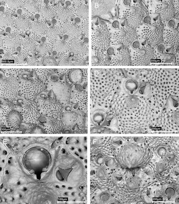
Schizoporella spinosa sp. nov. (A) (paratype MNCN-25.03/3782) general view; (B) (holotype) margin of the colony; (C) (paratype MNCN-25.03/3783) ovicellate and non-ovicellate zooids, and a well calcified zooid; (D) (holotype) an autozooid; (E) (paratype MNCN-25.03/3782) primary orifice and avicularium; (F) (holotype) ovicell and avicularia.

Measurements (in mm) of Schizoporella spinosa sp. nov. (holotype).

Min, minimum; Max, maximum, SD, standard deviation.
TYPE MATERIAL
Holotype: MNCN-25.03/3781: Station 2. 37°02079′N 008°18655′W, 19 m, December 2007.
Paratypes: MNCN-25.03/3781-3791: Station 2. 37°02079′N 008°18655′W, 19 m, December 2007. Twenty colonies.
COMPARATIVE MATERIAL EXAMINED
Schizoporella magnifica Hincks, 1886: NHM-1899.5.1.1076: Adriatic. Hincks Coll. type.
Schizoporella hesperia Hayward & Ryland, Reference Hayward and Ryland1995: NHM-1985.1.10.18A: Lundy Island. Hayward Coll. holotype.
DIAGNOSIS
Colony encrusting. Autozooids oval to round hexagonal. Primary orifice drop-shaped with small U-shaped sinus flanked by characteristic shoulders. Two–three constant stout spines. Suboral umbo. Triangular avicularia single or paired; proximal end level with the sinus, distal end below distal rim of orifice.
ETYMOLOGY
The name spinosa refers to the constant presence of oral spines.
DESCRIPTION
Colony encrusting, unilaminar, forming small irregular beige patches. Autozooids oval to round hexagonal, in alternating linear series separated by distinct sutures. Frontal wall convex, heavily calcified, evenly perforated by conspicuous round pores; with a median suboral stout umbo. In older autozooids pores may even disappear. Primary orifice drop-shaped; with a small, narrow, U-shaped sinus flanked by characteristic shoulders. Two-three (sometimes four) stout, hollow spines, sometimes as long as autozooid length; basis of spines dark brown in fresh material. Avicularia single or paired, occasionally absent; a third avicularia sometimes present. Proximal end level with the sinus, distal end below distal rim of orifice. Rostrum triangular, acute to frontal plane, or even nearly perpendicular; disto-laterally directed, slightly hooked distally. Ovicell prominent, globular, with irregular ridges and two–three series of marginal pores, and a few scattered pores, developing one or two small frontal umbos.
REMARKS
The genus Schizoporella is well defined, and many species have been described worldwide. Hayward & Ryland (Reference Hayward and Ryland1995) have re-described the species from British coasts, but many others certainly exist on other European coasts.
No other European species of the genus Schizoporella present the same combination of characters as S. spinosa sp. nov. More specifically, none of these present a drop-shaped orifice with a small U-shaped sinus flanked by shoulders, nor spines throughout the whole colony.
The sinus of the present species is similar to that of Schizoporella magnifica Hincks, 1886, recently re-described by Hayward & McKinney (Reference Hayward and McKinney2002). In addition, Ryland (Reference Ryland1968) reported the existence of three or four avicularia in some zooids in this species, as well as the presence of three oral spines on marginal zooids in some of the colonies collected at Skomer (Wales), characters also observed in our material. However, the differences between the two species are evident: the shape of the orifice, development of the suboral umbo and the position of the avicularia.
The position of the avicularia is similar to that of Schizoporella hesperia Hayward & Ryland, Reference Hayward and Ryland1995 a species that also has a suboral umbo, but other characteristics are different, such as the shape of orifice and sinus, ovicell, and development of spines.
Finally, Schizoporella dunkeri (Reuss, 1848), a species that may show also some similarities, can be easily distinguished by the D-shaped orifice, the absence of spines, and the shape and position of the avicularia, longer and placed more proximally.
Fenestrulina inesae sp. nov. (A–C: paratype MNCN-25.03/3797). (A) Margin of the colony, showing dietellae; (B) primary orifice; (C) group of zooids, one with simple pores (up right); (D) (holotype) ovicellate zooids.

Measurements (in mm) of Fenestrulina inesae sp. nov. (holotype).

DOA, distance orifice–ascopore; Min, minimum; Max, maximum; SD, standard deviation.
TYPE MATERIAL
Holotype: MNCN-25.03/3796: Station 3. 37°02423N 008°19576′W, 20 m, December 2007.
Paratypes: MNCN-25.03/3772, 3774, 3780, 3792, 3797, 3798: Station 2. 37°02079′ N 008°18655′W, 19 m, December 2007. Seven colonies.
COMPARATIVE MATERIAL EXAMINED
Fenestrulina asturiasensis Álvarez, Reference Álvarez1992: MNCN-25.03/187: holotype. Cape Peñas (northern Iberian Peninsula).
Fenestrulina barrosoi Álvarez, 1993: MNCN-25.03/280: holotype. Isla de Alborán, 45–52 m.
DIAGNOSIS
Primary orifice D shaped, nearly as long as wide. Three distal spines. Frontal wall evenly perforated by stellate pores. Ascopore level with the frontal surface.
ETYMOLOGY
This species is dedicated to Ms Inés Reverter González, daughter of O.R.-G.
DESCRIPTION
Colony encrusting, unilaminar, forming whitish irregular crusts. Autozooids in alternating series, typically hexagonal, separated by distinct grooves. Frontal wall smooth, convex, evenly perforated by some 60 stellate pores (0.025 mm diameter), except in a small area proximal to ascopore; two or three rows of pores between orifice and ascopore. Pores initially simple, soon filled by three to five (usually four) radii joined centrally. Two pairs of pores distal to orifice, each pair in a depression. Primary orifice wide, D shaped; three (sometimes two or four) distal spines; in ovicellate zooids only the two lateral spines are sometimes visible. Ascopore in the centre of the zooid, with smooth rim reniform to heart-shaped; lumen crescentic to horseshoe shaped, with small delicate denticulations. Large basal pore-chambers. Ovicell globular, prominent, with a smooth surface and a single peripheral series of some 15 basal pores. Ancestrula not known.
REMARKS
Until recently, only two species of the genus Fenestrulina were known in European waters: the presumed widespread Fenestrulina malusii (Audouin, 1826) and the Mediterranean Fenestrulina joannae (Calvet, 1902). In the last 25 years, almost 35 new species of the genus have been described throughout the world, although only two of these in European waters, and more specifically Iberian waters: Fenestrulina asturiasensis Álvarez, Reference Álvarez1992 and Fenestrulina barrosoi Álvarez, 1993. It is clear, as reported by several authors (Álvarez, Reference Álvarez1992; Hayward & Ryland, Reference Hayward and Ryland1999), that a thorough revision of this genus is required, and that many of the previous reports of F. malussii probably correspond to different as yet undescribed species. However, such a study is outside the scope of the present paper.
Fenestrulina inesae sp. nov. is easily distinguished from the other four species of Fenestrulina present in Iberian waters. Both F. joannae and F. asturiasensis have simple pores; in addition the former has an orbicular ascopore situated on an umbo, and six to eight oral spines, whereas in the latter the orifice is wider than long and has four or five distal spines. The other two Iberian species, F. barrosoi and F. malusii, also have stellate pores, although fewer and situated at the margins of the zooid; in addition the first has three to six spines (usually four or five) with the proximal pair particularly thickened, whereas the different forms that have been cited as F. malusii in Europe have a thickened and raised ascopore rim.
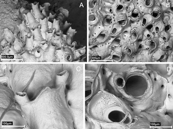
Celleporina derungsi sp. nov. (holotype). (A) Margin of the colony; (B) view of the colony, showing avicularia and ovicells; (C) autozooid and lateral avicularia; (D) primary orifice and vicarious avicularia.

Measurements (in mm) of Celleporina derungsi sp. nov. (holotype).

Min, minimum; Max, maximum; SD, standard deviation. Autozooids measured at the edge of the colony.
TYPE MATERIAL
Holotype: MNCN-25.03/3792: Station 2. 37°02079′N 008°18655′W, 19 m, December 2007. One colony marked with a black arrow.
Paratypes: MNCN-25.03/3792: Station 2. 37°02079′N, 008°18655′W, 19 m, December 2007. Six colonies with the holotype.
MNCN-25.03/3774, 3793-3795: Station 2. 37°02079′N 008°18655′W, 19 m, December 2007. Four colonies.
MNCN-25.03/3793: Station 3. 37°02423N 008°19576′W, 20 m, December 2007. Two colonies.
COMPARATIVE MATERIAL EXAMINED
MNHN-10233: Cellepora costazii, Tiboulen de Pomegnes, Marseille. Gautier Coll.
MNHN-10234: Cellepora costazii. Niolon (Marseille) (empty tube) Gautier Coll.
MNHN-10235: Cellepora costazii. Port Man (Marseille). Gautier Coll.
DIAGNOSIS
Tall tubular peristome, with a median triangular projection and flanked by two small avicularia. Vicarious avicularia broadly spatulate. Primary orifice orbicular, with broad, shallow sinus almost straight, occupying most of the proximal width.
ETYMOLOGY
This species is dedicated to Mr Manuel Derungs, grandfather of J.S.
DESCRIPTION
Colony unilaminar, forming small circular domed patches. Autozooids oval, vitreous, slightly rough, with widely spaced but distinct marginal pores. Primary orifice orbicular, with a broad, shallow sinus almost straight, occupying most of the proximal width. Orifice completely surrounded by a tall tubular peristome distally directed, slightly flared apically, and frequently with a median triangular projection. Avicularia paired, situated laterally on the peristome rim, their columnar cystids running over the lateral margins of the peristome; oval, with toothed distal rim, slightly acute to the apertural plane and directed outwards. Vicarious avicularia frequent, broadly spatulate, with a palatal foramen occupying half the rostrum and a slender crossbar without columella. Ovicell hemispherical, closing distally the peristome. Frontal tabula narrow, perforate by irregular peripheral pores and a few scattered central pores.
REMARKS
This species is very similar to Celleporina tubulosa (Hincks), a species cited on very few occasions; it was originally described by Hincks (Reference Hincks1880) as a variety of Cellepora costazii, but neither the original material nor the type locality are known. Hayward & Ryland (Reference Hayward and Ryland1999) described a specimen from south-west England that appears to correspond well to the description made by Hincks. Celleporina derungsi sp. nov. differs from this description by a more orbicular orifice, with an almost straight sinus, by the presence of a median triangular projection on the edge of the peristome, which is shorter, and by a wider spatulate avicularium.
The present species also appears to differ from the Mediterranean material reported by several authors as C. tubulosa. The citations by Zabala (Reference Zabala1986) and Zabala & Maluquer (Reference Zabala and Maluquer1988) may correspond to C. tubulosa, although the primary orifice must be examined. The citation by Gautier (Reference Gautier1962 as Celleporina hassallii var. tubulosa) presents more problems, as this author only includes a brief diagnosis. We have revised 3 original samples (MNHN-10233, MNHN-10234 and MNHN-10235) labelled by Gautier as Cellepora costazii and originating from Marseille, which may correspond to this record. Sample 10234 was empty, whereas the material of the other two samples presents abundant vicarious avicularia with parallel edges, and the autozooids have not an authentic peristome, but rather a highly developed suboral border, delimited by two lateral avicularia; in addition the primary orifice is transversally elliptical (Figure 7). This material appears to be very similar to that figured as Celleporina hassallii (Johnston, 1847) by Zabala (Reference Zabala1986, pl. 21 figures C & D). We consider that this material, very different from C. tubulosa and from C. derungsi sp. nov., must correspond to an undescribed species of the genus Celleporina. Finally, Hayward & McKinney (Reference Hayward and McKinney2002) report a specimen from the northern Adriatic as C. tubulosa, although the narrow rostrum on the vicarious avicularia and the position of the peristomial avicularia lead us to doubt the identification; nonetheless, this material is clearly different from that collected from the Algarve.
Celleporina sp. (A, B, ‘Celleporina costazii’ MNHN-10233). (A) Margin of the colony; (B) view of the peristomes; (C, D, ‘Celleporina costazii’ MNHN-10235); (C) primary orifice; (D) group of vicarious avicularia.

REVISED DIAGNOSIS
Colony encrusting. Zooids erect; frontal surface smooth, inflated, with areolar marginal pores. Primary orifice semicircular: anter denticulate, poster straight. Condyles small. Opercula with strong marginal sclerites. No spines. Peristome well developed, not obscuring the orifice. Dimorphic suboral adventitious avicularia: small and oval, or large and triangular. Uncinate process poorly developed. Ovicell smooth, imperforate, not closed by the operculum; ectooecium membranous.
Type species: Cellepora sardonica Waters, Reference Waters1879.
TYPE MATERIAL
Lectotype of Cellepora sardonica Waters, Reference Waters1879 (designated here): MM-H.1186.5995: Naples, 23/11/1875.
Paralectotypes of Cellepora sardonica Waters, Reference Waters1879 (designated here): MM-H.1186.6095: Naples, 22/12/1875. MM-H.1186.6096: Naples, 23/12/1875. MM-H.1186.6097: Naples, 30/12/1875. MM-H.1186.6098: Naples, 22/12/1875, opercula. MM-H.1186.10024: Naples, 1875.
OTHER MATERIAL EXAMINED
Cellepora sardonica Waters: MM-H.1186.5996: Capri. MM-H.1186.6099: Madeira. MM-H.1186.6100: Madeira, opercula. MM-H.1186.6101: Villefranche, slide. MM-H.1186.6102: Villefranche, slide. MM-H.1186.6103: Villefranche, slide. MM-H.1186.6104: Oran, opercula.
REMARKS
This genus was created by Barroso (Reference Barroso1926) for the species Cellepora sardonica Waters, which is therefore the type species of the genus. However, a year later (Barroso, Reference Barroso1927) the author made a more complete diagnosis of the genus Dentiporella, although still rather scant. Gautier (Reference Gautier1962) rejected the validity of this genus as he considered that the characters indicated did not justify the creation of a new genus. This author placed C. sardonica in the genus Celleporaria; however, this genus after the re-description by Harmer (Reference Harmer1957) does not present denticulation in the anter, so his decision does not seem appropriate. Zabala (Reference Zabala1986) recovered the genus Dentiporella, with some reservations, and made a new, more detailed diagnosis, although in our opinion with some errors, as this author interpreted the large triangular suboral avicularium of C. sardonica as vicarious, whereas we believe that it is actually adventitious.
We have revised the original material of C. sardonica, which consists of 13 samples deposited in the MM (Figure 8A–D), and we have selected a lectotype and paralectotypes.
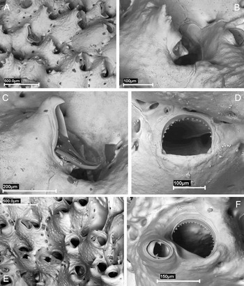
(A–C) Dentiporella sardonica (lectotype). (A) View of the colony; (B) oval suboral avicularium; (C) triangular suboral avicularium; (D) Dentiporella sardonica (MM-H.1186.6096), primary orifice; (E, F) Rhynchozoon revelatus Hayward & McKinney; (E) view of the colony; (F) primary orifice and suboral avicularium.

In our opinion, Dentiporella is a valid genus, which fits perfectly within the family Phidoloporidae. This genus shows similarities to the genus Rhynchozoon Hincks, as proved by the recent description of Rhynchozoon revelatus Hayward & McKinney, Reference Hayward and McKinney2002 (Figure 8E, F) a species considered here to be a synonym of D. sardonica (see below). However, Dentiporella differs from Rhynchozoon by a straight proximal edge of the orifice, without sinus; small rounded condyles; the absence of spines; and by the dimorphic suboral avicularium, with a poorly developed uncinate process.
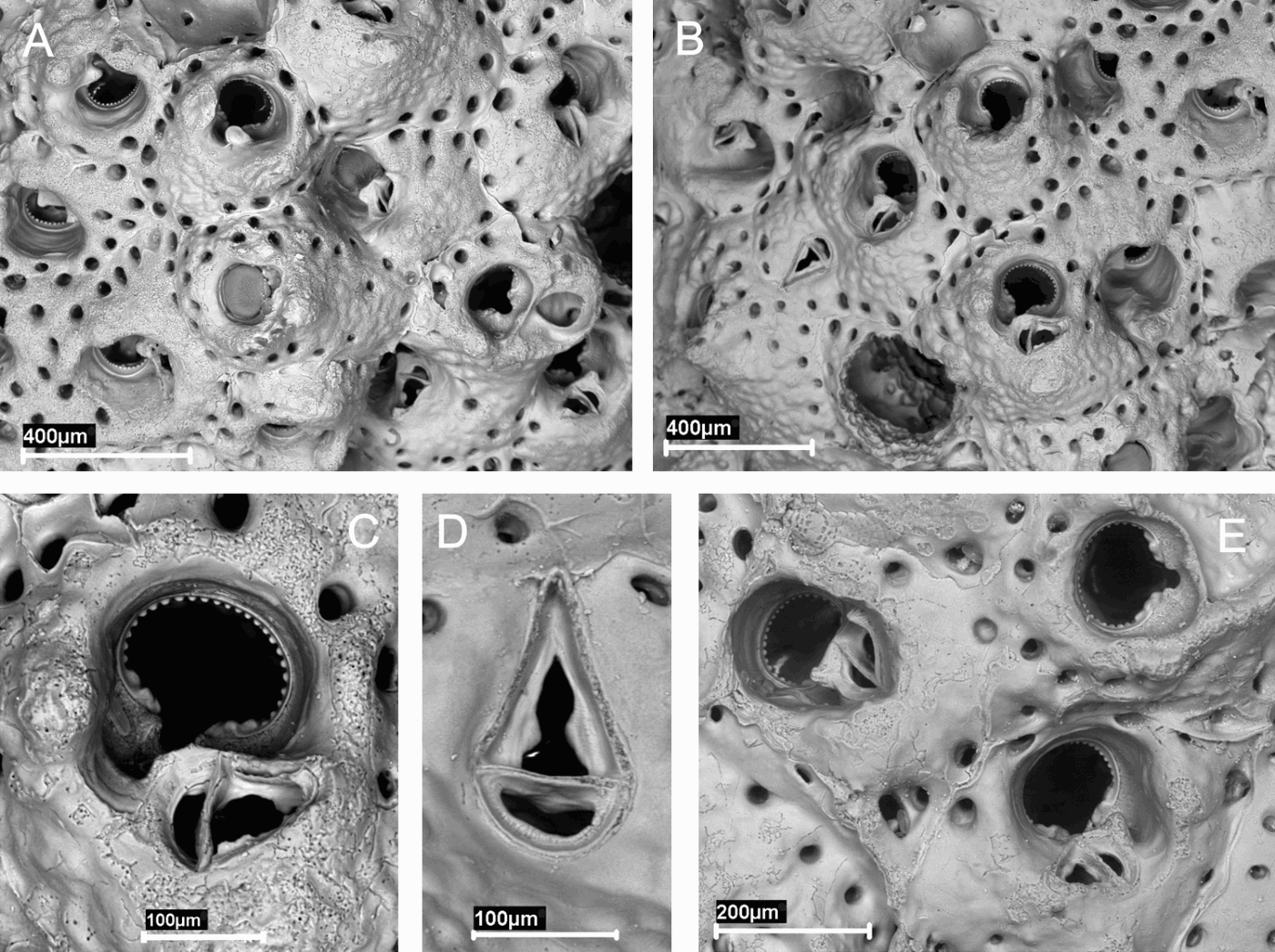
Dentiporella saldanhai sp. nov. (A–E, paratype MNCN-25.03/3773). (A) Group of zooids, some ovicellate, and frontal avicularia (left); (B) group of zooids with marginal pores and frontal avicularia; (C) frontal view of a group of zooids; (D) primary orifice with triangular suboral avicularium; (E) primary orifice with oval suboral avicularium; (F) (holotype) ovicellate zooids and suboral avicularia.

Measurements (in mm) of Dentiporella saldanhai sp. nov. (holotype).

DO, distance between midpoints of adjacent orifices; Min, minimum; Max, maximum; SD, standard deviation.
TYPE MATERIAL
Holotype: MNCN-25.03/3772: Station 3. 37°02423N 008°19576′W, 20 m, December 2007.
Paratypes: MNCN-25.03/3773, 3774: Station 2. 37°02079′N 008°18655′W, 19 m, December 2007. Two colonies.
COMPARATIVE MATERIAL EXAMINED
Dentiporella sardonica: original material from Waters Coll. (see above material revised for the genus). MNHN-4045: Oran, Calvet Coll. MNHN-4057: Oran, Calvet Coll. MNHN-6034: Corse, Calvet Coll. MNHN-8727: Antedon, 1960, st. 1692, Gautier Coll. MNHN-8741: Bonifacio, Gautier Coll. MNHN-10257: Marseille, Gautier Coll. MNHN-10258: Marseille, Gautier Coll. MNHN-10259: Marseille, Gautier Coll. MNHN-10260: Marseille, Gautier Coll.
Rhynchozoon revelatus Hayward & McKinney: five colonies from Rovinj (northern Adriatic).
DIAGNOSIS
Primary orifice widely D-shaped, as wide as long, with 14–19 denticles; thick and deep peristome, with a stout, columnar suboral umbo. Dimorphic suboral adventitious avicularia; sporadic small frontal oval avicularia.
ETYMOLOGY
This species is dedicated to Professor Luiz Saldanha, famous Portuguese zoologist.
DESCRIPTION
Colony multilaminar, forming extensive irregular crusts, cream-coloured. Autozooids with thick frontal shield finely granular, bordered by a single series of few marginal pores, reduced in later ontogeny. Primary orifice D-shaped, as wide as long. Distal rim with 14–19 thin and widely spaced denticles. Proximal rim straight to shallowly concave; small rounded condyles in the proximolateral corners. No oral spines. Peristome thick and deep, defining an orbicular secondary orifice but not obscuring the primary one. A stout, columnar, median suboral umbo, spiky at the apex when not broken; distally, the umbo presents an avicularium, facing towards the orifice and directed upwards. The avicularium may be either small, with oval rostrum, and a poorly developed columella in the crossbar, placed at the basis of the umbo; or large, with triangular rostrum distally hooked; both without palate. A short uncinate process projects from the base of avicularia, facing a shallow lateral notch in the peristome. Sporadically, small frontal oval avicularia budded from the marginal pores. Ovicell globular, with smooth surface, flat frontally, projecting vertically above peristome rim. Frontal ectooecium partly membranous. Embryos cream-coloured.
REMARKS
Dentiporella saldanhai sp. nov. is very similar to Dentiporella sardonica (Waters, Reference Waters1879) and to the recently described Adriatic species Rhynchozoon revelatus Hayward & McKinney, Reference Hayward and McKinney2002.
We have revised the type material of C. sardonica, deposited in the MM (Figure 8A–D), as well as some material cited by Gautier (Reference Gautier1962) deposited in the MNHN. We have also revised five colonies of R. revelatus from the northern Adriatic, sent to us by Dr McKinney (Figure 8E, F). According to the original description (Hayward & McKinney, Reference Hayward and McKinney2002), the type material of this species is deposited in the American Museum of Natural History. However, the staff of the museum has informed us that the material is not deposited in their collections and they do not know where it is located. Furthermore, we have not been able to gain access to the paratypes reported to be deposited in the Centre for Marine Research-Rovinj. It is possible that registration numbers were designated for the type material, but that the material was never deposited. However, the original description of the species is very complete and corresponds perfectly to the material owned by Dr McKinney. Finally, we have also examined several photographs of Mediterranean material, sent to us by Dr Chimenz, and labelled as D. sardonica and R. revelatus; some of these colonies were originally identified by Dr J.G. Harmelin and Dr P.J. Hayward (Chimenz, personal communication, 24 June 2008). We have found that apart from the expected variability between different colonies there is also a large degree of variability between different parts of the same colony, which affects the orifice, development of the peristome and of the umbo, as well as the orientation and position of the avicularia. However, we have not been able to identify any character that enables us to differentiate R. revelatus and C. sardonica. We therefore believe that Rhynchozoon revelatus Hayward & McKinney, Reference Hayward and McKinney2002 must be considered as a junior synonym of Cellepora sardonica Waters, Reference Waters1879.
Dentiporella saldanhai sp. nov. differs from D. sardonica by its primary orifice, that is as long as wide and which has 14–19 denticles and moderately developed condyles, whereas in the latter species the orifice is clearly wider than long, has 20–22 denticles and poorly developed condyles. In addition, D. saldanhai sp. nov. may sporadically present small frontal oval avicularia, not present in D. sardonica.
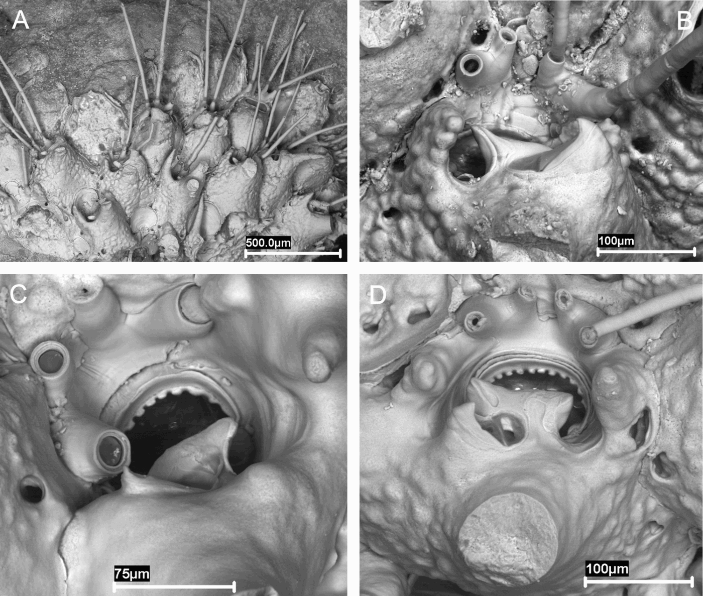
Rhynchozoon celestinoi sp. nov. (A, D: holotype). (A) View of the colony; (B) group of zooids with a frontal avicularium; (C) primary orifice and suboral avicularium; (D) frontal avicularium; (E) (paratype MNCN-25.03/3770) group of zooids.
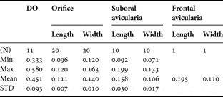
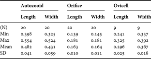
Measurements (in mm) of Rhynchozoon celestinoi sp. nov. (holotype).

DO, distance between midpoints of adjacent orifices; Min, minimum; Max, maximum; SD, standard deviation.
TYPE MATERIAL
Holotype: MNCN-25.03/3769: Station 3. 37°02423N 008°1957′6W, 20 m, December 2007.
Paratypes: MNCN-25.03/3770, 3771: Station 3, 37°02423N 008°19576′ W, 20 m, December 2007. Two colonies.
COMPARATIVE MATERIAL EXAMINED
Rhynchozoon neapolitanum Gautier, Reference Gautier1962: MNHN-11007. Type. Station 1732. Bonifacio (Corse). MNHN-11008. Type. Station 561. Port de Pantellaria (Tunisie).
DIAGNOSIS
Primary orifice elliptical, with a narrow sinus delimited by two strong, warty condyles. Median suboral avicularia present in most zooids; frontal avicularia triangular, infrequent.
ETYMOLOGY
This species is dedicated to Mr Celestino Souto, grandfather of J.S.
DESCRIPTION
Colonies encrusting, developing frontally budded, massive nodular form. Autozooids with vitreous, nodular frontal shield, bordered by single series of conspicuous marginal rounded pores. Primary orifice elliptical, wider than long. Distal and lateral rim with thick and closely spaced denticulations. Proximal border with a small, quadrate sinus, accentuated by warty condyles. A deep asymmetric peristome developing through ontogeny, not obscuring the primary orifice; oral spines and knobs absent. Median suboral avicularium present in most zooids, acute to frontal plane and laterally directed; rostrum broadly triangular, distally hooked. The avicularium is slightly displaced laterally, while its proximal end projects an uncinate process defining a lateral pseudosinus. Frontal avicularia infrequent; rostrum triangular, directed outwards. Ovicell not known.
REMARKS
Although the presence of a sinus is one of the descriptive characteristics of the genus Rhynchozoon, the sinus is particularly marked in this species, as most have a wide, shallow sinus.
The species described here presents some similarities to the Mediterranean species Rhynchozoon neapolitanum Gautier, Reference Gautier1962, the type material of which we have examined (MNHN-11007 and MNHN-11008). However, there are remarkable differences: the orifice of R. celestinoi sp. nov. is elliptical, wider than long and has a small, quadrate sinus and large warty condyles, whereas in R. neapolitanum the orifice is more orbicular and tends to be as long as wide, the sinus is proportionally shorter and narrow, and the condyles smaller and faceted. The development of the peristome also differs in the two species, as in R. neapolitanum there are distal and lateral knobs and two stout proximal processes, which do not appear in R. celestinoi sp. nov. Finally, the suboral avicularium is fairly frequent in R. celestinoi sp. nov., whereas the frontal avicularium occurs sporadically; on the contrary, in R. neapolitanum the suboral avicularia are scarce, whereas the frontal avicularia may be abundant, and also have a well developed cystid.
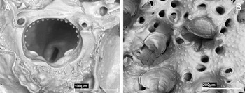
Of the remaining European species of the genus Rhynchozoon, none present characteristics that may be confused with R. celestinoi sp. nov. The type species of the genus, in theory the most cited from the European Atlantic coasts, and which was also found in the present study, is Rhynchozoon bispinosum (Johnston, 1847) (Figure 11). Its orifice differs from the orifice of the species described here, by a wide, shallow sinus, and a very different ratio between the width and length. Moreover, the morphology of the frontal wall is different in both species and the knobs that surround the orifice in R. bispinosum do not exist in R. celestinoi sp. nov.
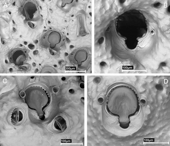
Rhynchozoon bispinosum. (A) Primary orifice (MNCN-25.03/3806, Station 3); (B) ovicellate zooids (MNCN-25.03/3792, Station 2).

Another European species with a marked sinus is Rhynchozoon sp. 1, cited by Hayward (Reference Hayward1974) from Chios (Aegean Sea), although it is much wider than the species described here. Zabala & Maluquer (Reference Zabala and Maluquer1988) consider Rhynchozoon sp. 1 to be very closely related to R. bispinosum. Nonetheless, there are many important differences from R. celestinoi sp. nov. As described by Hayward (Reference Hayward1974), the frontal wall of this species presents many knobs, not present in R. celestinoi sp. nov. In addition, the suboral avicularium observed in Hayward (Reference Hayward1974: figure 8A), does not resemble that present in the species described here.
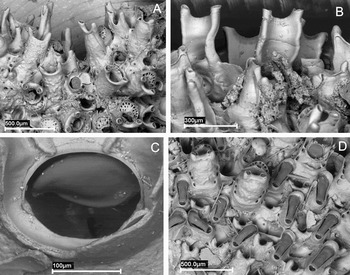
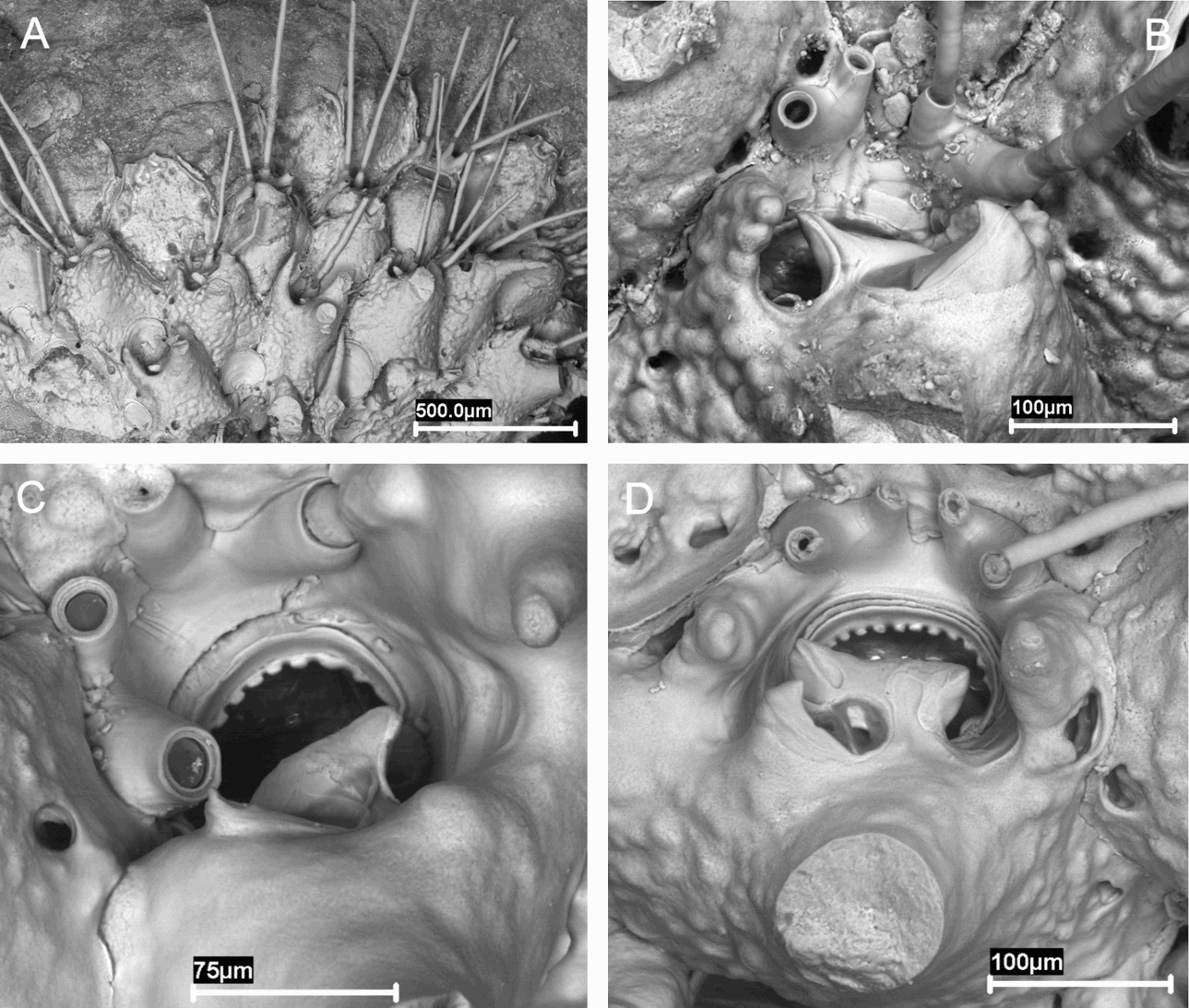
Rhynchozoon rosae sp. nov. (A, B, paratype MNCN-25.03/3759). (A) Margin of the colony showing oral spines; (B) suboral avicularium; (C, D, paratype MNCN-25.03/3760); (C) primary orifice partially obscured by uncinate process; (D) orifice showing one condyle (right), and some knobs.
Measurements (in mm) of Rhynchozoon rosae sp. nov. (holotype).
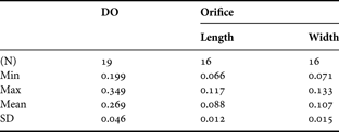
DO, distance between midpoints of adjacent orifices; Min, minimum; Max, maximum; SD, standard deviation.
TYPE MATERIAL
Holotype: MNCN-25.03/3758: Station 2. 37°02079′N 008°18655′W, 19 m, December 2007.
Paratypes: MNCN-25.03/3759-3768: Station 2. 37°02079′N, 008°18655′W, 19 m, December 2007. Ten colonies.
DIAGNOSIS
Small circular colonies. Autozooids with large peripheral pores. Primary orifice wider than long, with poster shallowly concave. Two distal pairs of stout spines.
ETYMOLOGY
This species is dedicated to Mrs Rosa Dopico, grandmother of J.S.
DESCRIPTION
Colonies forming small circular patches on maërl. Autozooids oval; frontal wall granular, with few large peripheral pores. Primary orifice wider than long; anter with thick, blunt denticulations; poster shallowly concave, with two lateral, rounded condyles, difficult to see. Peristome enclosing the orifice proximally, developing as a big suboral knob; the distal rim of the peristome formed by the thick bases of four stout spines, united in two pairs. Avicularia medio-proximal; rostrum triangular, hooked distally. Its proximal end projects an uncinate process defining a lateral pseudosinus. Frontal avicularia and ovicells not present in the studied material.
REMARKS
Only one European species of the genus Rhynchozoon has four spines, R. quadrispinatum Zabala & Maluquer, Reference Zabala and Maluquer1988. The original description of this species is extremely vague, but we have examined the type material of the species. Our material shows enough differences to be considered as a different species. Both species have four spines, but these are long and thick, especially at the base in R. rosae sp. nov., whereas in R. quadrispinatum they are slender and fragile (Zabala, personal communication, July 2008). Moreover, in the description of R. quadrispinatum no reference is made to the presence of knobs, although some fine broken knobs appear in the figure, which are abundant in our species. Finally, R. quadrispinatum has only been found in submarine caves in the Balearics (Zabala, personal communication, July 2008).
Stephanollona armata (MNCN-25.03/3801, Station 3). (A–D) Variability of the primary orifice and avicularia on one single colony.

MATERIAL EXAMINED
MNCN-25.03/3791, 3792: Station 2, six colonies. MNCN-25.03/3793, 3794, 37800-3803: Station 3, five colonies.
COMPARATIVE MATERIAL EXAMINED
NHM-1899.5.1.973: Lepralia armata. Holotype. Hincks Coll.
REMARKS
The material from Portugal that we have studied consists of 11 colonies, which corresponds well to the holotype of Lepralia armata Hincks, Reference Hincks1862 deposited in the NHM, and with the descriptions made of this species (e.g. Gautier, Reference Gautier1962; Zabala, Reference Zabala1986; Hayward & Ryland, Reference Hayward and Ryland1999). However, some details in both our material and the holotype (Figure 14) have not been described in the literature consulted: firstly, the constant presence of a thick quadrate denticle in the apertural bar in both the small oval avicularium, and in the spatulate avicularium; secondly, the serrated distal rim of the avicularia, a detail that can only be observed by SEM.
Stephanollona armata (holotype). (A) Group of zooids and spatulate avicularia; (B) ovicellate zooids.

Several authors have indicated a great variation in the appearance of the colonies with age and degree of calcification: e.g. disordered zooids and deeply immersed orifices. We have observed the extent to which this species varies, as the primary orifice in the same colony may present a very different appearance, especially as regards the shape of the sinus (Figure 13). The position of the oval avicularia may also vary considerably, from clearly distolateral at the edges of the colonies, to suboral in the most calcified zones.
Stephanollona contracta (MNCN-25.03/3798, Station 1). (A) Group of ovicellate zooids; (B) group of zooids and a spatulate avicularia; (C) ovicellate zooid; (D) primary orifice and avicularium.

Measurements (in mm) of Stephanollona contracta (waters).

Min, minimum; Max, maximum; SD, standard deviation.
MATERIAL EXAMINED
MNCN-25.03/3799: Station 1, one colony on a Serpulidae.
DESCRIPTION
Colony encrusting, unilaminar to multilaminar, whitish in colour. Autozooids irregular shaped. Nodular frontal shield imperforate, except by a single series of large areolar marginal pores. Primary orifice roughly orbicular, longer than wide, with lateral walls parallel. Anter surrounded by a rim that extends down to the small condyles, those marking the opening of a broad, rounded sinus; some 18 thick and closely spaced denticles in the lateral and distal borders. Proximally, the primary orifice presents a rounded rim of smooth calcification, sometimes interrupted by a blunt projection of the frontal shield. Four to six hollow spines surround the distal half of the orifice; spines articulate at base, long (even as long as zooidal length), progressively narrow, with transversal rims marking successive regenerations. Small avicularia, frequently absent, with finely serrate oval rostrum. Placed latero-proximal to orifice, raised at an angle, and orientated proximolateraly. Sporadically, large adventitious spatulate avicularia, parallel-sided, with hooked rostrum. Subtriangular foramen, cross-bar with a small denticle. Ovicell globular, nodular, not closed by the operculum. Proximally presents a semielliptical area partially calcified, with a distal fenestra.
REMARKS
Lepralia contracta was described from Madeira by Waters (Reference Waters1899). The phylogenetic position of this species has been uncertain for many years, and the species has been included in different genera, although at present it is usually included in the genus Stephanollona Duvergier, 1920.
This species was later cited by many authors from diverse geographical areas, although many of these must correspond to other closely related species; in fact, it is now considered that S. contracta represents a widely distributed species complex (Winston, Reference Winston2005). However, the main problem is the lack of type material for this species. The original material of L. contracta was first deposited at the Museo do Seminario do Funchal, and later in the Jardim Botânico de Madeira. However, it seems that the type material has been lost. We were only able to study a photograph deposited in the NHM, assumed to be of this material and labelled as type material, but this was not sufficient to characterize the species. It is therefore not possible at present to define S. contracta correctly.
Our material is similar to that cited by Cook (Reference Cook1964) and Arístegui Ruiz (Reference Arístegui Ruiz1984) as Cleidochasma contractum. However, there are some differences: both authors indicate the presence of four to eight spines in their material, whereas in our material there are four to six oral spines. On the other hand, the sinus figured by Arístegui Ruiz (Reference Arístegui Ruiz1984: figure 36d, e; pl. 21, figures 3 & 4) appears to be narrower. Finally, both authors indicate that the ovicell has a large semi-circular area on the front; in our material the ovicell has a small frontal fenestra, although this may have originally been larger but became gradually reduced due to calcification (Figure 15 A, C).
This species has previously been reported for the Iberian Peninsula by d'Hondt (Reference d'Hondt1988), specifically at a depth of 200 m close to Estepona (Andalusia); however, the original material (MNHN-14947) actually belongs to S. armata, and thus the present citation of S. contracta represents the first for this species in continental European waters.
DISCUSSION
As already indicated in the Introduction, the literature referring to Bryozoa from the Portuguese coasts is relatively scarce. Of the 57 species identified during the present study, eight are new to science, and 16 more are new to the Portuguese coasts. In other words 42% of the species examined in the present study were not previously known in the area, which demonstrates the scarce knowledge of the Portuguese fauna, and also of the Iberian fauna in general.
In our opinion, the fact that in supposedly well known genera, widely reported from European waters, such as Schizoporella, Fenestrulina, Hippoporina or Celleporina, new species are still being described, demonstrates the profound lack of knowledge of the European bryozoological fauna. In light of the present results, and those of many other recent authors, it is evident that the detailed study of new material will reveal that there are still many new species to be discovered in European waters. Study of the material deposited in collections is especially revealing, as on not a few occasions prior identifications have proved to be incorrect, either because of simple mistakes, or more often because the concept of a single species was not the same for different authors. The correct characterization of species by the designation of types and their complete description according to current standards, often lacking in older studies, is therefore fundamental; it is the only way to confirm later citations. The general distributions of species, even those well-known, must be considered with caution, as they may actually correspond to several species with more restricted distributions.
Revision of the original material of Mollia multijuncta has enabled us to designate a lectotype and clarify its specific status. The material collected from Portuguese waters shows enough differences to consider it as a new species, which has, however, been cited at least once with another name. The aspect of the autozooids of the different species of Mollia and other similar genera appears to vary relatively little among species. However, the ovicell appears to be a much more diverse character and therefore very important for characterizing and distinguishing species. A thorough revision of material previously cited as Mollia, Rosseliana and Steraechmella, and of other similar genera is required in order to establish new relationships between them.
The redescription of the genus Dentiporella from the original material of C. sardonica, and the designation of a lectotype must be emphasized. Rhynchozoon revelatus is here considered as a junior synonym. The material from Portugal corresponds to a different species, D. saldanhai sp. nov., which represents the second of this genus.
The descriptions of new species of the genera Hippoporina (H. teresae sp. nov.), Schizoporella (S. spinosa sp. nov.), Fenestrulina (F. inesae sp. nov.) and Celleporina (C. derungsi sp. nov.) are especially interesting as these genera are supposed to be well known in European waters, and several species of the latter three have been described in recent years. Study of the collection material of the genera Hippoporina and Celleporina also suggests that unknown species remain to be described, and that a thorough revision of both genera is required.
The genus Fenestrulina is highly diverse in Iberian waters, as it is represented by five well differentiated species, whereas in European waters only one or two species are known. However, a revision of collection material will certainly show a much greater biodiversity.
Finally, the morphology of the genus Rhynchozoon makes it very difficult to study, although use of SEM is enabling better characterization of the species. The type species of the genus, R. bispinosum, as well as two new species have been found in Portuguese waters. This represents a significant increase in the biodiversity of this genus in Europe.
However, we believe that it would be an exaggeration to try to split all widely distributed species into several species of more limited distribution.
It has been stressed that as bryozoans are sessile organisms that produce short-lived non-planktotrophic larvae, their capacity for dispersion is rather limited (e.g. Taylor, Reference Taylor1988; Goldson et al., Reference Goldson, Hughes and Gliddon2001; Watts & Thorpe, Reference Watts and Thorpe2006; Berning & Kukliński, Reference Berning and Kukliński2008). This appears to contradict the existence of species with a wide distribution. However, we consider that the results of the present study, and those of many other authors, show that some species are actually widely distributed, however paradoxical this may seem. This apparent contradiction can be explained by other mechanisms of dispersion like step-by-step colonization and artificial introductions. Moreover, some species of bryozoans produce non-planktotrophic larvae that are able to be carried by the currents of water to a rather large distance from the laying place (Uly & d'Hondt, Reference Uly and d'Hondt1988; d'Hondt & Goyffon, Reference d'Hondt and Goyffon1992, Reference d'Hondt and Goyffon1993).
A certain degree of flexibility would be expected in widely distributed species, which would be reflected in intercolonial geographical variability, but not necessarily in differences of taxonomic relevance. It is also essential to take into account the importance of intracolonial variability. However, such variations, which may be highly significant, are not usually reflected in the traditional descriptions, other than the biometric limits of the measurements; this is partly because of the need for brevity and conciseness in the descriptions and figures, and partly because of the general idea that some characters are constant, such as the primary orifice. We have found that in species such as D. sardonica and particularly in S. armata, the characters are so variable within a colony, especially the shape of the orifice, that on viewing different photographs of the same colony, it could easily be believed that they correspond to different species (see Figure 13B–D). This must be taken into account when differentiating cryptic species.
Different authors (e.g. Ekman, Reference Ekman1967; López de la Cuadra & García-Gómez, Reference López de la Cuadra, García-Gómez, Hayward, Ryland and Taylor1994; Carballo et al., Reference Carballo, Naranjo and García-Gómez1997) consider the Strait of Gibraltar as the limit of three biogeographical regions: the Mediterranean region (to the east), the Lusitanian region (to the north) and the Mauretanian region (to the south). The sampling locations in the present study are found to the north-west of the Gulf of Cadiz, and are therefore included in the Lusitanian region. This is reflected by the presence of many Lusitanian or Lusitanian–Boreal species, in accordance with the data provided by López de la Cuadra & García-Gómez (Reference López de la Cuadra, García-Gómez, Hayward, Ryland and Taylor1994), although there are also many species of assumed wide distribution in both the Atlantic Ocean and Mediterranean Sea.
However, we have also found many colonies of Myriapora truncata (Pallas, 1766) and Schizomavella mamillata (Hincks, Reference Hincks1880) (Figure 16). These well-known species are considered as possibly endemic to the Mediterranean (López de la Cuadra & García-Gómez, Reference López de la Cuadra, García-Gómez, Hayward, Ryland and Taylor1994; Hayward & McKinney, Reference Hayward and McKinney2002) which indicates the influence of this region on the sampling area. Even other citations for both species further to the north and previously considered doubtful may be valid.
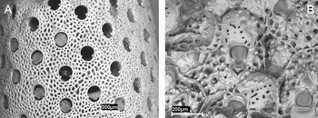
(A) Myriapora truncata (MNCN-25.03/3804, Station 3); (B) Schizomavella mamillata (MNCN-25.03/3805, Station 3).

The presence of these species in Atlantic waters provides more data relative to the transfer of species from shallow Mediterranean waters to the Atlantic via the Strait of Gibraltar. The deep water bryozoan fauna at both sides of the Strait of Gibraltar was studied during the Balgim sampling survey by Harmelin & d'Hondt (Reference Harmelin and d'Hondt1993). A great similarity was observed between the benthonic fauna from the Mediterranean Sea and that from the Atlantic Ocean, but only in the regions that are clearly influenced by the Mediterranean outflow; this in turn represents a barrier for the entry of Atlantic benthonic species to the Mediterranean Sea. However, it would be expected that the superficial Atlantic current will represent a barrier to the exit of species from the Mediterranean; therefore the presence of shallow Mediterranean species on the western side of the Strait of Gibraltar is contradictory to the general circulation of currents in the region (Gascard & Richez, Reference Gascard and Richez1985). The same problem has been indicated by Carballo et al. (Reference Carballo, Naranjo and García-Gómez1997) in a study of littoral sponges, as species endemic to the Mediterranean Sea were found on the other side of the Strait of Gibraltar. The authors speculate that these species are not actually endemic to the Mediterranean Sea, but originate from tropical or subtropical waters—where they have been overlooked as these regions have been less well studied. Later they have extended to the north and entered the Mediterranean Sea during past geological eras. In the present case, the hypothesis of a meridional origin for the species is unlikely; although these fauna are comparatively less well studied, several studies on Bryozoa have been carried out on the Atlantic coast of north Africa (e.g. Canu & Bassler, Reference Canu and Bassler1925, Reference Canu and Bassler1928; d'Hondt, Reference d'Hondt1978; Arístegui Ruiz, Reference Arístegui Ruiz1984), and the presence of species that are abundant on both sides of the Strait of Gibraltar would probably have been detected. We believe that the presence of shallow water Mediterranean species on the other side of the Strait of Gibraltar may be due to step-by-step colonization, aided by wind driven movement of surface water masses in the opposite direction to the general circulation. This distribution may also be relictual reflecting an ancient distribution when the currents through the Straits were different or a pre-Messinian situation. The importance of the dispersion of species as a result of anthropogenic activities, increasingly observed in the case of several species of Bryozoa and other marine organisms, must also be considered.
Finally, the study region is influenced by warmer waters from the Mauretanian region, as shown by the presence of Stephanollona contracta, previously unknown in continental European waters.
ACKNOWLEDGEMENTS
We are grateful to M. Spencer Jones (NHM), and to J.-L. d'Hondt and P. Lozouet (MNHN) for valuable assistance during our visits and loan of material. We also wish to thank H. McGhie and R. Machin (MM), and F. Yagüe (MNCN) for the loan of material, and also F.K. McKinney and C.M. López de la Cuadra for helpful comments and sending of personal material. Many other people have contributed to this work through their comments, namely D.P. Gordon, C. Chimenz, P.J. Hayward, M. Zabala, H. McGhie, and finally the three anonymous referees. We also wish to thank I. Bárbara, V. Peña and P. Díaz (Universidade de A Coruña), P. Neves and E. Berecibar (Universidade do Algarve), and M. Rodrígues (Centro de Mergullo ‘Dive Spot’) for their collaboration in the sampling collection. This work was supported by project ‘Fauna Ibérica: Briozoos I (Ctenostomados y Queilostomados Anascos)’ (CGL2006-04167), co-financed by the Ministerio de Ciencia e Innovación (Spanish Government) and FEDER. J.S. and O.R.G. gratefully acknowledge two Synthesys grants (FR-TAF-3988 and GB-TAF-4454 respectively), which enabled visits to the MNHN and the NHM. The Synthesys Project (http://www.synthesys.info/) is financed by European Community Research Infrastructure Action under the FP6 ‘Structuring the European Research Area’ Programme. Thanks are also due to M. Barreiro (Servizo de Microscopía Electrónica, Universidade de Santiago de Compostela) for the SEM photographs.