Introduction
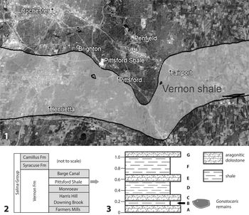
The Pittsford Shale Member of the upper Silurian Vernon Formation (Ludlow) is a poorly exposed eurypterid-dominated Lagerstätte from localities just south of Rochester, New York. It was originally described by Sarle (Reference Sarle1903) on the basis of material excavated during the deepening of a now-abandoned section of the Erie Canal in 1897–1898 at Spring House, near the border of Pittsford and Brighton, New York (Fig. 1; 43°6'29.4''N, 77°32'41.4''W). The fauna of the Pittsford Shale was originally described by Clarke (Reference Clarke1903) and Sarle (Reference Sarle1903) and reviewed by Ruedemann (Reference Ruedemann1916a). The results of extensive additional collecting at a dump pile from the canal were summarized by Ruedemann (Reference Ruedemann1920), including a faunal list.
Locality information for the specimens from the Pittsford Shale localities. (1) Map of Pittsford Shale localities. Banded area is the mapped distribution of the Vernon Shale in the area. Based on the current New York State Geological Survey Map. (2) Stratigraphic context of the Pittsford Shale within the Vernon Formation and Salina Group. Modified from Vrazo et al. (Reference Vrazo, Brett and Ciurca2017). (3) General stratigraphic column of the Pittsford Shale representing the stratigraphic interval where Gonatocaris decora remains are generally found (redrawn from Ciurca and Tetlie, Reference Ciurca and Tetlie2007). Letters represent the ‘sections’ in Ciurca and Tetlie (Reference Ciurca and Tetlie2007). Scale is in meters.

An important secondary component of the eurypterid-dominated fauna is the unusual phyllocarid Gonatocaris decora (Clarke, Reference Clarke1901), originally described from a very limited number of specimens, none complete. This has hampered our understanding of the form and its place within the Phyllocarida (Collette and Hagadorn, Reference Collette and Hagadorn2010). The collections of Plotnick and Samuel Ciurca, especially the latter, have greatly added to the material available and allow a redescription of the form. We were also able to obtain photographs of British specimens originally assigned to the same genus and previously illustrated only with line drawings. Here, we redescribe the species and genus, clarify issues surrounding genus names and species assignments, and discuss aspects of its functional morphology, ontogeny, and paleoecology.
Geological setting
The Pittsford Shale is known from a very limited number of localities in the vicinity of Pittsford, New York, a suburb of Rochester (Ciurca, Reference Ciurca1980, Reference Ciurca2010). There is little or no surface exposure, with most of what we know based on material exposed during excavations for buildings or the Erie Canal. The section containing the fossiliferous beds was originally described by Sarle (Reference Sarle1903) and updated by Ciurca and Tetlie (Reference Ciurca and Tetlie2007). The eurypterids and other fossils come from a dense olive-green to black shale, somewhat fissile and with minor pyrite, which disintegrates when exposed to water.
The unit is currently the Pittsford Shale Member of the Vernon Formation, Salina Group (Ciurca and Tetlie, Reference Ciurca and Tetlie2007; Vrazo et al., Reference Vrazo, Brett and Ciurca2017), and is composed of a series of interbedded dolostones and fissile black, gray, and green shales that contain pelecypods, phyllocarids, and common eurypterids (Ciurca, Reference Ciurca2010). Mud cracks and possible microbialites suggest a series of shallowing and desiccation events (Ciurca, Reference Ciurca1990, Reference Ciurca2010). On the basis of conodonts (Polygnathoides siluricus Branson and Mehl, Reference Branson and Mehl1933) and acritarchs (Dilatisphaera williereae Lister, Reference Lister1970), Miller et al. (Reference Miller, Brett and Kleffner1989) suggested an early Ludlow age for the unit. Vrazo et al. (Reference Vrazo, Brett and Ciurca2017) also gave a Ludlow age. In the current Silurian timescale, the presence of P. siluricus reliably indicates an early Ludfordian age within the Ludlow Epoch (Aldridge and Schönlaub, Reference Aldridge, Schönlaub, Holland and Bassett1989). Although the acritarchs suggest a Gorstian age, D. willeriereae is only doubtfully known younger than the Llandovery (Martin, Reference Martin, Holland and Bassett1989). The Vernon Formation is older than the classic ‘Bertie Waterlime’ eurypterid Lagerstätten of the same region (Kluessendorf, Reference Kluessendorf1994; Vrazo et al., Reference Vrazo, Brett and Ciurca2017).
On the basis of the lithology and the associated fauna, Plotnick (Reference Plotnick, Boucot and Lawson1999) suggested a very shallow marginal marine setting, possibly lagoonal. He assigned it to Benthic Assemblage 1 of Boucot (Reference Boucot1975). This assignment is supported by the terrestrial spores listed by Miller et al. (Reference Miller, Brett and Kleffner1989) and is consistent with the interpretation of Vrazo et al. (Reference Vrazo, Brett and Ciurca2017), with the Pittsford fossils occurring just below facies indicating subaerial exposure.
Associated fauna
The relative rarity and commonness of Pittsford shale animals was assessed by Ruedemann (Reference Ruedemann1920). His results can be compared with the list of museum specimens, mostly collected by Ciurca, downloaded from the IDigBio Portal (https://www.idigbio.org/portal). The fauna of the Pittsford Shale is dominated by the eurypterids Hughmilleria socialis Sarle, Reference Sarle1903 and Eurypterus pittfordensis Sarle, Reference Sarle1903. Other much less-common eurypterids include Mixopterus multispinosus (Clarke and Ruedemann, Reference Clarke and Ruedemann1912), Erettopterus osiliensis (Schmidt, Reference Schmidt1883) (originally Pterygotus monroensis Sarle, Reference Sarle1903; Ciurca and Tetlie, Reference Ciurca and Tetlie2007), and Carcinosoma spiniferum Kjellesvig-Waering and Heubusch, Reference Kjellesvig-Waering and Heubusch1962. An extremely rare species is the synziphosurine Pseudoniscus roosevelti Clarke, Reference Clarke1901. Among the crustaceans, the ostracode Leperditia scalaris (Jones, Reference Jones1858) is relatively common. Gonatocaris decora is a relatively abundant phyllocarid, whereas the phyllocarid Ceratiocaris praecedens Clarke, Reference Clarke1901 is very rare. Non-arthropod taxa include the common brachiopod Lingula semina Ruedemann, Reference Ruedemann1916b, the common bivalve Pterinea poststriata Ruedemann, Reference Ruedemann1916b, and very rare cephalopods Hexamoceras cf. H. chadwicki Ruedemann, Reference Ruedemann1920 and Orthoceras sp. In addition, Miller et al. (Reference Miller, Brett and Kleffner1989) identified conodonts, acritarchs, and terrestrial spores. We have also identified the presence of a paulinitid scolecodont (probably Kettnerites [personal communication, M. Eriksson, 2005]. Specimens are deposited at the Yale Peabody Museum [YPM IP 536166, 536167, and 536188]).
History and status of Emmelezoe and Gonatocaris
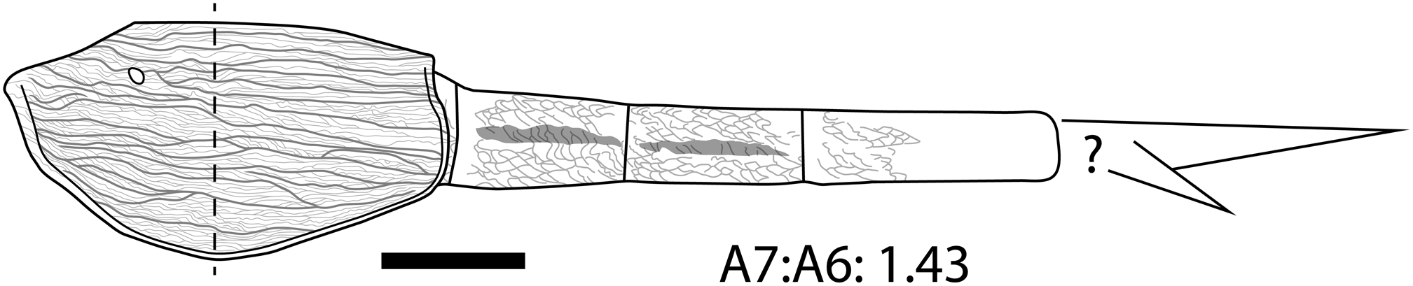
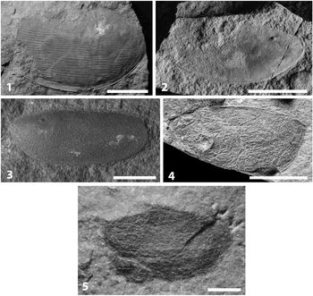
Gonatocaris decora is a very distinctive species of phyllocarid crustacean (Fig. 2) that was originally assigned to the genus Emmelezoe Etheridge, Woodard, and Jones, Reference Etheridge, Woodard and Jones1887. Gonatocaris decora and the genera Emmelezoe and Gonatocaris have had a complex nomenclatural history. Etheridge et al. (Reference Etheridge, Woodard and Jones1886) remarked on the presence of an ‘eye spot’ in some British specimens of Ceratiocaris elliptica M'Coy, Reference M'Coy1849, a feature they considered to be of generic-level importance in distinguishing these from other Ceratiocaris species. They listed these as Ceratiocaris ?elliptica, under the open nomenclatural heading ‘C. Genera distinct from Ceratiocaris’ in 1886. A year later, Etheridge et al. (Reference Etheridge, Woodard and Jones1887) erected the genus Emmelezoe for these specimens, characterized by a distinct ‘ocular tubercle’ thought to be “equivalent to the eye” (Etheridge et al., Reference Etheridge, Woodard and Jones1887, p. 232). Four species were assigned to Emmelezoe and formally described by Ethridge et al. (Reference Etheridge, Woodard and Jones1887, p. 232–233): E. elliptica (type), E. crassistriata, E. tenuistriata, and E. maccoyiana (see Fig. 3 for original types). Each of these designations was based on single specimens that had been previously identified as Ceratiocaris elliptica and were distinguished from each other by possession of coarse, fine, or somewhat intermediate striae of the carapace valves, respectively. However, the descriptions were unaccompanied by formal diagnoses.
General morphology of Gonatocaris decora. The dashed line indicates the greatest width of the carapace valve. Note the straight posteroventral carapace margin and the bluntly rounded carapace horn. Abdominal length estimated from the closely associated carapace and abdomen on the small slab NYSM 4427—possibly an exuviae of a single individual. Scale for this composite drawing based on NYSM 4427; for that carapace valve, the scale bar = 1 cm.

Emmelezoe type specimens; all have been referred back to Ceratiocaris. Note the presence of the ‘ocular tubercle.’ (1) Emmelezoe crassistriata, GSM 7457, British Geological Survey. (2) Emmelezoe maccoyiana, GSM 7470, British Geological Survey. (3) Emmelezoe elliptica, SM A 16644, Sedgwick Museum of Earth Sciences, University of Cambridge. (4) Emmelezoe tenuistriata, In. 43897, Natural History Museum, London. (5) Emmelezoe minnuta, NYSM 13503/1, New York State Museum. (1–4) Scale bars = 1 cm; (5) scale bar = 1 mm.

There is some confusion regarding authorship and the first publication of the genus Emmelezoe. Jones (Reference Jones1886) published a nearly identical copy of Etheridge et al. (Reference Etheridge, Woodard and Jones1887) under his name exclusively in September of 1886 in Geological Magazine. Thus, it would seem that Jones (Reference Jones1886) would be the first printed publication of Emmelezoe, because Etheridge et al. (Reference Etheridge, Woodard and Jones1887) was a conference reading (read 8 September before the 56th meeting of the British Association for the Advancement of Science, Birmingham, 1886) and was not published as a conference volume until the following year. However, Jones (Reference Jones1886) references the 1886 conference reading in his paper. Thus, the first formal mention of Emmelezoe was the conference reading in 1886 by Ethridge et al., which was published as a printed volume in 1887, and not Jones (Reference Jones1886).
In 1901, Clarke described some new phyllocarid material from the ‘Salina Beds’ (now the Salina Group) from a collection of material sourced from near Pittsford, New York (Clarke, Reference Clarke1901). Clarke (Reference Clarke1901) assigned this material to Emmelezoe on the basis of the possession of an “ocular tubercle” (p. 96) and assigned this material to E. decora. Clarke's material was substantially different from previously described examples attributed to Emmelezoe because of the extremely coarse ornament of the carapace valves, the much more robust nature of the carapace cuticle, the valve shape, the apparent lack of a rostral plate, and the characteristics of the very rare abdomen.
Subsequent study by Rolfe (Reference Rolfe and Moore1969) referred the type species (Emmelezoe elliptica) back to Ceratiocaris, and thus it became a junior synonym; however, a discussion is warranted here to clear up some issues surrounding historical use of characters related to diagnoses. The main morphological character on which the type species (and genus Emmelezoe) was based was the ‘eye spot’ or ocular node. However, an anterior tubercle or node is frequently present in valid Ceratiocaris species and is likely produced by a robustly sclerotized mandibular process that underlies and disrupts the relatively thin valve cuticle as it drapes over the remains post-burial or during taphonomy (Salter, Reference Salter1860; Rolfe and Burnaby, Reference Rolfe and Burnaby1961; Rolfe, Reference Rolfe1962; Collette and Rudkin, Reference Collette and Rudkin2010). In addition, the four species that were transferred to or erected by Etheridge et al. (Reference Etheridge, Woodard and Jones1887) all possess a distinctly ceratiocaridid carapace outline and ornament, E. elliptica and E. tenuistriata both possess a rostral plate, and characteristics of the abdominal somites, telsons, and furcal rami are all otherwise indistinguishable from species of Ceratiocaris (Rolfe, Reference Rolfe and Moore1969; Collette and Hagadorn, Reference Collette and Hagadorn2010).
Ruedemann (Reference Ruedemann1925) subsequently identified a single tiny carapace (4 mm long and 2 mm wide) in the younger Bertie Group from Buffalo, New York, as Emmelezoe and erected the species E. minuta, although he indicated that he lacked sufficient material for a full description. A reexamination of this specimen reveals only that it is suggestive of a ceratiocaridid or gonatocaridid in outline; unfortunately, the lack of detail on the carapace valve surface necessitates an open nomenclatural assignment (see Fig. 3.5).
In 1929, the genus Gonatocaris was erected by Gürich to differentiate Clarke's (Reference Clarke1901) highly ornamented ‘Emmelezoe’ specimens from the Etheridge et al. (Reference Etheridge, Woodard and Jones1887) Ceratiocaris-like ‘Emmelezoe’ material, with the Pittsford shale species being referred to Gonatocaris decora. Gürich (Reference Gürich1929) also provided a clearer basis for differentiating Gonatocaris from other Ceratiocarididae in addition to the distinct ‘ocular tubercle’ and the coarse longitudinal ribbing. These characteristics were a wider (ventral?) margin than in Ceratiocaris, a blunt rounded angle on the ventral margin just anterior to the midpoint (Ceratiocaris is evenly rounded in this respect), and a relatively straight posteroventral margin (the same margin in Ceratiocaris is slightly curved).
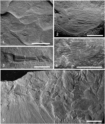
Curiously, Gürich referred Emmelezoe crassistriata (Etheridge et al., Reference Etheridge, Woodard and Jones1887; Fig. 3.1) to Ruedemann's (Reference Ruedemann1925) E. minuta, although E. crassistriata would have had taxonomic priority at that time. In addition, given the similar morphological characteristics evident in Ruedemann's (Reference Ruedemann1925) drawing, E. minuta should have been synonymized with G. decora; according to Ruedemann's description and drawing, E. minuta is smaller but otherwise very similar to G. decora (compare Ruedemann, Reference Ruedemann1925, plate 24, fig. 4 with the lectotypes of G. decora [Fig. 4.1, 4.2] and with the actual specimen of E. minuta [Fig. 3.5]). However, it appears that Ruedemann's interpretation of the surface ornament of E. minuta was exaggerated—an examination of the E. minuta type material indicates that it is largely devoid of the ornament drawn by Ruedemann (Reference Ruedemann1925) (see Fig. 3.5).
Gonatocaris decora from the upper Silurian Salina Group of New York State. (1) An accumulation of well-preserved, intact carapace valves, NYSM 4427. (2) A well-preserved left carapace valve illustrating the coarse and fine anastomosing striations of the species, NYSM 4428. (3) An abdomen preserving at least four somites and with well-preserved scale-like ornament, NYSM 4434. (4) A carapace valve with a possible furcal ramus or telson preserved, YPM IP 536187. (5) A slab preserving an accumulation of whole carapace valves and fragments, YPM IP 535963. (2–4) Scale bars = 5 mm; (1, 5) scale bars = 1 cm.

Moreover, the ventral margins in all of the Etheridge et al. (Reference Etheridge, Woodard and Jones1887) material is evenly rounded, and there is no hint of the blunt angle posterior of the ventral midpoint or the straight posteroventral margin necessary for a Gonatocaris diagnosis. Rolfe (Reference Rolfe and Moore1969) referred E. elliptica back to Ceratiocaris, making the genus Emmelezoe, and all referred members, junior synonyms of Ceratiocaris. Thus, Collette and Hagadorn (Reference Collette and Hagadorn2010) erred by retaining E. decora within Emmelezoe.
Materials and methods
The Pittsford Shale collections of Sarle (Reference Sarle1903), Clarke (Reference Clarke1901), and Ruedemann (Reference Ruedemann1920) are housed in the New York State Museum. One of the authors (RP) made a small collection in 1979. Extensive collections were made over several decades by Samuel J. Ciurca (Ciurca, Reference Ciurca2010), mostly in building excavation sites. His extensive collections, as well as those of Plotnick, are now housed at the Yale Peabody Museum.
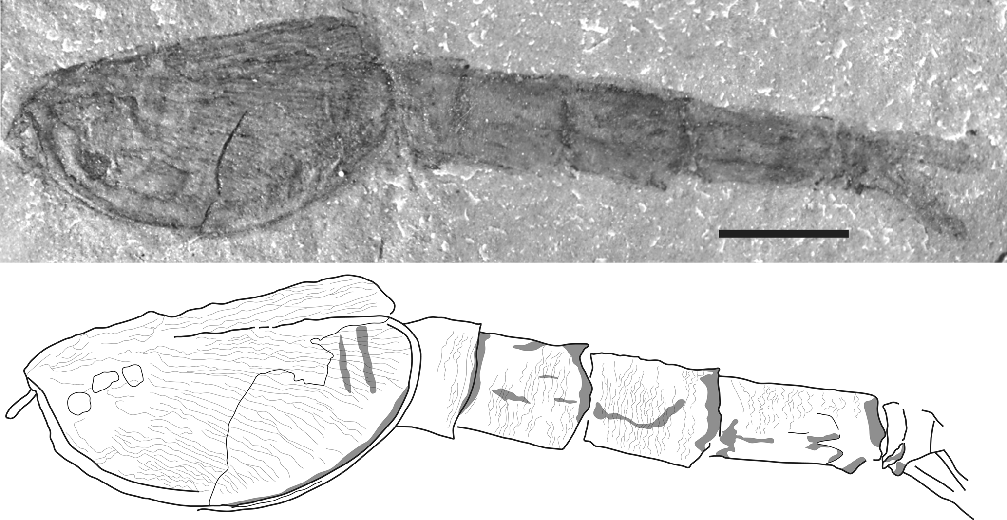
All phyllocarid specimens imaged by the authors were photographed using a Nikon D-3100 digital camera equipped with a Nikkor 105 mm f2.8 macro lens mounted on a copy stand. Lighting was provided by copy-stand lighting and a studio strobe. All line drawings (e.g., Fig. 5) were done by importing photographs into Adobe Illustrator as background layers, tracing features on a new layer, and then removing the photographic image. Photographs were processed for color and contrast in Adobe Photoshop. The Natural History Museum (London), the Sedgwick Museum of Earth Sciences, and the British Geological Survey, provided high-quality photographs of British ‘Emmelezoe’ specimens to compare with G. decora (Fig. 3).
Photograph and interpretive drawing of a small (possible juvenile) specimen of Ceratiocaris from the Bertie group, Ridgemount Quarry, Ontario, collected and currently in the personal collection of John Spina. Note the elongated abdomen. Scale bar = 5 mm.

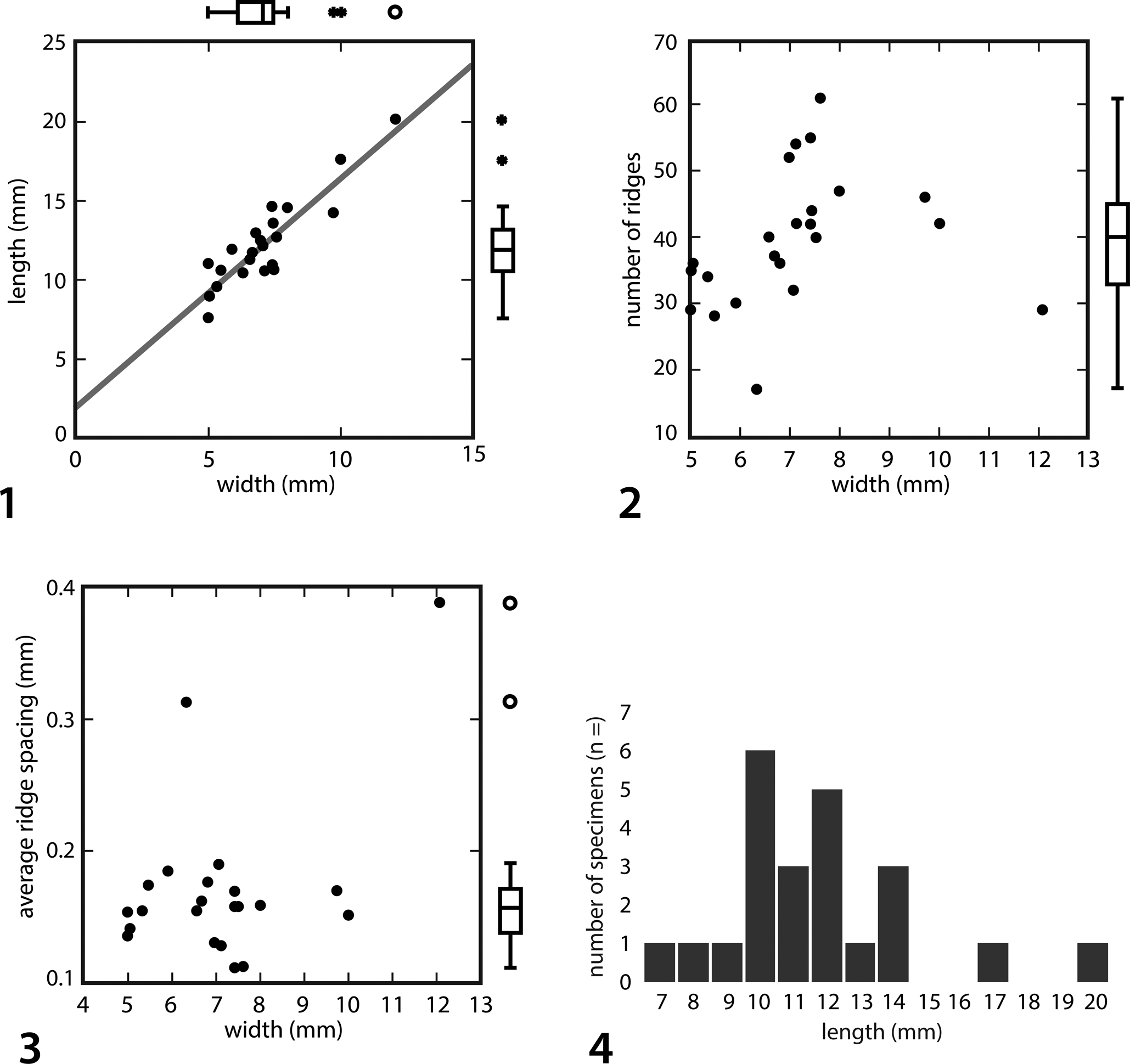
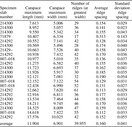
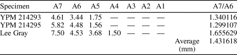
A suite of valves was photographed and measured by using a Leica M205C Stereomicroscope equipped with a Leica DFC495 Digital Microscope Camera and Leica Application Suite (LAS) imaging software. Maximum length and width of each carapace valve was determined, and the number and spacing between carapace ridges were measured across a transect through the carapace's widest area (Table 1). Bivariate analyses were made to determine whether there was allometric growth of the carapace (Fig. 6.1) and whether the number and/or spacing of the ridges changes with size (Fig. 6.2, 6.3). Additional measurements were made of partially articulated abdominal remains where at least the posteriormost three segments were articulated (Table 2). Statistical analyses were done using Microsoft Excel and SYSTAT software.
Morphometric analyses of carapace valves of Gonatocaris decora. (1) Bivariate plot of carapace valve length to width; n = 23. Line is the least squares fit to the data; r = 0.898. (2) Bivariate plot of carapace width versus the total number of ridges across the widest part of the carapace valve; n = 23. (3) Bivariate plot of carapace width versus the average spacing of the ridges; n = 23. (4) Histogram of the length of complete carapace valves studied in this paper binned by number of occurrences per 1 mm bin.

Measurements of Gonatocaris decora individual carapace valves.

Measurements of Gonatocaris decora abdominal remains where at least the three posteriormost abdominal segments were articulated. Values represent the maximum length (mm) of each abdominal segment from the posterior (A7) forward (A6, A5, etc.). Missing values (—) represent a lack of that segment in the fossil remains.

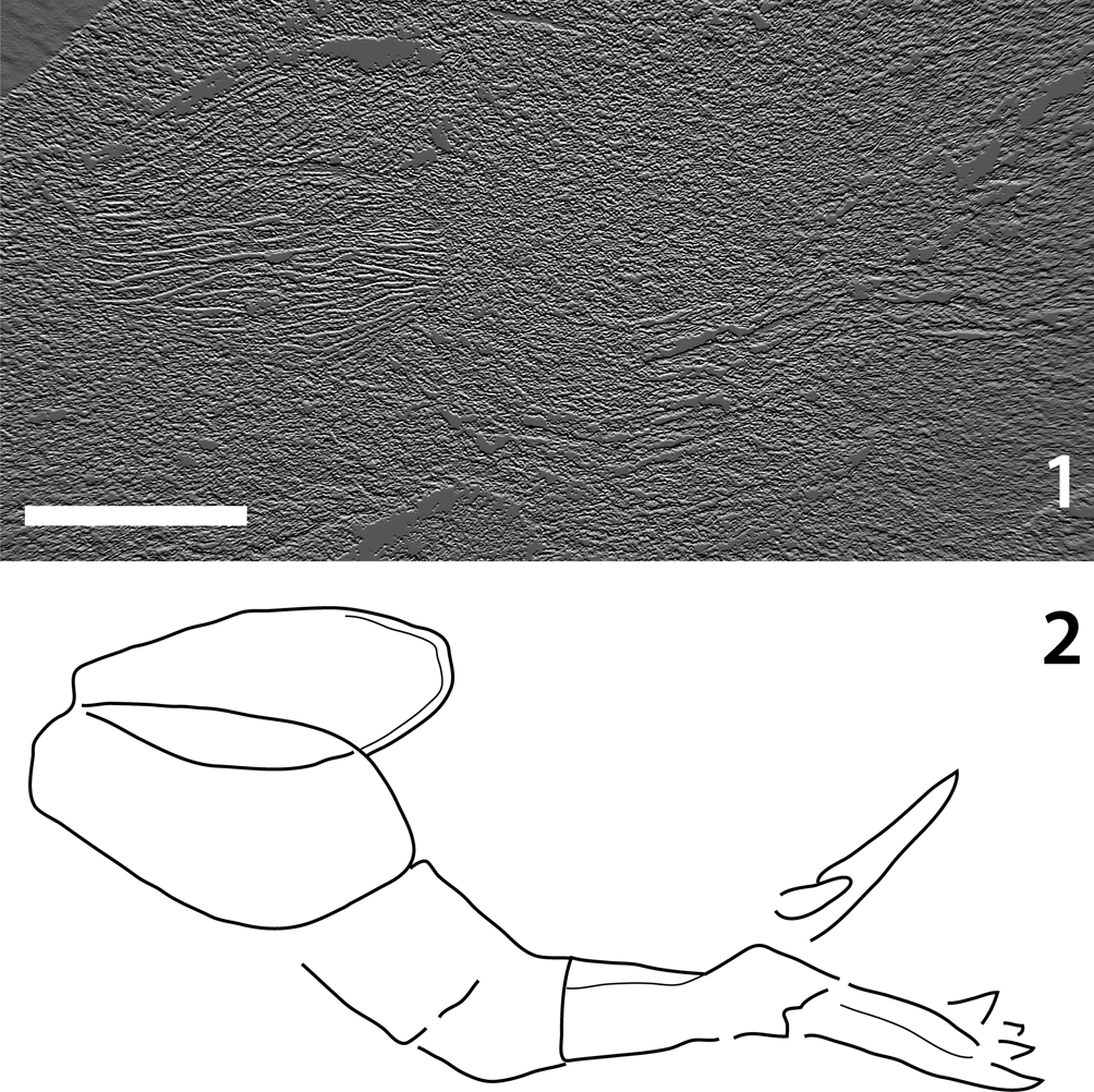
Much of the material preserving Gonatocaris remains is thin shales that preserve substantial relief only on the carapace valves—due, presumably, to the structural support lent by the carapace ornamentation. However, when preserved, the abdominal and possible furcal remains are usually extremely flattened, preserving little original convexity. We employed directional difference filtering, also known as an ‘emboss filter,’ to help elucidate tenuous topographic information. This software technique works by emphasizing edges in a particular direction in a digital photograph in a manner similar to using oblique lighting with a physical sample. Each pixel in the image is analyzed and replaced by either a light-, a dark-, or a middle-gray pixel depending on the color change and contrast ratio between adjacent pixels in the original image (Umbaugh, Reference Umbaugh2005). By changing the directional component of the software filter, edges perpendicular to filter direction are enhanced. By performing this analytical technique in all four cardinal directions on an image, all low-relief edges present in the image can be enhanced and retained as a separate layer. Each image layer can then be recombined to retrieve a grayscale image where all edges have been directionally enhanced. We applied this technique to a photograph of NSYM 4427 (Fig. 7).
Close association of nearly articulated right and left carapace valves of Gonatocaris decora. Partial abdomen and possible furca of Gonatocaris decora on small slab NYSM 4427. (1) A jpeg photo of NYSM 4427 that has been processed using directional difference filtering. This image was processed from the N (upper), E (right), S (bottom), and W (left) directions and then the layers combined to bring out as many edges as possible. (2) An interpreted line drawing of the additional morphology shown in (1). Scale bar = 5 mm.

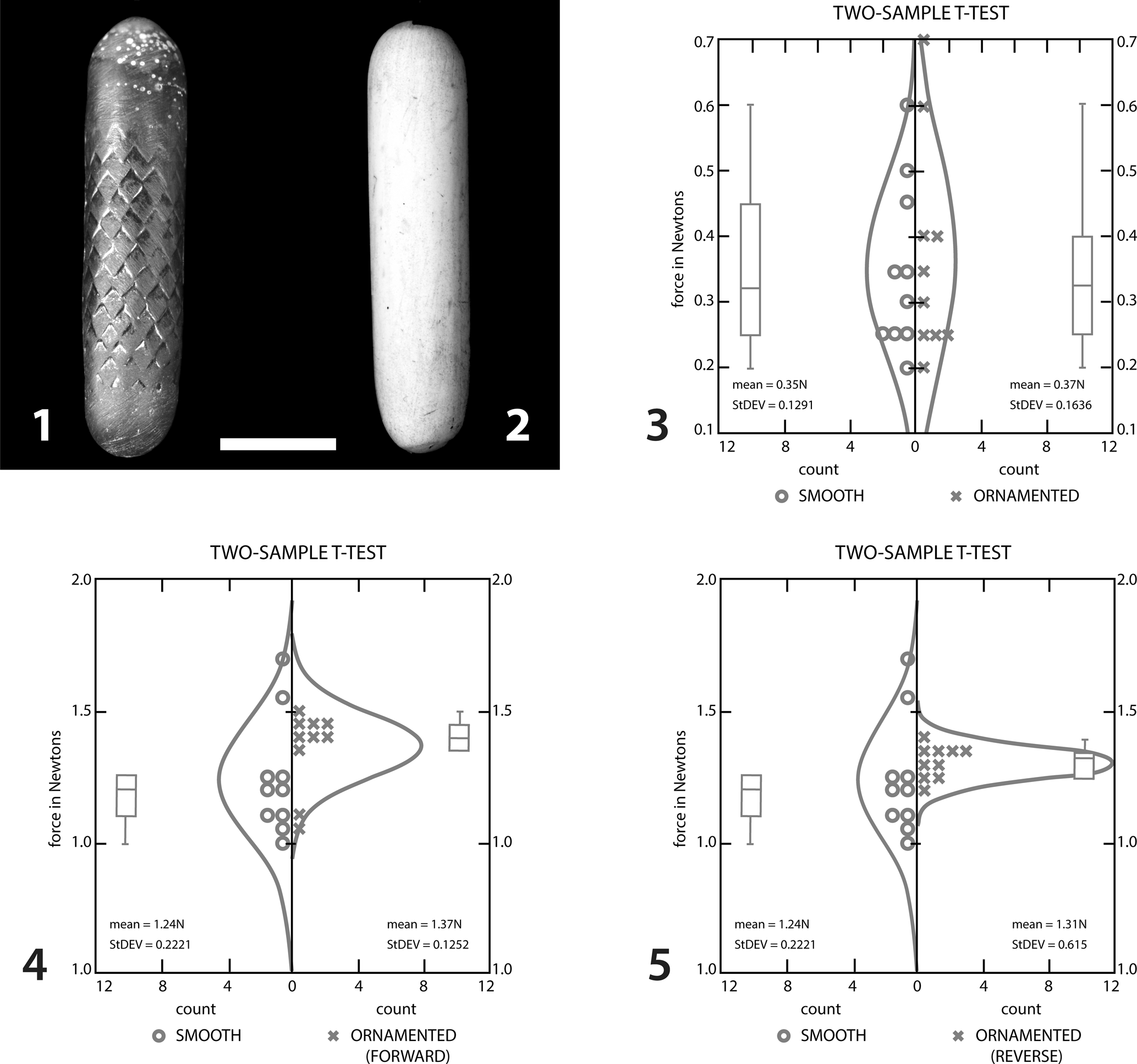
We used a simplified model of the abdomen to determine whether the sculpture played a role in resisting movement of the abdomen through sediment, as suggested for extant Nebalia and archaeostracan Ceratiocaris species (Vannier et al., Reference Vannier, Boissy and Racheboeuf1997; Collette and Rudkin, Reference Collette and Rudkin2010). The model was constructed of white copy paper that had been cut into a series of approximately 0.5 × 2 inch strips and a zig-zag pattern cut into one edge. A series of 15 of these strips were glued together then rolled onto a pencil as a form and glued together using an internal spline. Polyform Products Sculpey brand heat-hardening polymer modeling clay was then used to plug both ends of the hollow model. A mold was then produced from the finished model using two-part silicone molding compound. This mold was then cast with Alumilite clear two-part epoxy casting resin to which a small amount of colorant had been added to facilitate photography; the finished casting is shown in Figure 8.1. An effaced version of this same model was produced using Sculpey brand heat-hardening polymer (Fig. 8.2) so that any effect of just the ornament could be measured using the experimental apparatus described in the following. Both models were buffed with 0000 steel wool to produce a smoothed surface and to minimize the effects of friction due to the different materials.
Models used in experiment to determine forces required to move ornamented versus unornamented sclerites through a mud substrate. (1) Ornamented model based on Gonatocaris and Ceratiocaris style ornamentation. (2) Effaced (unornamented) model. (3–5) Box plot, dot density, and density plots for the maximum force in Newtons to fully insert the two models into various substrates; 10 runs for each experiment. (3) Models pushed into saturated kaolinite substrate; values are not significantly different (t test, p = 0.765). (4) Models pushed into saturated fine sand; model 1 pushed into sediment in ‘normal’ orientation (points of scales facing opposite to direction of movement); values indicate significant difference at this grain size (t test, p = 0.1289). (5) Models pushed into saturated fine sand; model 1 pushed into sediment in ‘opposite’ direction (points of scales facing direction of movement); values indicate significant difference at this grain size (t test, p = 0.3587). (1, 2) Scale bar = 1 cm.

Forces required to push both models into saturated kaolinite and fine sand substrates were measured using a Chatillon DFIS-10 digital force gauge attached to a Chatillon TCM-200 motorized test stand (see Plotnick and Bauer, Reference Plotnick, Bauer, Hembree, Platt and Smith2014 for additional details). Both models were pushed into the sediments at a constant rate of 5.08 cm per minute (2 inches per minute); force was applied vertically by a flat disc that had been hot glued to the posterior end of each model. Attempts to pull models through sediments were unsuccessful (very low to no force readings could be recovered by instruments). Models were pushed in until they were fully inserted into the mud or fine sand. The maximum force in Newtons was recorded; data were analyzed using SYSTAT software. Three experiments were conducted: (1) models inserted in ‘normal’ orientation (where the ornamented model had the raised scale ornament facing posteriorly) in saturated kaolinite mud, (2) models inserted in ‘normal’ orientation in saturated fine sand, and (3) models inserted in ‘reverse’ orientation in saturated fine sand. Ten runs were done for each experiment; results are summarized in Figures 8.3–8.5.
Repositories and institutional abbreviations
Specimens described and illustrated here are housed in collections of the British Geological Survey (GSM), London, England; Natural History Museum (NHM), London, England; New York State Museum (NYSM), Albany, New York; Sedgwick Museum of Earth Sciences (SM), University of Cambridge, Cambridge, England; and Yale Peabody Museum (YPM), New Haven, Connecticut.
Systematic paleontology
Crustacea Brünnich, Reference Brünnich1772
Phyllocarida Packard, Reference Packard1879
Family Ceratiocarididae Salter, Reference Salter1860
Genus Gonatocaris Gürich, Reference Gürich1929
Type species
Gonatocaris decora (Clarke, Reference Clarke1901).
Other species
G. nankingensis (P'an, Reference P'an1962); Gonatocaris sp. Zong et al., Reference Zong, Liu, Wei and Gong2017.
Diagnosis
As per type species.
Gonatocaris decora (Clarke, Reference Clarke1901)
Figures 4, 7
- Reference Gürich1929
Gonatocaris decora Gürich, p. 60, text-figure 5.9.
- Reference Rolfe1969
Gonatocaris decora; Rolfe, p. R316, fig. 138.
- Reference Clarke1901
Emmelezoe decora Clarke, p. 95, pl. 2, figs. 4–11, pl. 3, figs. 1–4.
- Reference Clarke and Ruedemann1903
Emmelezoe decora; Clarke and Ruedemann, p. 705.
- Reference Clarke1907
Emmelezoe decora; Clarke, p. 310.
- Reference Grabau and Shimer1910
Emmelezoe decora; Grabau and Shimer, p. 379, fig. 1683.
- Reference Bassler1915
Emmelezoe decora; Bassler, p. 476.
- 1919
Emmelzoe decora (incorrect generic spelling); Ruedemann, p. 206.
- Reference Schuchert and Schuchert1943
Emmelezoe decora; Schuchert, p. 70.
- Reference Leutze1959
Emmelzoe decora (incorrect generic spelling); Leutze, p. 36.
- Reference Collette and Hagadorn2010
Emmelezoe decora; Collette and Hagadorn, p. 807, fig. 6.9, 6.10.
Types
Clarke (Reference Clarke1901) did not designate a type specimen in the original description of this species; Collette and Hagadorn designated NYSM 4427 as the lectotype and NYSM 4428–4430, 4434–4436 as paralectotypes.
Emended diagnosis
Carapace subquadrate to subovate; hinge line flat or slightly convex, narrow thickened ventral margin present. Anterior carapace horn rounded, weakly developed. Posterior carapace margin very gently convex to slightly sinuous. Greatest width of carapace valves occurs anterior of carapace midpoint; anteroventral and posteroventral margins separated by a blunt, rounded angle. Anteroventral margin flat or slightly concave; posteroventral margin past approximately carapace midline flat. Carapace ornamented with prominent raised, wavy, anastomosing longitudinal striations of two distinct scales; fewer larger ridge-like striae with smaller striae occupying the space between. Anterior node present; median dorsal plate absent. Portion of abdomen that protrudes from carapace exceptionally long (after Clarke, Reference Clarke1901).
Occurrence
Upper Silurian (Ludlow, Ludfordian) Salina Group Beds, Erie Canal, Pittsford, New York.
Remarks
The presence of a flat or gently convex hinge line, a thickened ventral carapace margin, raised striae ornament of the carapace valves, elongated abdomen, greatly elongated posteriormost abdominal somite, and abdominal scale ornamentation (see Figs. 2, 4.1–4.5) are all characters that indicate a close relationship between Gonatocaris and Ceratiocaris. However, even considering these similarities, the differences between these genera are substantial. In Gonatocaris, the ratio of the length of the abdomen to carapace valves appears to be greater than that of Ceratiocaris, although fully articulated abdomens are rarely preserved in Gonatocaris. The maximum width of the carapace valves in Gonatocaris occurs before the longitudinal midline (see Figs. 2, 4.2); in Ceratiocaris, the valves are generally widest closer to the midline or posterior to it (Gürich, Reference Gürich1929). Likewise, the very straight (not evenly curved as in Ceratiocaris) posteroventral margin of the carapace valves is diagnostic for Gonatocaris (see Figs. 2, 4.2, 4.4). No rostral plate is known in Gonatocaris, and the ‘carapace horn’ is very rounded and unlike the acuminate horn in Ceratiocaris. Carapace ornamentation is also unique in Gonatocaris valves. Two distinct scales of raised longitudinal striations occur on the valve surfaces whereas in Ceratiocaris, striae are generally much more uniform in height (compare Figs. 2, 4). The more robust, topographically higher relief, striations are ridge-like and anastomose less frequently than the smaller-scale striations. Finer wavy, raised striae that frequently anastomose occur between the coarser ridges.
Although others have noted minor differences between possible juvenile archaeostracans (e.g., Broda et al., Reference Broda, Collette and Budil2018), the consistent morphological differences in carapace outline, morphometric ratios, abdominal length, and ornamentation of both the carapace and abdominal somites between specimens of Gonatocaris and Ceratiocaris preclude the former as a juvenile of the latter. In addition, the length-to-width ratio of carapace valves examined in this study exhibits strong isometry and thus a probable lack of significant change in carapace shape between juveniles and adults. Moreover, the fairly normal size–frequency histogram of the analyzed group (see Fig. 6.4) suggests patterns typical for arthropod molt associations: a large number of intermediate individuals due to rapid growth as instars approach adulthood, followed by many fewer large molts due to the slowing of molts following adulthood (e.g., Hartnoll and Bryant, Reference Hartnoll and Bryant1990). The lack of very small carapaces could be due to winnowing away of very small molts by currents or juveniles may have had different ecological niches.
Unfortunately, no demonstrably fully articulated specimens of G. decora are currently known. One of the specimens that Clarke figured in 1901 (NYSM 4427) appears to be an articulated abdomen protruding from a carapace valve (Figs. 4.1, 7); however, this abdomen protrudes from the anterior of a nearly articulated left and right carapace valve. This specimen may still be an articulated molt with the abdomen in this orientation due to contortions necessary in casting off the old exoskeleton and is still important in establishing a firm linkage between isolated purported abdomens and carapace valves of G. decora.
When directional difference filtering is applied to a photograph of NSYM 4427 (Clarke's Reference Clarke1901 slab with the abdomen protruding from the anterior of a carapace), additional, tentative morphology of the abdomen and possible furcae not visible in oblique lighting become visible (see Fig. 7). The abdomen in this specimen is flattened, and thus the somites appear to have a distorted, wider appearance than those of other Gonatocaris abdomens. At the posterior end of the abdomen, there appears to be a rather large Y-shaped structure that is much flatter than the previous parts of the abdomen. These may represent furcal rami.
Possible furcae of Gonatocaris decora are exceptionally rare. A possible furcal ramus or a telson of G. decora occurs on a small slab with an individual valve (YPM IP 536187, Fig. 4.4). This small fragment is approximately 1 mm in length, is elongate triangular in shape, and is broken along the wider margin. It appears to have a rounded distal terminus, thickened lateral margins, and at least three grooves on the surface; the axes of the grooves converge toward the distal terminal end of the structure. It is possible that this structure represents either a furca or a telson of G. decora. Directional difference filtering (see the following) suggests that furcae were slender and elongate (see Fig. 7); thus, this may be a shorter telson, a fragment of a carapace, or a nonrelated fossil.
Clarke did figure a structure that he believed might be part of a rostral plate (Clarke, Reference Clarke1901, p. 96, pl. 3, fig. 3), describing the structure as a “small, leaflike plate among the carapaces, which is marked by a venation unlike anything seen elsewhere on these bodies.” However, Clarke did not indicate which slab this structure is located on, and in Clarke's published woodcut, the specimen appears indistinguishable from a fragment of ornamented E. decora carapace. In reexamination of Clarke's (Reference Clarke1901) original material, and of additional material housed in the collections of the Yale Peabody Museum, neither author was able to detect a structure that we would interpret as a rostral plate.
Discussion
Distribution of Gonatocaris
In addition to the remains from the Pittsford Shale, fossils assignable to Gonatocaris are known from the Silurian of central China. In the Fentuo Biota of Wuhan, an unnamed species of Gonatocaris occurs in the diverse Assemblage 1 together with conulariids, crinoids, sponges, and trilobites (Zong et al., Reference Zong, Liu, Wei and Gong2017). The Fentuo biota is also considered to have inhabited a shallow-water estuarine or littoral paleoenvironment (Zong et al., Reference Zong, Liu, Wei and Gong2017). G. nankingensis, also from the Fentuo region, was collected in 1956 and described as the remains of a fish of the suborder Cyathaspida (P'an, Reference P'an1962). It is possible, due to the geographic proximity and significant morphological similarity, that the unnamed Assemblage 1 Gonatocaris and G. nankingensis are conspecific. The Gonatocaris remains from the Fentuo Biota are remarkably similar to G. decora from New York (compare Fig. 4 with Zong et al., Reference Zong, Liu, Wei and Gong2017, fig. 3J–3M).
Affinities of Gonatocaris
In their review of Paleozoic archaeostracan phyllocarids, Collette and Hagadorn (Reference Collette and Hagadorn2010) placed ‘Emmelezoe’ (= Gonatocaris) within the Ceratiocaridina, as the sister group of a clade composed of Ceratiocaris and Warneticaris Racheboeuf, Reference Racheboeuf1994. Among archaeostracan taxa, this placement appears to be the least problematic; both Ceratiocaris + Warneticaris and Gonatocaris possess longitudinally striate carapace valves with distinct ventral and posterior margins, Gonatocaris valves possess a marked anterior carapace node (i.e., ‘optic node’) while Ceratiocaris species often also have a node- or bump-like protuberance in this position on the carapace, and the scale-like abdominal ornamentations of both are very similar. Indeed, most species of Ceratiocaris possess either scale-like or fingerprint-like ornament of abdominal somites, longitudinal striations of the carapace valves, ‘nodes’ at the anterior of the carapace, or a combination of all three of these characters (compare the outline and surface ornament in Fig. 4 with Collette and Hagadorn, Reference Collette and Hagadorn2010, fig. 4 [key to carapace shape and ornament of well-known Ceratiocaris species]). Gonatocaris and Ceratiocaris also both possess a greatly elongated seventh (posteriormost) abdominal segment, as do many other archaeostracan and leptostracan taxa. However, the ratio of the length of abdominal segment 7 (A7) to that of abdominal segment 6 (A6) in reasonably well-articulated abdominal remains is fairly low (1.43) in comparison with this ratio in ceratiocaridids (1.59–2.15 for Ceratiocaris species and 2.23–2.53 in Warneticaris species). In addition to the A7:A6 ratio, there are other problems with this assignment as well: Gonatocaris has a more blunt, nearly perpendicular anterior margin without any real carapace ‘horn,’ and no rostral plate is currently known; the carapace outline in well-preserved species such as C. papilio Salter in Murchison, Reference Murchison1859 and C. harpago Poschmann, Bergmann, and Kühl, Reference Poschmann, Bergmann and Kühl2018 have their greatest widths past the carapace midline (Rolfe, Reference Rolfe and Moore1969; Poschmann et al., Reference Poschmann, Bergmann and Kühl2018). Perhaps most problematic in assessing relatedness between Ceratiocaris and Gonatocaris, however, is the paucity of complete and fully articulated abdominal remains, and particularly of rami and telsons; without articulated furcal remains, direct comparisons are difficult.
Rolfecaris lethiersi Racheboeuf, Crasqauin, and Brussa, Reference Racheboeuf, Crasqauin and Brussa2009 also appears to be superficially similar to members of Gonatocaris. This taxon is from the Acoite Formation, Tetragraptus phyllograptoides–T. akzharensis zones, (early Floian) 30 km WNW of Purmamarca, Jujuy, Argentina (Racheboeuf et al., Reference Racheboeuf, Crasqauin and Brussa2009). Rolfecaris lethiersi was tentatively placed within Ceratiocarididae due to several inconsistencies with the diagnosis of this family, including the more-oval general outline, the apparent lack of a rostral plate, and a lack of a carapace horn. It was noted by Racheboeuf et al. (Reference Racheboeuf, Crasqauin and Brussa2009) that the ornament was quite similar to that of Gonatocaris but that R. lethiersi lacked large (‘optic’) tubercles. The lack of tubercles is not necessarily problematic for placement within Gonaticaris; significant evidence exists within thin-shelled taxa (notably Ceratiocaris) to suggest that the ‘optic tubercle’ is likely an artefact of a relatively thin, pliable cuticle draping over a more robustly mineralized mandibular process (Rolfe, Reference Rolfe1962; Collette and Rudkin, Reference Collette and Rudkin2010). However, Rolfecaris differs from Gonatocaris in more significant ways. It occurs significantly earlier than all other known occurrences of Gonatocaris in the Lower Ordovician, Rolfecaris may have had a very short abdomen with only the furca protruding from the posterior margin of the valves, and the valves are much more rounded and significantly thinner than in Gonatocaris (Racheboeuf et al., Reference Racheboeuf, Crasqauin and Brussa2009). Due to these differences, it is unlikely that Rolfecaris is closely related to Gonatocaris.
Taphonomy of Gonatocaris
There are two major aspects of the preservation of Gonatocaris: the lack of articulated remains, including the absence of furcae, and the dominance of isolated carapaces, including the formation of dense accumulations (Fig. 4.1, 4.5). We can suggest four alternative scenarios for this type of preservation.
(1) Gonatocaris was largely nektonic, and individuals and molts disarticulated in the water column. Carapace valves, abdomens, and telsons would subsequently undergo differential transportation due to differences in their shapes and masses and how these interact with prevailing ocean currents as was hypothesized for accumulations of single sclerites of Caryocaris by Collette and Hagadorn (Reference Collette and Hagadorn2010). Caryocaris valves, however, are most often found in black shales, indicating a more open-water planktonic habit (Vannier et al., Reference Vannier, Racheboeuf, Brussa, Williams, Rushton, Servais and Siveter2003), while the lithology and fauna of the Pittsford Shale Member indicate a shallow-water setting. Molted sclerites are far less robustly held together than are dead individuals and thus are much more easily disarticulated given even slight currents. It is possible that currents could have allowed for differential transport of disarticulated sclerites where those with thinner cuticle and wing-like processes such as abdomens and furcae might be transported farther than more robustly sclerotized parts such as carapace valves.
(2) Nonmineralized abdomens of dead animals or of molts were composed of thin cuticle and were easily disarticulated and swept away by slight currents while the valves were more robust and/or were more hydrodynamically stable on the shallow seafloor. This is not supported by the preserved remains.
(3) The specimens represent dead individuals (not molts), and predators or scavengers removed fleshy abdominal and thoracic remains while the carapace valves were left because of their lack of edible tissues; this is the ‘peel-and-eat’ hypothesis. A similar scenario for the formation of dense accumulations of bivalved arthropod remains in the Cambrian Maotianshan Shale was described by Vannier and Chen (Reference Vannier and Chen2005).
(4) It is possible that juveniles and/or adult individuals congregated for protection or underwent exuviation at or near the same time. The complete lack of very small individuals (under 7 mm carapace length) could indicate that juveniles were excluded from groups, congregations came together for copulation, or that adult and juvenile cohorts inhabited different ecological niches or had a different life habit (i.e., benthic/planktonic). Mass molting assemblages of arthropods are common in the fossil record (e.g., Daley and Drage, Reference Daley and Drage2016).
Paleogeographic significance of Gonatocaris distribution
The paleogeographic distribution of Gonatocaris species is intriguing considering the traditional, widely separated paleogeographic placement of South China and eastern Laurentia during the Silurian. There are three possible scenarios that might explain Gonatocaris remains in these localities: (1) paleocurrents existed at this time that were conducive to larval dispersals across large ocean basins between Laurentia and Gondwana, (2) these areas were located more proximally than previously interpreted, or (3) supposedly related taxa are the result of convergence.
The emended diagnosis (herein) for Gonatocaris places the Fentou phyllocarid remains firmly within this genus because those carapace valves have multiscale raised striae and a distinct angle on the ventral margin; abdominal remains have the raised scale ornament and elongate posterior segments typical of the genus (Zong et al., Reference Zong, Liu, Wei and Gong2017). Possession of these characters make it unlikely that these occurrences are due to convergence.
Likewise, robust paleomagnetic data link the South China block with Australia from the Cambrian through the Silurian (Opdyke et al., Reference Opdyke, Huang, Xu, Zhang and Kent1987; Han et al., Reference Han, Yang, Tong and Jing2015), so it is unlikely that a more proximal geographic relationship existed between Laurentia and South China.
In most paleogeographic reconstructions of the late Silurian, Laurentia is located near the center of a large Panthalassic Ocean basin, straddling the equator with Baltica approaching from the East as the Iapetus subducts away prior to the start of the Caledonian Orogeny (Cocks and Torsvik, Reference Cocks and Torsvik2002; Torsvik and Cocks, Reference Torsvik and Cocks2013). The South China Block, in these same reconstructions, is positioned on the eastern margin of the Panthalassic, mostly to the north of the equator, and located in a position to the western margin of and just slightly outboard of core Gondwana (see Torsvik and Cocks, Reference Torsvik and Cocks2013, fig. 2.16). Thus, these paleocontinents were widely separated from each other and each contains a Gonatocaris taxon that could be considered conspecific. Yet Gonatocaris remains have not been found outside of northern South China or eastern Laurentia.
Ocean paleocurrent reconstructions for the Silurian suggest two warm-water surface currents in the northern hemisphere that could allow dispersal across an ocean basin: a north equatorial gyre centered between Baltica (to the northeast of Laurentia) and western Gondwana and a second, much larger, ocean-scale clockwise current extending from the western margin of Laurentia toward the eastern side of Gondwana (Chen et al., Reference Chen, Männik and Fan2017). A dispersal from South China or Laurentia on either of these currents is problematic because neither hypothesized current flows directly to either locality (see Chen et al., Reference Chen, Männik and Fan2017, fig. 12). Such a dispersal is complicated, additionally, by the extreme rarity of Gonatocaris remains: a dispersal from South China to Laurentia would be expected to leave a record in Baltica, Siberia, Greenland, and Kazakhstan; a dispersal from eastern Laurentia to South China would be expected to leave a record more broadly in North America, Australia, and Malaysia (see Chen et al., Reference Chen, Männik and Fan2017, fig. 12). However, no record of Gonatocaris exists outside of New York and South China. Such a distribution is striking and unusual because it would seem to argue for a more proximal location of these paleogeographic areas. Other Silurian phyllocarids, Ceratiocaris, for example, occur over a much more cosmopolitan range (North America, Scotland, the Czech Republic, Italy, and North Africa; Whitfield, Reference Whitfield1896; Rolfe, Reference Rolfe1962; Chlupáč, Reference Chlupáč1963; Gourvennec et al., Reference Gourvennec1994; Gnolli and Serventi, Reference Gnoli and Serventi2005).
One possible alternative is purely taphonomic; the lithology and environment of the Pittsford Shale is virtually unique in the context of contemporaneous eurypterid-dominated localities, especially in North America (Plotnick, Reference Plotnick, Boucot and Lawson1999). Although these localities occasionally contain phyllocarids, this is usually Ceratiocaris. The empirical distribution of Gonatocaris may be an artifact of the lack of appropriate environments in the record.
Paleobiology of Gonatocaris decora
As discussed by Collette and Hagadorn (Reference Collette and Hagadorn2010), the greatly elongated length of the distal parts of the Gonatocaris abdomen is unique, especially in comparison with adult Ceratiocaris specimens, and thus may be a derived character for the genus. However, a problem with this interpretation is the relatively consistent size of the known individuals of G. decora; it is unknown, for example, whether these represent adults or juveniles. Interestingly, a juvenile Ceratiocaris specimen from the Bertie group, Ridgemount Quarry, Ontario, collected by John Spina (Fig. 5) shows a similar elongated abdomen. Given that Gonatocaris is smaller than the typical Ceratiocaris, it is possible that the former genus may have arisen via paedomorphosis. A recent study by Luque et al. (Reference Luque2019) has emphasized the importance of heterochrony in crustacean evolution.
As is typical with crustaceans (Clayton, Reference Clayton1990), the carapace valves of Gonatocaris decora show isometric growth (Fig. 6.1). With the notable exception of two outliers, the number of ridges generally increases whereas average spacing between the ridges remains more or less constant (Fig. 6.2, 6.3). This suggests that, during growth, additional ridges were added in a regular way. The most striking outlier from this pattern is the largest individual, which has relatively wide spacing between the ridges. Patterns of isometric growth, together with the fairly normal distribution of the studied carapace remains, make an argument for the studied remains representing a sample of an adult population and thus not a cohort of juveniles of species found elsewhere in the Vernon Formation or Salina Group.
Ornamentation in Gonatocaris
One of the most distinctive features of Gonatocaris is its robust ornamentation. Gonatocaris carapace valves are ornamented with coarse- and fine-scale parallel anastomosing ridges in which a number of finer-scale ridges occur between coarser-scale ridges (see Figs. 2, 4). Abdominal ornamentation consists of imbricated, overlapping, triangular scale-like ornament in which the pointed end of each scale is slightly raised and points anteriorly (Fig. 2). Note that in contrast to the statement by Thomas (Reference Thomas1988), the exoskeleton is not calcified.
Similar styles of ornamentation have been discussed in other archaeostracan taxa, most notably in the genus Ceratiocaris (Rolfe, Reference Rolfe1962, Reference Rolfe1963; Collette and Hagadorn, Reference Collette and Hagadorn2010; Collette and Rudkin, Reference Collette and Rudkin2010). Nearly all Ceratiocaris remains that are well preserved possess parallel, straight to slightly wavy ridge ornament on the cuticle of the carapace valves and triangular scale-like ornamentation on the abdominal cuticle (Rolfe, Reference Rolfe1962; Vannier et al., Reference Vannier, Boissy and Racheboeuf1997; Collette and Rudkin, Reference Collette and Rudkin2010). Various authors interpret the pointed end of each scale as being oriented anteriorly (Rolfe, Reference Rolfe1962; Vannier et al., Reference Vannier, Boissy and Racheboeuf1997) or posteriorly (Collette and Rudkin, Reference Collette and Rudkin2010), but this may be due to taphonomy.
There are several possible functional explanations for sculpturing of exoskeletons of arthropods with ornament similar to that of Gonatocaris. Benson (Reference Benson1981) suggested that external structures could stiffen the shell of ostracodes with thin cuticles and help prevent bending. Similarly, the carapace ornamentation of Gonatocaris could have stiffened the exoskeleton. This stiffening may have provided resistance to crushing in the predator-rich environment of the Pittsford Shale. Another hypothesis that merits investigation is the possibility that the higher-relief ridge ornament of the carapace, which is oriented approximately horizontally in life position, may have helped in hydrodynamically stabilizing the organism during active swimming.
Similarly, Collette and Rudkin (Reference Collette and Rudkin2010) posited that if Ceratiocaris was infaunal, the ornament of the abdominal somites may have increased the friction between the abdomen and the sediment, thereby making the animal more difficult to dislodge by a tail-first pulling motion produced by a predator. An alternative but related hypothesis might be that such scale-like ornamentation, with the slight posterior raised area of each scale acting as a brake, may have made forward motion through the sediment easier by helping to prevent back slippage as the limbs performed sediment manipulation and/or burrowing tasks (as suggested for Nebalia by Vannier et al., Reference Vannier, Boissy and Racheboeuf1997 and for Ceratiocaris by Collette and Rudkin, Reference Collette and Rudkin2010); a similar function may have been achieved by the trigonal ornamentation of Gonatocaris. Among extant invertebrates, it has been demonstrated that, so long as the ornamentation of the cuticle scales to local grain size in preferred sedimentary environments, raised cuticular structures can be highly effective at increasing friction (Seilacher, Reference Seilacher1972; Savazzi, Reference Savazzi1985). Finally, in extant leptostracan phyllocarids, trigonal micro-ornament that is smaller than sediments in preferred muddy environments has been interpreted as a mechanism for detecting velocity or as a means for breaking up a turbulent boundary layer around a swimming arthropod (Vannier et al., Reference Vannier, Boissy and Racheboeuf1997).
To determine whether the micro-ornament may be an adaptation to aid in burrowing, the force required to move a single elongated model somite was determined experimentally with multiple sediment grain sizes. The models (see Fig. 8.1, 8.2) were subjected to being pushed through saturated kaolinite clay and through fine sand to simulate burrowing activity using an identical experimental apparatus.
The mean force required to push the unornamented model into saturated kaolinite substrate was 0.35 newtons (N); the range was from 0.2 to 0.6 N; standard deviation was 0.129. The mean force required for the model with raised scales in ‘normal’ orientation with pointed ends of scales facing posterior (see Fig. 8.3) was 0.37 N; the range was 0.2 to 0.7 N; standard deviation was 0.164. A two-sample t test failed to reject the null hypothesis of no difference between the two models (p = 0.765).
The mean force required to push the unornamented model into saturated fine sand substrate was 1.24 N; the range was from 1.0 to 1.7 N; standard deviation was 0.222. The mean force required for the model with raised scales in ‘normal’ orientation with pointed ends of scales facing posterior (see Fig. 8.4) was 1.37 N; the range was 1.1 to 1.5 N; standard deviation was 0.125. The mean force required for the model with raised scales in ‘reverse’ orientation with pointed ends of scales facing forward (see Fig. 8.5) was 1.31 N; the range was 1.2 to 1.4 N; standard deviation was 0.0615.
The similarity in mean forces required to push both models through mud, and the lack of statistical significance in the two-sample t test, both indicate that the ornament was not likely derived to help ease burrowing activity through very fine-grained or muddy sediments. However, some trends did become evident when the same experiments were repeated using a fine sand substrate (0.125–0.25 mm grain size). The force required to move forward through the sediment was significantly greater (mean = 1.37 N) in the ornamented ‘normal’ experiment compared with the unornamented model (mean = 1.21 N) (two-sample t test, p = 0.1289). When the model was pushed into the sediment backward (‘reverse’) to simulate being pulled from the substrate (e.g., by a predator), the force required had a narrower range, a lower standard deviation (0.0615), but a lower mean value (mean = 1.31 N) (two-sample t test, p = 0.3587). Both the ‘normal’ and ‘reverse’ ornamented model experiments have statistically significant greater resistance to motion through a fine-sand-sized saturated sediment when compared with the effaced model. This greater resistance (0.1–0.16 N, mean) would necessitate more effort to dislodge a phyllocarid with this type of ornament in a sediment with grain sizes that are similar to the size of the cuticular ornament (see Fig. 8.3–8.5). The experiments, however, do not provide strong support for this functional interpretation.
Plausible explanations for the abdominal ornamentation that warrant further consideration include defense against predation and as a means of decreasing hydrodynamic drag while swimming. Further modeling should be able to aid in assessing these ideas.
Life habits
The small size of individuals, the unusual paleogeographic distribution of the genus, and the accumulation of ‘sorted’ sclerites all argue for a nektonic lifestyle. However, such an assertion is difficult to assess with certainty when there are no preserved limbs and only incomplete abdomens and practically no furcae preserved.
Accumulations of carapace valves largely without associated abdominal remains support a molt origin for these accumulations, perhaps sorted by currents. Other mass associations of arthropod remains have been attributed to coprolites, but in those instances the sclerites were compacted and broken (e.g., Vannier and Chen, Reference Vannier and Chen2005) whereas masses of Gonatocaris carapace valves are generally not as tightly clustered and are largely unbroken.
In modern malacostracans that utilize brood care, mating is timed to occur following ecdysis so that molting does not occur while the female is brooding (Nelson, Reference Nelson1991). Modern leptostracan phyllocarids utilize the interior of the carapace as a brood chamber to protect embryos within the developing eggs (Vetter, Reference Vetter1996; Shu et al., Reference Shu, Vannier, Luo, Chen, Zhang and Hu1999). Some Paleozoic malacostracans are known to have used the interior of the carapace valves as a brood chamber also, including Waptia fieldiensis (Caron and Vannier, Reference Caron and Vannier2016) and the Silurian ostracod Nymphatelina gravida (Siveter et al., Reference Siveter, Siveter, Sutton and Briggs2007). Given the overall similarity in tagmosis noted between archaeostracans and leptostracans (e.g., Bergstrom et al., Reference Bergström, Briggs, Dahl, Rolfe and Stürmer1987), it is possible that archaeostracans may have also practiced brood care and timed mating to coincide with molting. Moreover, the similarity in size of individuals of Gonatocaris in these associated clusters and lack of very small (less than 7 mm) individuals indicates that members of these associations may have been a similar age. Unfortunately, it is not possible to assess adulthood or sex in archaeostracan phyllocarids solely on the basis of the size or geometry of the carapace as can be done in some leptostracan taxa. However, a size–frequency histogram of specimens examined during this study indicates a possible bi- or multimodal distribution, indicating two or more distinct size modes (see Fig. 6.4; note that overall the histogram could indicate a ‘normal’ distribution with sampling error or a bimodal distribution reflecting a true size–frequency difference in the local population). Such a bimodal size–frequency distribution could indicate differing sizes for adulthood in males and females (e.g., horseshoe crabs, where the female is distinctly bigger than males). It is also possible that the bi- or multimodal distribution could be indicating a series of distinct instars or molt stages. In this scenario, missing smaller individuals (which would be expected to occur in much greater abundance due to juvenile mortality) could have inhabited different ecological niches or had different lifestyles (nektonic larvae and juveniles, benthic adult). The extreme rarity of abdominal remains also precludes an analysis of sexual differences in this species.
Conclusions
A reexamination of the original type material of Emmelezoe confirms that all specimens originally transferred to this genus are variants of Ceratiocaris species and that Emmelezoe is currently an empty genus based on a morphological feature that may be taphonomic (= optic node).
The revised systematic position of Gonatocaris supports a sister-group position for this genus with Ceratiocaris on the basis of possession in both groups of a thin, flexible carapace with raised striae, an abdomen with trigonal scale-like ornament, and an elongated A7:A6 ratio of the abdominal somites. The revised diagnosis for Gonatocaris indicates that the following characters are useful in distinguishing Gonatocaris from Ceratiocaris: two distinct scales of raised, largely horizontal ridges and striae of the carapace valves; a distinct acute angle separating the anteroventral and posteroventral carapace margins; the maximum width (height) of the carapace occurring anterior to the carapace midpoint; a distinct and always-present anterior carapace node; a generally longer abdomen; and a ratio of abdominal somites 7 to 6 less than that of Ceratiocaris.
Statistical analyses of the size of clustered Gonatocaris valves indicate isometric growth in G. decora, while it is most likely that the robust ornamentation of valves and abdomen were an adaptation in response to a predator-rich Bertie paleoenvironment or possibly a means of resisting predatory dislodgement from a sandy substrate. Clusters of individual valves with isolated, partially articulated and disarticulated abdominal remains are indicative of molt assemblages, while clustering may have been due to current sorting or to group mating. The small size and unusual paleogeographic distribution of this genus (occurring in South China and eastern Laurentia and nowhere else) argues for a planktonic or nektonic lifestyle for this genus, while a lack of very small individuals (less than 7 mm in length) in clustered remains argues for a markedly different lifestyle in larvae and adults. Sexual differences cannot be recognized within this species.
Acknowledgments
We thank two anonymous reviewers for helpful comments that improved an earlier version of this manuscript. JHC thanks C. Mellish (Natural History Museum, London), L. Amati (New York State Museum), M. Riley (Sedgwick Museum of Earth Sciences, Cambridge University), P. Shepherd, and S. Harris (British Geological Survey) for help in locating and photographing type specimens. Thanks to R. Zong for photographs of Chinese Gonatocaris specimens from the Fentuo Biota; thanks to J. Spina for specimen loan. REP thanks C. Rathjen (UIC) for measuring the valves and ridge spacing, C. Easterday for high-quality photographs of the NYSM type specimens, and S. Butts (Yale Peabody Museum) for access to collections and facilities. M. Eriksson identified the scolecodonts.