Introduction
Helminth parasites are extraordinarily abundant, ubiquitous and diverse, and a full understanding of biodiversity would be impossible without considering them. They wield more influence in the marine ecosystem than their size and cryptic nature would suggest; they cause disease, regulate host populations, alter the composition of natural communities and are strong agents of natural selection (Marcogliese, Reference Marcogliese2005). Unfortunately though, necessary baseline data on what parasitic helminths occur in particular host species or groups, is often incomplete.
New Zealand is home to over one third (14 of ~40 spp.) of shags and cormorants (Pelecaniformes: Phalacrocoracidae) (Kennedy & Spencer, Reference Kennedy and Spencer2014), with ten species being endemic to New Zealand and its surrounding islands (Tennyson, Reference Shamsi, Briand and Justine2010). Taxonomy of the family has been reviewed using molecular techniques by Kennedy & Spencer (Reference Kennedy and Spencer2014), and we follow their classification here. In addition, recently the Stewart Island shag has been convincingly shown to consist of two species, the Otago (Leucocarbo chalconotus (G.R. Gray)) and Foveaux (Leucocarbo stewarti (Ogilvie-Grant)) shags (Rawlence et al., Reference Presswell, García-Varela and Smales2016). The terms ‘cormorant’ and ‘shag’ are often used interchangeably depending upon local common usage, and the distinction has no taxonomic basis (Kennedy & Spencer, Reference Kennedy and Spencer2014). Traditionally, the term ‘shag’ has been used for phalacrocoracid birds in New Zealand, so we adopt the usage herein. For the common names of species from elsewhere in the world we use the term most usually applied.
Four species of shag occur in Otago, in the south-east of the South Island, New Zealand: little pied shag Microcarbo melanoleucos brevirostris (Gould), Otago shag L. chalconotus and spotted shag Phalacrocorax punctatus (Sparrman) are marine phalacrocoracids found around the coast, and the black shag Phalacrocorax carbo novaehollandiae (Stephens) (a subspecies of the cosmopolitan Great cormorant) is found more often inland in freshwater or estuarine habitats. Other species occur as vagrants in the region, including the Auckland Island shag Leucocarbo colensoi (Buller) of which a single specimen is reported in this paper. Phalacrocoracids are important as marine environmental indicators (Parsons et al., Reference Parsons, Mitchell, Butler, Ratcliffe, Frederiksen, Foster and Reid2008; Dias et al., Reference Dias, Morais, Leopold, Campos and Antunes2012), so all aspects of their ecology are of interest, including their parasite fauna, which is directly connected with prey choice and availability, population distribution and migratory behaviour (Sheehan et al., Reference Sándor, Molnár, Gibson, Székely, Majoros and Cech2016).
The parasite fauna of New Zealand native birds is still relatively little studied, and only seven of the 14 species of shag are recorded as hosts in the parasite literature (McKenna, Reference Mawson2010). In this study, we present new data on the helminths from shags of the Otago region, along with scanning electron micrographs, new records, illustrations, descriptions where appropriate and genetic sequence data.
Materials and methods
Morphological data
A total of 61 shags (one Auckland Island, three black, 17 Otago, six little pied, 34 spotted) was collected from different locations around Otago between 1994 and 2020. Deceased birds were donated frozen by the Dunedin Wildlife Hospital, Department of Conservation or found dead as beach washup or roadkill. Birds were defrosted and gastrointestinal tracts removed. Helminths were collected and preserved in 70% ethanol for whole-mount, 96% ethanol for genetic analyses and 4% buffered formalin for scanning electron microscopy (SEM) photography.
Acanthocephalan specimens were cleared in beechwood creosote, and nematodes were cleared in lactophenol as temporary mounts for light microscopy. Acanthocephalan, cestode and trematode specimens were stained using acetic acid iron carmine, dehydrated in an ethanol series, cleared in clove oil and permanently mounted with Canada balsam.
Measurements were made using ImageJ software (Wayne Rasband, NIH, USA) from photographs taken on an Olympus BX51 compound microscope mounted with DP25 camera attachment. All measurements are in micrometres unless otherwise indicated, and in descriptions are given as range followed by mean in parentheses, where numbers permit. Drawings were made with the aid of a drawing tube mounted on an Olympus compound microscope.
Specimens were identified to the lowest taxon possible. Taxa unfamiliar to the authors were identified as far as possible using morphological keys such as Khalil et al. (Reference Khalil, Jones and Bray1994), Anderson et al. (Reference Anderson, Chabaud and Willmott2009), Gibson et al. (Reference Gibson, Jones and Bray2001), Bray et al. (Reference Bray, Gibson and Jones2008) and original species descriptions. Some of the material reported in this paper was described by us in previous papers (Blasco-Costa et al., Reference Blasco-Costa, Poulin and Presswell2016; Hernández-Orts et al., Reference Hernández-Orts, Smales, Pinacho-Pinacho, García-Varela and Presswell2017; Presswell et al., Reference Presswell, García-Varela and Smales2018).
Specimens of each species, when available, were chosen for SEM. These specimens were fixed in 2.5% glutaraldehyde in 0.1 M phosphate buffer, then post-fixed in 1% osmium tetroxide and dehydrated through a gradient series of ethanols, critical-point dried in a CPD030 BalTec critical-point dryer (BalTec AG, Balzers, Liechtenstein) using carbon dioxide, mounted on aluminium stubs and sputter coated with gold/palladium (60:40) to a thickness of 10 nm in an Emitech K575X Peltier-cooled high-resolution sputter coater (EM Technologies, Ashford, Kent, UK). The specimens were viewed with a JEOL 6700 F field emission scanning electron microscope (JEOL Ltd., Tokyo, Japan) or Zeiss Sigma VP variable-pressure scanning electron microscope (Carl Zeiss Inc., Oberkocken, Germany) at the Otago Centre for Electron Microscopy (OCEM, University of Otago, New Zealand). Voucher specimens of two species were deposited in South Australia Museum under accession numbers AHC 49074 – 49075.
Molecular data and analysis
Specimens of each species, when numbers and condition allowed, were chosen for DNA sequencing. Genomic DNA was extracted using the DNeasy® Blood & Tissue Kit (Qiagen, Hilden, Germany) according to the manufacturer's protocol. For acanthocephalans, cestodes and trematodes, a partial fragment of 28S gene was amplified using T16 and T30 primers (Harper & Saunders, Reference Harper and Saunders2001) and conditions of 94°C for 5 min, 38 cycles of 94°C for 30 sec, 45°C for 30 sec and 72°C for 2 min, and 72°C for 7 min. For nematodes, the 18S and internal transcribed spacer (ITS)1 and ITS2 genes were targeted using Nem18SF and Nem18SR, SS1 and NC13R, and SS2 and NC2, respectively, using conditions from Wood et al. (Reference Singh2013) and Shamsi et al. (Reference Shamsi, Norman, Gasser and Beveridge2008). All polymerase chain reaction (PCR) products were cleaned using EXOSAP-TM™ Express PCR Product Cleanup Reagent (USB Corporation, Cleveland, OH, USA), following manufacturer's instructions. Sanger sequencing by capillary electrophoresis was performed by the Genetic Analysis Service, Department of Anatomy, University of Otago (Dunedin, New Zealand).
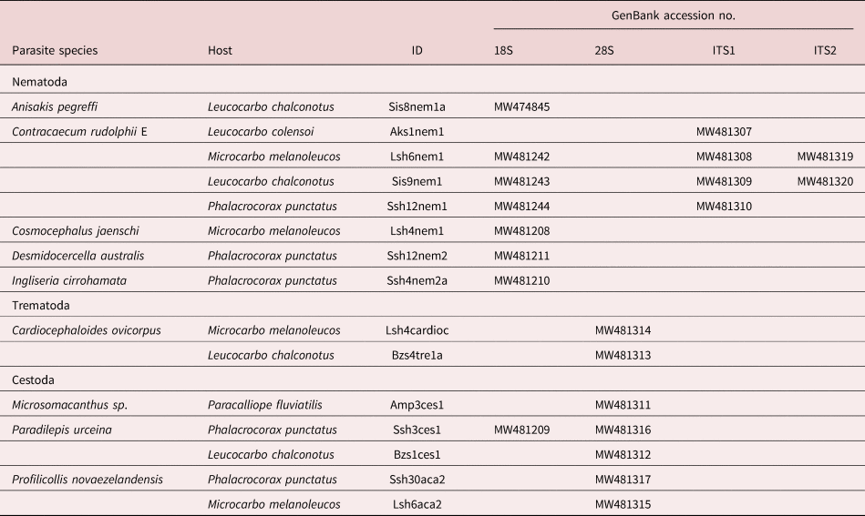
Sequences were used in BLASTn searches on GenBank® to establish a preliminary identification. Sequences were imported into Geneious Prime® version 1.2 (https://www.geneious.com), trimmed using the trim function with default parameters and manually edited for incorrect or ambiguous base calls. A contiguous sequence was assembled for each sequence, alignments were created separately for each species sequenced and genetic divergences were calculated in MEGA version 8 (Tamura et al. Reference Tamura, Dudley, Nei and Kumar2007), comparing between closely related species to confirm identity. Newly generated sequences were submitted to GenBank, listed in table 1.
GenBank accession numbers of newly generated sequences of various parasites from shags in Otago, New Zealand.

Results
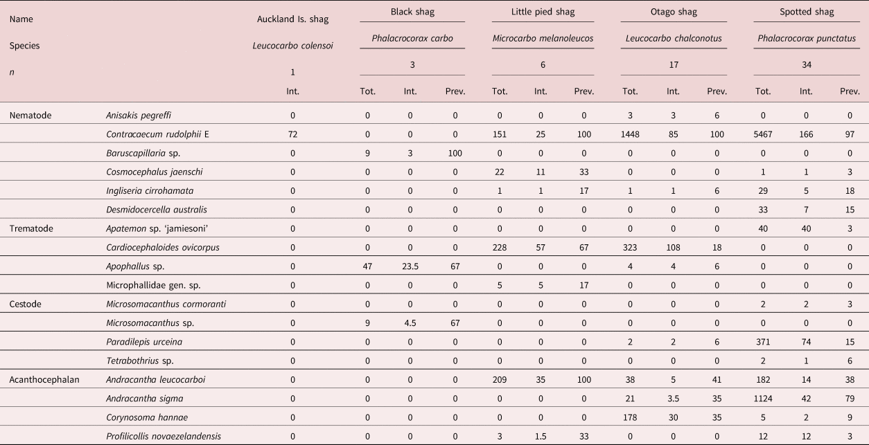
In total,10,043 helminths were recovered from five species of shag. Eighteen parasite species were identified: six nematodes, four trematodes, four cestodes and four acanthocephalans. All shag individuals were infected with at least one species of parasite. The spotted shag harboured the greatest diversity of gastrointestinal helminths with 12 species. The most prevalent parasite was Contracaecum rudolphii E, which was found in all marine shag species and 99% of individuals. The parasite with the highest mean intensity was C. rudolphii E, infecting spotted shags at an average of 166 individuals per host (prevalence and intensity data are summarized in table 2).
Prevalence and intensity data of helminths found in shags from Otago.

Tot., total number of parasites in all birds; Int., mean intensity among parasitized birds; Prev., % prevalence among all birds.
The Otago shag specimens included two different morphotypes, the bronze morph and the pied morph. We observed no differences in number or species of parasites between them and their data were combined. In the following we provide a list of the helminth parasites found in five species of shag from around Otago, along with host identities, collection data, infection site, brief descriptions and remarks on their genetic affinities, life cycles and other data.
Systematics
Phylum: Nematoda
Order: Ascaridida:
Superfamily: Ascaridoidea
Anisakidae
Anisakis pegreffii Campana-Rouget & Biocca, 1955
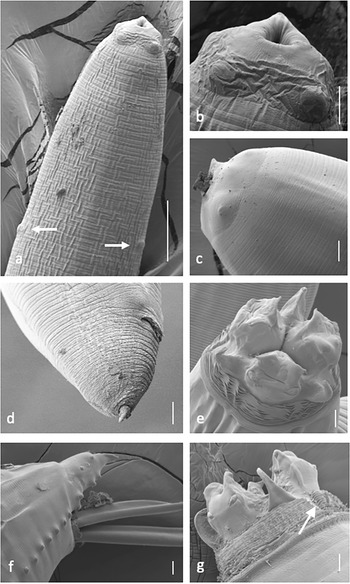
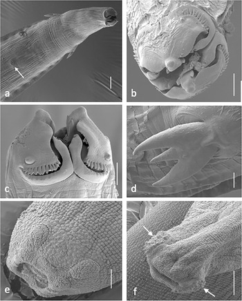
Anisakis pegreffii Campana-Rouget & Biocca, 1955 (fig. 1a-d)
(a) Anisakis pegreffi L3 anterior end showing deirids (arrows). (b) Anisakis pegreffi L3 anterior end. (c) Anisakis pegreffi L3 showing boring tooth. (d) Anisakis pegreffi L3 female posterior end showing anus and mucron. (e) Contracaecum rudolphii E adult anterior end, en face view, showing variable bifurcation of interlabia. (f) Contracaecum rudolphii E adult male posterior showing caudal papillae and spicules. (g) Contracaecum rudolphii E adult anterior end, lateral view, showing V-shaped lateral region with no striations (arrow), and dorsal lip with two papillae. Scale bars: (a) 100 μm; (b–f) 20 μm.

Synonym: Anisakis simplex A. of Nascetti, Reference Mutafchiev, Halajian and Georgiev1983
Three nematodes were found in the stomach of a single Otago shag, that conformed to the morphology of Anisakis sp. Type-1 third-stage larva (L3) (sensu Berland, Reference Berland1961) based on the location of the excretory pore, simple ventriculus and presence of a mucron on the tail. The specimens ranged from 20 to 24 mm in length, with a maximum body width of 320–460 μm and an oesophagus length of c. 2 mm. Characteristic features are indicated in fig. 1a–d.
Remarks
Anisakis pegreffii is one of three currently recognized sibling species, all formerly known as Anisakis simplex (Rudolphi, 1809): A. simplex sensu stricto, A. pegreffii and Anisakis berlandi Mattiucci, Cipriani, Webb, Paoletti, Marcer, Bellisario, Gibson, Nascetti, Reference Mattiucci, Nascetti and Clanchi2014 (=A. Simplex sp. C) (Mattiucci et al., Reference Mattiucci, Paoletti and Webb1997, Reference Mattiucci, Cipriani, Webb, Paoletti, Marcer, Bellisario, Gibson and Nascetti2009, Reference Mattiucci, Nascetti and Clanchi2014; Nadler et al., Reference Moravec, Scholz and Našincová2005; Shamsi et al., Reference Shamsi, Briand and Justine2017).
In a BLASTn search, 18S sequence (803 bp) produced 100% identity with both A. pegreffii and A. simplex, from a conger eel and shark, respectively (Li et al., Reference Li, Lü, Nadler, Gibson, Zhang, Chen, Zhao and Guo2018). Genus Anisakis is a difficult and poorly resolved genus and many records occur under the name A. simplex, although this has been shown to be a complex of species (Mattiucci et al., Reference Mattiucci, Nascetti and Clanchi2014). Given that it is possible the sample of A. simplex was misidentified and that A. pegreffii has been found in New Zealand waters, we therefore assign our specimens to A. pegreffii.
Contracaecum rudolphii E. of Shamsi et al., Reference Shamsi, Briand and Justine2009
Contracaecum rudolphii E. of Shamsi et al., 2009 (fig. 1e–g).
By far the most abundant parasite in the shags examined, C. rudolphii E (Shamsi et al., Reference Shamsi, Briand and Justine2009) was found in 100% of Otago and little shags, and 97% of spotted shags, as well as the single Auckland Island shag. Specimens occurred sometimes in large numbers (>500 individuals in three spotted shags) and were mainly concentrated in the stomach. New infections were sometimes found in the oesophagus and decaying worms were encountered in the upper intestine. Infections included L3 and fourth-stage larvae, and adults, with a large size range from 5 to 45 mm. Males smaller than females, and metrics agree with those given by Shamsi et al. (Reference Shamsi, Briand and Justine2009) for C. rudolphii E from a pied cormorant, Phalacrocorax varius Gmelin, in Australia. Characteristic features are indicated in fig. 1e–g.
Remarks
There are possibly over 100 species in the genus Contracaecum (Shamsi, Reference Rawlence, Scofield and Spencer2019), and differentiation between closely related species is hindered by inadequate descriptions in the early literature, rendering the validity of some species questionable. Contracaecum rudolphii Hartwich, 1964 (syn. C. spiculigerum Rudolphi, 1809) has been found in cormorants and pelicans, and has a global distribution. However, molecular studies have found C. rudolphii sensu lato to consist of several sibling species, with seven species now recognized: C. rudolphii sensu stricto and C. rudophii A to F (Li et al., Reference Li, D'Amelio, Paggi, He, Gasser, Lun, Abollo, Turchetto and Zhu2005; Zhu et al., Reference Stokell2007; Shamsi et al., Reference Shamsi, Briand and Justine2009; d’Amelio et al., Reference D'Amelio, Cavallero, Dronen, Barros and Paggi2012; Mattiucci et al., Reference Mattiucci, Paoletti and Webb2020).
Specimens from one little shag, the Auckland Island shag, two Otago shags and four spotted shags were taken for DNA sequencing. The 18S sequences (758–838 bp) and ITS1 and ITS2 sequences (ITS1: 468–480 bp; ITS2: 379 bp) were identical between hosts, and all showed a 100% match with C. rudolphii E ITS1 and ITS2 (GenBank numbers FM210257 and FM210269). There are currently no sequences of the 18S marker available for comparison on GenBank for C. rudolphii E.
Order: Enoplida
Superfamily: Trichinelloidea
Trichuridae
Baruscapillaria sp.
A total of nine specimens of a hair-like nematode were found in the stomachs of all three black shags (table 2), of which two were male, four were female and three were immature. In having a male with caudal alae absent, pseudobursa with two small, rounded lobes with ventral projections, a long single spicule with aspinous spicule sheath, and presence of bacillary bands, the specimens conformed to the genus Baruscapillaria (Gibbons, 2010), a genus commonly found in phalacrocoracids.
Description
Adult. Sexually dimorphic, males up to 10.2 mm and females up to 16.4 mm in length. Filiform, length:width up to 1:230 (females) and 1:340 (males). Oesophagus length 48–56% of body length (BL).
Male. Pseudobursa consists of two rounded lobes. Single spicule (840 μm long; n = 1) well-sclerotized, smooth, simple at tip and expanded at proximal end. Spicular sheath aspinous, and transversely striated.
Female. Vulva simple with no swelling or appendage, opening flush against the body. Situated about 50% length of body. Egg size 46–48 × 21–27.
Remarks
Two species of the genus are known to parasitize the small intestine of the same shag species, Phalacrocorax carbo, from the northern hemisphere: Baruscapillaria carbonis (Dubinin & Dubinina, 1940) and Baruscapillaria rudolphii Moravec, Scholz & Našincová, Reference Moravec, Scholz and Našincová1994. Baruscapillaria carbonis is characterized mainly by a well-developed membranous bursa in the male, consisting of five distinct lobes, and a spicule length of 1.9–2.3 mm; gravid females have a long vulvar appendage (Frantová, Reference Frantová2001). Males of B. rudolphii have reduced, bi-lobed membranous bursa and the spicule is 0.9–1.3 mm long; the vulvar appendage is absent in gravid females (Moravec et al., Reference Moravec and Scholz1994). Using those features that are visible, these specimens appear to be morphologically closest to the description of B. rudolphii, described from the Czech Republic (Moravec et al., Reference Moravec and Scholz1994). However, in our specimens the length of the spicule and the egg size are slightly smaller. Until more specimens are available allowing scanning electron micrographs to be obtained, we refrain from describing them as new species. Extraction of DNA was not successful.
Order: Spirurida
Superfamily: Acuarioidea
Acuariidae
Cosmocephalus jaenschi Johnston & Mawson, 1941
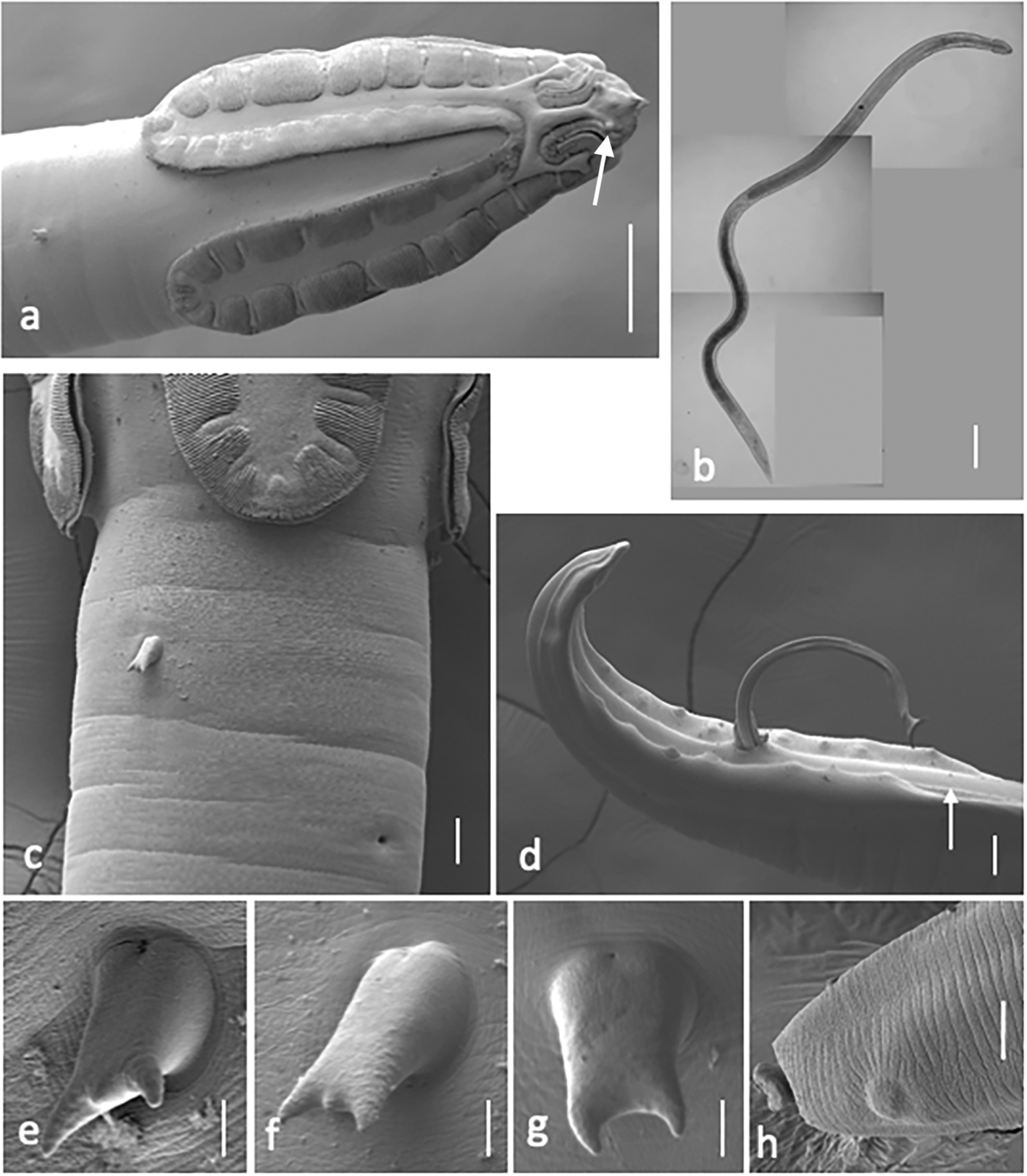
Cosmocephalus jaenschi Johnston & Mawson, 1941 (fig. 2a–h).
(a) Cosmocephalus jaenschi anterior end, lateral view, showing cordons, cephalic papillae (arrow) and position of deirid. (b) Cosmocephalus jaenschi female whole worm. (c) Cosmocephalus jaenschi ventro-lateral view showing deirid and excretory pore. (d) Cosmocephalus jaenschi male posterior end showing caudal papillae, spicules and ventral cuticular ridge (arrow). (e–g) Cosmocephalus jaenschi deirids: (e, f) tricuspid; (g) bicuspid. (h) Cosmocephalus jaenschi female, posterior end of tail showing phasmid and button-like projection. Scale bars: (a)100 μm; (b) 1 mm; (c, d) 20 μm; (e–g) 2 μm; (h) 10 μm.

A total of 22 specimens of this species was found in the proventriculus of two little pied shags, of which eight were male, eight were female and six were L3 larvae, and a single gravid female was found in a spotted shag (table 2). In having recurrent cordons forming a simple arch, convex anteriorly, these specimens were attributable to genus Cosmocephalus, and following the key given by Mutafchiev et al. (Reference Moravec, Prokopic and Shlikas2010) they could be assigned with confidence to C. jaenschi. This species was described from males only, and females have not been hitherto described. Therefore, we present a brief description of our male specimens and a more complete description of our female specimens, as a supplement to the original description (Johnston & Mawson, Reference Johnston and Mawson1941) and redescription (Mutafchiev et al., Reference Moravec, Prokopic and Shlikas2010).
Description
Male. Based on two mounted and two SEM specimens from little pied shag. BL 10.5–11.0 mm. Maximum body width 266–271. Cordons extending 280–344 from anterior end (cordons/BL 0.025–0.028), recurrent and anastomosing laterally 85 from anterior end. Buccal cavity 370–387 long. Muscular oesophagus 841–911 long. Glandular oesophagus 3.1–3.5 mm long. Muscular/glandular oesophagus 0.26–0.27; total oesophagus/BL 0.38–0.40. Deirids bicuspid or tricuspid, salient, 399–415 from anterior end. Excretory pore 475–500 from anterior end. Nerve ring 405 from anterior end. Lateral alae extending from slightly posterior to deirids to about middle body. Tail 233–325 long. Caudal alae 536 long; caudal alae length/BL 0.049–0.051. Single ventral cuticular ridge present, situated anterior to cloaca (fig. 2d). Caudal papillae, four precloacal and five postcloacal pairs. Left spicule 765–824, with prominent projection on its distal end. Right spicule 147–153 long. Left spicule 5–6 times longer than right.
Female. Based on four mounted specimens and one SEM specimen from little pied shag. BL 14.2–16.8 mm (14.6 mm). Maximum body width 337–387 (366) about mid-body. Anterior end with two triangular pseudolabia, each with single amphid and pair of prominent papillae. Pair of swellings situated dorsally and ventrally at base of pseudolabia. Cordons extending 372–571 (446) from anterior end (cordons/BL 0.025–0.035), recurrent and anastomosing laterally 145–149 (147) from anterior end; scalloped on both ascending and descending cordons; longitudinal cuticular ridge along outer rims of cordons, delimiting deep longitudinal groove. Maximum width of cordons 44–57 (50.3). Buccal cavity 459–583 (510) long. Muscular oesophagus 760–866 (803) long. Glandular oesophagus 2152–2442 (2297) long. Muscular/glandular oesophagus 0.31–0.36; total oesophagus/BL 0.19–0.20. Deirids 18 long, bicuspid or tricuspid, salient (fig. 2e–g), 444–695 (522) from anterior end. Postdeirids c. 8 long, bicuspid or tricuspid, situated around termination of alae, 2.4 mm from posterior end of tail (n = 1). Excretory pore posterior to deirids, 528–787 (634) from anterior body end. Nerve ring surrounding anterior part of muscular oesophagus, 517–630 (575) from anterior end. Lateral alae begin slightly posterior to deirid, extending to slightly anterior to vulva. Vulva situated 41–49% BL; 5.9–8.2 (6.9) mm from anterior end. Vagina vera short, directed slightly posteriorly, vagina uterinа longer with muscular walls. Posterior extremity of tail with button-like projection (fig. 2h); paired phasmids subventral, near to tip of tail. Eggs oval, 36–39 × 18–22 (n = 20).
Remarks
The morphometrics of our male specimens concur with all of those reported in the original description (Johnston & Mawson, Reference Johnston and Mawson1941) and redescription (Mutafchiev et al., Reference Moravec, Prokopic and Shlikas2010) of this species, with the exception of the ‘head’ length and distance from the anterior of the deirids, nerve ring and excretory pore. Our specimens were somewhat longitudinally contracted in the anterior part, probably causing underestimation of the relevant measurements.
In New Zealand, C. jaenschi has previously been reported from a little pied shag (Johnston & Mawson, Reference Johnston and Mawson1944). The specific identity of Cosmocephalus sp. recorded from an Auckland Island shag (Johnston & Mawson, Reference Johnston and Mawson1953) is not known.
We have provided DNA sequence for the 18S gene (783 bp) on GenBank (table 1), which constitutes the first sequence available for specimens from this genus. Voucher specimens of females have been deposited with the South Australian museum.
Ingliseria cirrohamata (Linstow, 1888) Gibson, 1968
Ingliseria cirrohamata (Linstow, 1888) Gibson, 1968 (fig. 3a–d).
(a) Ingliseria cirrohamata anterior end, ventral view, showing deirids, excretory pore (arrow) and lateral alae. (b) Ingliseria cirrohamata anterior end, en face view. (c) Ingliseria cirrohamata anterior end, ventral view showing cephalic collarette. (d) Ingliseria cirrohamata tricuspid deirid. (e) Desmidocercella australis anterior end showing papillae and mouth opening. (f) Desmidocercella australis female posterior end showing cushion-like punctate papillae near tip (arrows). Scale bars: (a) 20 μm; (b, c, f) 10 μm; (d) 2 μm; (e) 5 μm.

Synonyms: Filaria (Spiroptera) cirrohamata Linstow, 1888; Streptocara cirrohamata (Linstow) Skrjabin, 1916)
This species was found in low numbers in the stomachs of six spotted, one little pied and one Otago shag. In having paired pseudolabia with dentiform processes, cephalic collarette interrupted by lateral lines, with scalloped cordons bearing about 15 teeth and large tricuspid deirids, these specimens could be assigned confidently to I. cirrohamata (Diaz et al., Reference Diaz, Fonteneau, Panisse, Cremonte and Navone2009).
Infections included immature specimens as well as males and gravid females, BL ranging from 4.0 to 15.5 mm. Males were smaller than females, and the morphology agreed with the description given by Diaz et al. (Reference Diaz, Fonteneau, Panisse, Cremonte and Navone2009). Despite some variation in metrics, our specimens agree with those given by Diaz et al. (Reference Diaz, Fonteneau, Panisse, Cremonte and Navone2009) for a range of records from Phalacrocorax spp. Characteristic features are indicated in fig. 3a–d.
Remarks
The only record of this species from New Zealand is from an Auckland Island shag in the Auckland Islands (Johnston & Mawson, Reference Johnston and Mawson1953). We have provided DNA sequence for the 18S gene marker (787 bp) on GenBank (table 1), which constitutes the first sequence available for specimens from this genus.
Order: Spirurida
Superfamily: Aproctoidea
Desmidocercidae
Desmidocercella australis Mawson, 1953
Desmidocercella australis Mawson, 1953 (fig. 3e, f).
A total of 33 worms was found in five spotted shags. These worms were found in the body cavity washings, and almost certainly originated in the air sacs. They are small, ventrally flexed, brown nematodes with a tubular buccal cavity with two lateral teeth bluntly tricuspid, spicules markedly different in size and shape and with large posterior caudal papillae covered by numerous denticles, all of which make them assignable to the family Desmidocercidae (Anderson et al., Reference Anderson, Chabaud and Willmott2009). In possessing only one preanal and two postanal papillae and a long spicule in excess of 550 μm, these specimens compare consistently with D. australis, which was described from an Imperial shag, Leucocarbo atriceps (King), from subantarctic Heard Island (Mawson, Reference Mattiucci, Cipriani, Webb, Paoletti, Marcer, Bellisario, Gibson and Nascetti1953).
This species was described from two males only, and females have not been hitherto described. Therefore, we present a brief description of our male specimens and a more complete description of our female specimens, as a supplement to the original description (Mawson, Reference Mattiucci, Cipriani, Webb, Paoletti, Marcer, Bellisario, Gibson and Nascetti1953).
Description
Male. Based on two mounted and one SEM specimens. BL 3.8–4.0 mm. Maximum body width 154–162. Cuticle with pronounced punctations. Buccal cavity 27–28 long. Muscular oesophagus 164–172 long. Glandular oesophagus 243–250 long; total oesophagus 11% BL. Deirids not observed. Nerve ring 147–197 from anterior end. Excretory pore 197–232 from anterior end. Tail 55–62 long. Caudal papillae, one precloacal and two postcloacal pairs. Pair of large, subterminal, lateral, highly punctate cushion-like papillae on tail; length 14 × width 19. Long spicule 596–600, with needle-like distal end. Short spicule 238–249 long with complex scooped end.
Female. Based on six mounted and one SEM specimens. BL 4.5–4.9 mm (4.7 mm); approximately 20% larger than males on average. Maximum body width 150–210 (186), about mid-body. Cuticle with pronounced punctations. Cephalic papillae eight large, rounded, heavily punctate papillae radiating in pairs from oral opening. Pair of large dome-shaped amphids at level of anterior cephalic papillae. Oral aperture oval, containing paired pseudolabia with poorly defined teeth (fig. 3e). Buccal cavity 27–30 (29) long. Muscular oesophagus 150–161 (155) long. Glandular oesophagus 279–290 (286) long; total oesophagus 10% BL. Small paired deirids 185–200 (n = 2) from anterior end. Nerve ring 146–205 (189) from anterior end. Vulva situated 56–57% BL; 2.6–2.7 mm from anterior end. Tail 59–67 (64) long. Anterior lip of anus with distinct cuticular swelling, 5–6 thick. Pair of large, subterminal, lateral, highly punctate cushion-like papillae on tail (fig. 3f); length 14× width 19. Eggs oval, 50–55 × 18–25 (52–21; n = 20).
Remarks
The morphometrics of our male specimens concur with all of those reported in the original description of this species (Mawson, Reference Mattiucci, Cipriani, Webb, Paoletti, Marcer, Bellisario, Gibson and Nascetti1953), in terms of BL, number of caudal papillae and size of spicules. We have provided DNA sequence for the 18S gene marker (903 bp) on GenBank (table 1), which constitutes the first sequence available for specimens from this genus. Voucher specimens have been deposited with the South Australian museum.
Class: Trematoda
Order: Diplostomida
Superfamily: Diplostomoidea
Strigeidae
Apatemon sp. ‘jamiesoni’ of Blasco-Costa et al., Reference Blasco-Costa, Poulin and Presswell2016
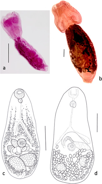
Apatemon sp. ‘jamiesoni’ of Blasco-Costa et al., 2016 (fig. 4a).
(a) Apatemon sp. ‘jamiesoni’ lateral view. (b) Cardiocephaloides ovicorpus ventral view. (c) Apophallus sp. composite drawing. (d) Microphallidae gen. sp. drawing. Scale bars: (a, b) 500 μm; (c, d) 100 μm.

Forty specimens of this strigeid were recovered from the intestine of a single spotted shag from Otago Harbour. The species was previously described without being formally named from a single specimen from a mallard duck, Anas platyrhynchos Linnaeus (Blasco-Costa et al., Reference Blasco-Costa, Poulin and Presswell2016). Although available at the time of publishing the 2016 paper, the specimens found in the spotted shag were not sufficiently well preserved to enable a formal description, but DNA extraction was successful and the 28S sequence was found to be identical to that of the mallard specimen. The measurements of the shag specimens also agreed with those given for the mallard specimen.
Remarks
In Blasco-Costa et al. (Reference Blasco-Costa, Poulin and Presswell2016) the authors provided a detailed description of the single specimen from the mallard, along with descriptions of the metacercaria and cercaria, but refrained from formally naming the species due to the poor quality of the remaining specimens from the spotted shag. DNA sequences from a shag specimen and from the mallard and intermediate hosts were submitted to GenBank with publication of the paper (Blasco-Costa et al., Reference Blasco-Costa, Poulin and Presswell2016) (28S ribosomal DNA (rDNA), KT334166–KT334169; ITS1-5.8S-ITS2, KT334170–KT334172; COI, KT334181–KT334182).
Cardiocephaloides ovicorpus Dubois & Angel, 1972 (fig. 4b)
This species was found, sometimes in large numbers, in the upper intestine of three Otago and four little pied shags. In having a bipartite body, with relatively small forebody, hindbody with neck region, large protrusible copulatory bursa and vitellarium confined to hindbody, these specimens were attributable to genus Cardiocephaloides (Dubois, Reference Dubois1970). Morphometrically, they were readily assignable to C. ovicorpus, which was described originally from little pied shags from Dunedin (Dubois & Angel, Reference Dubois and Angel1972). Specimens ranged from new infections with little hindbody development, to fully gravid worms 4.7–6.7 mm long.
Remarks
The original description of C. ovicorpus was comprehensive and no additions are needed here. Most specimens of C. ovicorpus were preserved in a relaxed state with the body oval and the copulatory bursa inverted. With the bursa everted, the worm acquires the more usual extended shape of Cardiocephaloides species. Dubois & Angel (Reference Dubois and Angel1972) described material from little pied shags in Dunedin and also from P. varius in South Australia in the same paper. This species has not been reported in the literature since its description, and our report from the Otago shags constitutes a new host record.
DNA sequence (28S rDNA) was identical for specimens from the two shag species, and a BLASTn search found sequences closest to Cardiocephaloides physalis from a Magellanic penguin, Spheniscus magellanicus (Forster), in Chile (Achatz et al., Reference Achatz, Pulis, González-Acuña and Tkach2020). The sequences from our material have been added to those on GenBank and represent the first available of this species.
Order: Plagiorchiida
Superfamily: Opisthorchioidea
Heterophyidae
Apophallus sp.
Apophallus sp. (fig. 4c).
Two out of three black shags were infected with this small trematode. All specimens had undergone a degree of lateral shrinkage in the forebody, giving them a club-like shape, and it was clear that, with the forebody not contracted, these worms would be an elongate pear shape. Other specimens found in a single Otago shag were in a better state of preservation and measurements were concurrent between the two sets of specimens.
In having a length less than 7 mm, pharynx present, prepharynx short, two entire parallel or oblique testes, ventrogenital sac with gonotyl, seminal vesicle elongate and S-shaped, vitellaria around margins of hindbody, extending into forebody and operculate thick-shelled eggs, these specimens conform to the diagnosis of family Heterophyidae. A combination of relevant features and apparent possession of two lateral gonotyls placed these specimens tentatively in the genus Apophallus, but preservation was not sufficiently good to observe the detail of the ventrogenital complex, so comparison with congeneric species is not attempted. A short description is given here for future comparison. As the black shag specimens were unnaturally attenuated, BL and width measures are given only for the Otago shag specimens.
Description
Based on seven specimens ex black shag and two specimens ex Otago shag. Small, pyriform, flat worms, forebody spinose. BL 347–402; forebody 192–232. Widest at mid-hindbody, 145–185. Oral sucker subterminal, round 34–48 × 34–49. Prepharynx absent. Pharynx elongate oval, 25–32 × 21–28. Ventral sucker mid-body length, round 36–45 × 36–43. Sucker length ratio 1:0.95. Ventrogenital sac, anterior, ventral to and slightly overlapping anterior of ventral sucker. Ovary round to triangular, 27–38 × 26–46. Seminal vesicle large, S-shaped, posterior to ovary. Testes entire, parallel or oblique, sub-round, 36–86 × 32–73 (left), 42–79 × 40–58 (right). Vitellaria around margins of hindbody, into forebody at least to caecal bifurcation, with pre-testicular bar across width. Uterus central, between ventral sucker and testes. Eggs large and few, 25–29 × 16–20.
Remarks
An important character in delimiting the taxa within Heterophyidae is the form of the ventrogenital complex. As these were not completely observable in our specimens, we have not attempted to assign them to a species. Only one heterophyid has been reported from New Zealand birds; Galactosomum otepotiense Presswell & Bennett, 2020. This is, therefore, the first record of a species of Apophallus for Otago and black shags, and for New Zealand. Sequencing of DNA was not successful, and collection of further specimens is clearly indicated to resolve the identity of this trematode.
Order: Plagiorchiida
Superfamily: Microphalloidea
Microphallidae gen. sp.
Microphallidae gen. sp. (fig. 4d).
Five specimens of trematode belonging to the Microphallidae were found in a single little pied shag. Although a weak ventral sucker was visible and some secondary genital structure was apparent, it was not possible to observe any accessory structures clearly in order to allocate the specimens to a genus. All specimens were mature and gravid. A brief description is provided for future comparison.
Description
Based on five stained and mounted specimens. Body linguiform, length 450–506, width 210–219. Oral sucker (OS) weak, subterminal, 42–51 × 38–42. Prepharynx 33–34. Pharynx round, 26–27 × 27. Oesophagus 58 long. Caeca not extending below level of ventral sucker, 155–147 (left) and 115–145 (right). Ventral sucker (VS) 39–42 × 32–42; VS:OS length ratio, 1:0.76–1.0. Cirrus sac wide and arching, 195–201 long, 44–49 wide; containing oval seminal vesicle and convoluted cirrus with small spines. Terminates with pore, level with and sinistral to ventral sucker. Ovary not observed. Testes oblique; edges obscured by eggs in uterus. Uterus fills area posterior to ventral sucker and between testes, full of eggs in all specimens. Vitellaria follicular, forming a ring surrounding posterior edge reaching to level with cirrus sac; internally not visible. Eggs 17–19 × 10–12.
Remarks
Although it is not possible to assign these specimens to a genus and species at this point, the combination of features which are visible allows us to state that they do not belong to a microphallid species already described from New Zealand. DNA sequencing was not successful.
Class: Cestoda
Order: Cyclophyllidea
Hymenolepididae
Microsomacanthus sp.
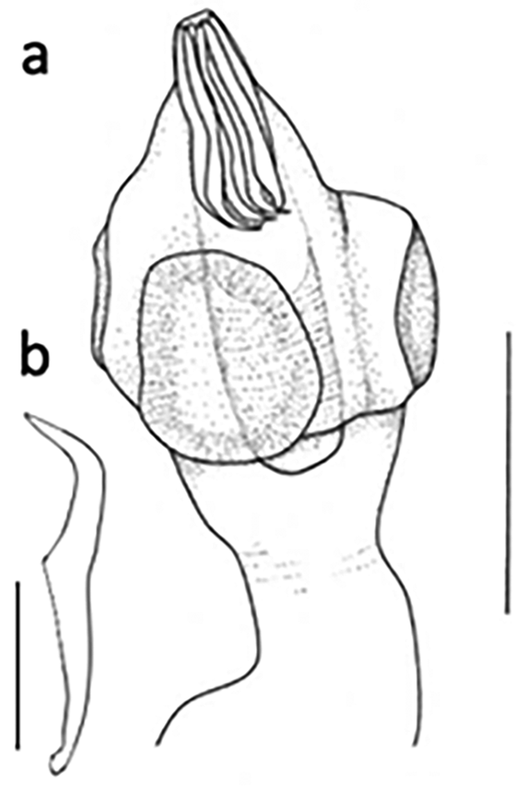
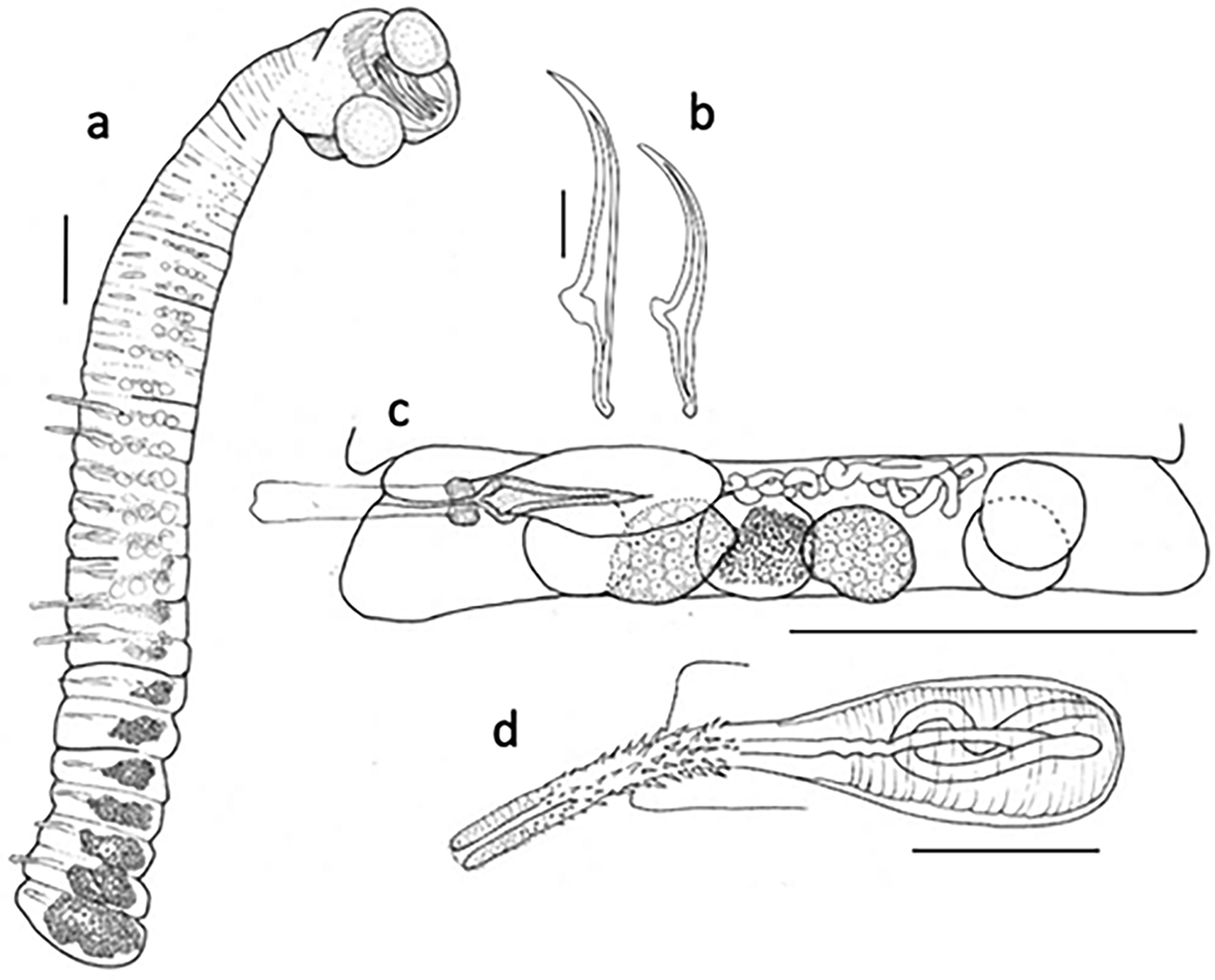
Microsomacanthus sp. (fig. 5a, b).
(a) Microsomacanthus sp. scolex. (b) Microsomacanthus sp. hook. Scale bars: (a) 100 μm; (b) 25 μm.

A total of nine specimens of this morphotype were found in two black shags. These tiny cestodes, while attributable to genus Microsomacanthus on the basis of the hook number, shape and size and the number of testes, could not be identified further to species. Two mounted specimens measured 960 and 1100 in length; 68 and 77 at widest. Scolex length 120, 130; width 93, 150. Suckers round to elongate oval 60 × 61 and 80 × 61. Rostellar apparatus muscular. Rostellar hooks diorchoid with short guard; 62 total length, handle 35, blade 27. Testes three in transverse row. Cirrus sac strongly muscular. Other internal organs not observed.
Remarks
A comparison of hook size with some 70 other Microsomacanthus spp. in the literature found just three species with hook length close to this material: Microsomacanthus diorchis (Fuhrmann, 1913) Lopez-Neyra, 1942 (hooks 50–64), Microsomacanthus floreata (Meggitt, 1930) Lopez-Neyra, 1942 (hooks 65–75), Microsomacanthus minimus Ryzhikov, 1965 (hooks 63–66). These can be differentiated by the shape of the rostellar hooks (M. diorchis and M. floreata) and the number of proglottides (maximum 22–25 in M. minimus) and it, therefore, appears that these specimens represent an undescribed species of Microsomacanthus. Further specimens of better clarity, along with genetic data, will enable description and naming of this species.
Microsomacanthus cormoranti (Ortlepp, 1938)
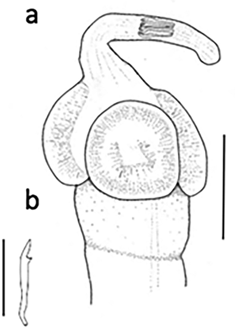
Microsomacanthus cormoranti (Ortlepp, 1938) (fig. 6a, b).
(a) Microsomacanthus cormoranti. (b) Microsomacanthus cormoranti rostellar hook. Scale bars: (a) 100 μm; (b) 25 μm.

Synonyms: Hymenolepis cormoranti Ortlepp, 1938; H. gyogonka Johri, 1941; H. childi Burt, 1940
Just two specimens of this species were found in a single spotted shag. This very small cestode with ten diorchoid rostellar hooks arranged in a single circle on the rostellum conforms to the description of genus Microsomacanthus. The better preserved specimen is 1.7 mm long and 65 wide at its widest point. Scolex about 150 wide with long, thin retractile rostellum, 143 long, hooks facing posteriorly when retracted. Rostellar hooks 26–27 long: handle 20, blade 7. Suckers unarmed, round or slightly longitudinal oval, length 80–90. Both specimens are complete, with a rounded terminal proglottid, but no internal organs are visible, despite staining, and they are presumed to be immature.
Remarks
Originally described from Microcarbo africanus (Gmelin) in Transvaal, mature specimens of M. cormoranti were said to have a scolex width of 130, sucker length of 54 and hooks 24–25 long (Ortlepp, Reference Nascetti1938). A subsequent record from Microcarbo melanoleucos (Vieillot) in South Australia (Clark, Reference Clark1957) described them as having a scolex width of up to 140, sucker length 40–50 and hooks 22 long. Their hooks and those of our specimens are the same diorchoid shape. The New Zealand specimens contain no mature segments, but based on the close resemblance of the scolices to those of the above records, we provisionally identify them as M. cormoranti. DNA extraction was not successful.
Gryporhynchidae
Paradilepis urceina Bona, 1975
Paradilepis urceina Bona, 1975 (fig. 7a–d).
(a) Paradilepis urceina whole worm. (b) Paradilepis urceina large and small rostellar hooks. (c) Paradilepis urceina mature proglottid showing carrot-shaped vagina. (d) Paradilepis urceina cirrus sac showing vas deferens. Scale bars: (a, c) 100 μm; (b, d) 25 μm.

One Otago shag and five spotted shag harboured specimens of this species. In having a cyclusteroid rostellar apparatus, rostellar hooks in two rows, four testes and a cirrus with rose-thorn spines, these specimens were attributable to genus Paradilepis. Morphometrically, they were readily assignable to P. urceina, which was described originally from a white ibis, Threskiornis molucca strictipennis (Gould) and a ‘blue heron’, which we assume to be Egretta novaehollandiae (Latham), both from South Australia (Bona, Reference Bona1975).
Specimens ranged from 1.8 to 2.7 mm in length, the largest proglottid 201 wide. Number of proglottides 36–47. Scolex width 230–275, with suckers 110–124 × 92–113. Rostellar hooks 20 in a double crown, of ‘urceus’ pattern; long hooks 126–130, short hooks 98–100. Length of rose-thorn spine field on cirrus 30–37. Cirrus sac length 53–96.
Remarks
All measurements fall within the ranges given by Bona (Reference Bona1975), except for total length, which is a little greater in the new specimens (1.8–2.7 vs. 0.85–1.3). Rostellar hooks are comparable in size (long hook, 126–130 vs. 123–136; short hook, 98–100 vs. 92–104) and are identical in shape. Sequences of 18S and 28S were obtained from these specimens and have been submitted to GenBank (see table 1). BLASTn searches using both of these markers returned species of Paradilepis, but none with a high similarity percentage.
Order: Tetrabothriidea
Tetrabothriidae
Tetrabothrius sp.
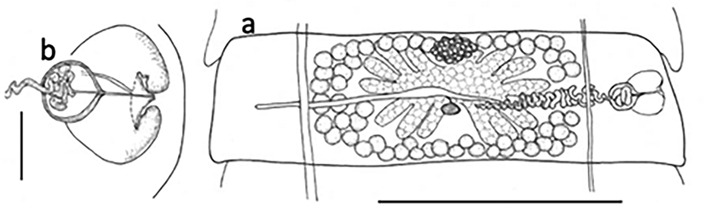
Tetrabothrius sp. (fig. 8a, b).
(a) Tetrabothrius sp. mature proglottid. (b) Tetrabothrius sp. genital atrium and cirrus sac. Scale bars: (a) 1 mm; (b) 100 μm.

The strobilae, without scoleces, of two specimens of this cestode were found in two spotted shags. In having craspedote proglottides with unilateral genital pores, a deep, muscular genital atrium, cirrus sac not extending beyond osmoregulatory canals, ovary multilobate extending to osmoregulatory canals, numerous testes and a spiny attenuated cirrus, these specimens were attributable to the genus Tetrabothrius. A brief description is given below based on proglottides from a stained and mounted specimen.
Description
Strobila more or less complete, ~130 mm long, 2 mm at widest. Strongly craspedote, genital pores unilateral. Genital opening about central or slightly anterior of central in proglottid.
Mature proglottid 377 × 1800, width:length, 1:4.7. Genital atrium round, 148 × 162. Cirrus sac small, sub-round, 82 um long (perpendicular to worm), 98 wide (parallel to length of worm), opens into genital atrium via a small conical papilla. Convoluted vas deferens arises around median line, with several coils inside cirrus sac; cirrus with minute spines. Testes numerous (~50), sub-round to oval, 52–58 × 42–45. Ovary with two sections each 130–150 wide, ~50 deep, in more mature segments digitiform lobes. Uterus tubular, not extending beyond osmoregulatory canals, nearer to anterior of proglottis than posterior, ~900 long and 80 deep.
Remarks
The morphology and measurements of the present specimens appear to fit well with the two Tetrabothrius species that are specific to shags, Tetrabothrius shinni and Tetrabothrius phalacrocoracis, and it is probable that these three species are closely related. No DNA sequences are yet available.
Phylum: Acanthocephala
Order: Polymorphida
Polymorphidae
Andracantha leucocarboi Presswell et al., 2018
This common acanthocephalan was found in the intestines of all little pied shags, eight Otago shags and 13 spotted shags. The species was previously described from some of the material reported herein (Presswell et al., Reference Parsons, Mitchell, Butler, Ratcliffe, Frederiksen, Foster and Reid2018), and gene sequences of 28S and cytochrome c oxidase 1 (CO1) were provided by the authors (28S, MF401623; CO1, MF527023-25). The type-specimens of this species were found in Otago shags, and the species was also recorded from spotted shags from coastal sites around Otago. This is the first report of A. leucocarboi since its description in 2018, and the first in little pied shags.
Andracantha sigma Presswell et al., 2018
Found in the intestines of six Otago shag and 27 spotted shags during this study, this species was also previously described from some of the material reported herein (Presswell et al., Reference Parsons, Mitchell, Butler, Ratcliffe, Frederiksen, Foster and Reid2018). Gene sequences for CO1 were provided by the authors (MF527026-33, MF527035). The same species infected little blue penguins, Eudyptula novaehollandiae Stephens (CO1, MF527034; 28S, MF401624). All specimens in the original study came from around the Otago coast. This is the first report of A. sigma since its description in 2018.
Corynosoma hannae Zdzitowiecki, 1984
This acanthocephalan was found in the intestine of six Otago and three spotted shags. The specimens exhibited all the morphological features of C. hannae; body shape, proboscis with pronounced dilation, proboscis hooks in 22 rows of 12–13 hooks per row, somatic spines extending to posterior body end, large genital spines in males. However, none were fully mature, as expected for a polymorphid acanthocephalan in a non-definitive host.
Remarks
The species C. hannae has been reported from these shag hosts before, as well as from yellow-eyed penguin, Megadyptes antipodes (Hombron & Jaquinot), although the definitive hosts are pinnipeds. In New Zealand, definitive hosts are New Zealand sea lion, Phocarctos hookeri (Gray), and long-nosed fur seal, Arctophoca forsteri (Lesson), and the life cycle has been demonstrated to include paratenic hosts New Zealand brill, Colistium guntheri (Hutton), and New Zealand sole, Peltorhamphus novaezeelandiae Günther (Hernández-Orts et al., Reference Hernández-Orts, Smales, Pinacho-Pinacho, García-Varela and Presswell2017). Representative CO1 sequences of C. hannae were submitted to GenBank by Hernández-Orts et al. (Reference Hernández-Orts, Smales, Pinacho-Pinacho, García-Varela and Presswell2017) under numbers KX957715–KX957726.
Profilicollis novaezelandensis Brockerhoff & Smales, 2002
This species of acanthocephalan was found infecting two little pied shags and one spotted shag. In having a long neck, trunk spines and a spheroid proboscis, they could be referred to the genus Profilicollis as resurrected by Nickol et al. et al. (Reference Nadler, D'Amelio, Dailey, Paggi, Siu and Sakanari1999), and confidently assigned to P. novaezelandensis following the original description (Brockerhoff & Smales, Reference Brockerhoff and Smales2002). Profilicollis novaezelandensis was described from the South Island pied oystercatcher, Haematopus ostralegus finschi Martens, in Canterbury, South Island.
Specimens were all juvenile, excysted or unexcysted cystacanths; 5–5.4 mm long, with a subspherical proboscis bearing 14–16 rows of 7–8 hooks.
Remarks
Hay et al. (Reference Hay, Jorge and Poulin2018) have supplied multiple DNA sequences for the CO1 marker from the Royal spoonbill, Platalea regia Gould, and crab, Hemigrapsus crenulatus (Milne Edwards) (GenBank MG602424–MG602475) and partial 28S from the crab (MG602357–MG602363). To these we add further 28S sequences from the little shag and spotted shag (table 1).
Discussion
We found a total of 18 endohelminth species infecting five species of shag around Otago, New Zealand: six nematodes (A. pegreffi, C. rudolphii E, Baruscapillaria sp., C. jaenschi, I. cirrohamata and D. australis), four trematodes (Apatemon sp. ‘jamiesoni’, C. ovicorpus, Apophallus sp. and Microphallidae gen. sp.), four cestodes (M. cormoranti, Microsomacanthus sp., P. urceina and Tetrabothrius sp.) and four acanthocephalans (A. leucocarboi, A. sigma, C. hannae and P. novaezelandensis). Previous to this study, there were no records of gastrointestinal cestodes or trematodes from shags in New Zealand. We report seven new host records for spotted shags, three for black shags, three for little pied shags and six for Otago shags. In the following we discuss the taxonomic histories, known biology and life cycles of the parasites.
No named species of Anisakis has been recorded for New Zealand shags. Anisakis sp. was listed by McKenna (Reference Mawson2010) from L. colensoi and Macquarie Island shags Leucocarbo purpurascens (Brandt), although it seems likely that this record originally referred to specimens of Terranova piscium, a marine mammal nematode, found as stomach contents in the shag (Johnston & Mawson, Reference Johnston and Mawson1953, Reference Johnston and Mawson1945). Our larval specimens are, therefore, the first reliable reports of Anisakis species in New Zealand shags. Anisakis pegreffi larvae have been found infecting bird hosts from Australia: a grey petrel, Procellaria cinerea (Gmelin), and a little penguin, Eudyptula minor (Forster), both confirmed by DNA identity (Shamsi et al., Reference Shamsi, Briand and Justine2017). Anisakis pegreffi has a worldwide distribution, and in New Zealand infects the pilot whale, Globicephala melas (Traill), as well as fish paratenic hosts: Parapercis colias (Bloch & Schneider), Pseudophycis bachus (Forster), Thyrsites atun (Euphrasen) and Hoplostethus atlanticus Collett, the second intermediate hosts being squid (Mattiucci et al., Reference Mattiucci, Paoletti and Webb1997). Anisakis pegreffii is one of the anisakid species that have extraordinary allergenic properties in humans, and are implicated in more than 20,000 cases of anisakiasis in the last 50 years (Llorens et al., Reference Llorens, Arcos and Robertson2018).
In New Zealand, C. rudolphii sensu lato (as C. spiculigerum) has historically been found in black and spotted shags in the brackish, coastal Lake Ellesmere (Stokell, Reference Shamsi, Gasser, Beveridge and Shabani1936), in little pied shags from Dunedin (Johnston & Mawson, Reference Johnston and Mawson1944) and in Auckland and Campbell Island shags (Johnston & Mawson, Reference Johnston and Mawson1943). It is not clear whether these records relate to C. rudolphii E or another of the species complex, although based on the host diversity, high prevalence and intensity of these nematodes in our samples, it seems likely. Black shags among our sample were not infected with C. rudolphii E, as they are generally freshwater feeders. As well as four out of five shag species reported herein, we have also found this species (confirmed by DNA sequence) in little blue penguins (E. novaehollandiae) from Otago. Species of the genus Contracaecum, typically parasites of fish-eating birds or marine mammals, infect a very broad range of invertebrate first intermediate hosts, with a variety of fish species acting as second intermediate or paratenic hosts (Shamsi, Reference Rawlence, Scofield and Spencer2019), and considerable work is needed to unravel specific identities and relationships within the genus.
This is the first record of any trichurid in shags and the first record of a species of Baruscapillaria for New Zealand. In fact, only three New Zealand marine bird species have been recorded as harbouring a species from the family Trichuridae and none of these have been identified or described, other than as ‘Capillaria sp.’ (McKenna, Reference Mawson2010). As members of the family have been recorded from terrestrial and freshwater birds, mammals, reptiles and fish in New Zealand (Hine et al., Reference Hine, Jones and Diggles2000; McKenna, Reference Mawson2003, Reference Mawson2010, Reference McKenna2012, Reference McKenna2018), there is clearly more work needed to elucidate the taxonomic identities and relationships of this group of nematodes. This genus of nematodes exhibits a monoxenous life cycle, thus requiring no intermediate hosts (Moravec et al., Reference McKenna1987).
Cosmocephalus jaenschi was described based on two male specimens from a black shag, P. carbo from Tailem Bend, Australia (Johnston & Mawson, Reference Johnston and Mawson1941) and recently redescribed from the same material in more detail, including the added observations of lateral alae and ventral cuticular ridge (Mutafchiev et al., Reference Moravec, Prokopic and Shlikas2010). Johnston & Mawson (Reference Johnston and Mawson1942) reported a single female specimen collected from Pelecanus conspicillatus Temminck as belonging to C. jaenschi, the identification of which was later questioned by Mutafchiev et al. (Reference Moravec, Prokopic and Shlikas2010). Indeed, when Mawson wrote of finding a single female in a whiskered tern, Chlidonias hybrida (Gould) from South Australia (Mawson, Reference Mawson1969), her list did not include that record, which suggests a retraction of the original identification. Mawson (Reference Mawson1969) did not describe or illustrate the tern specimen, and, therefore, our account comprises the first description of the female of this species. Delimitation of species in this genus has, in part, relied upon the form of the deirids, or cervical papillae. Johnston & Mawson's (Reference Johnston and Mawson1941) male type-specimens had tricuspid deirids, but the female reported from a little pied shag in Dunedin (Johnston & Mawson, Reference Johnston and Mawson1944) had bicuspid deirids. As we found both forms in our sample of specimens that were otherwise indistinguishable, we suggest that the number of cusps on the deirids may be more variable than previously considered (fig. 2e–g). The life cycle of Cosmocephalus species is reported to include amphipods and fish as first and second intermediate hosts, but specific intermediate hosts for the New Zealand specimens have not yet been found. Among the seven species of Cosmocephalus (Mutafchiev, 2010), the site of infection within the bird host may be the oesophagus or the stomach. We can confirm that C. jaenschi falls into the latter group, as we found all of our specimens in the birds’ stomachs.
Ingliseria cirrohamata has mostly been recorded from hosts belonging to the Phalacrocoracidae in the subantarctic region (Johnston & Mawson, Reference Johnston and Mawson1945, Reference Johnston and Mawson1953; Gibson, Reference Gibson1968; Diaz et al., Reference Diaz, Fonteneau, Panisse, Cremonte and Navone2009). The veracity of reports from freshwater birds in Eurasia is questioned by other authors (Gibson, Reference Gibson1968; Diaz et al., Reference Diaz, Fonteneau, Panisse, Cremonte and Navone2009), and until more data are available it seems that a circumpolar distribution confined to the shags is the reliable distribution for this species. Nothing is known of the life cycle. This study represents new host records for the three shags and a new geographical record for mainland New Zealand.
The original description of D. australis included two males from the airways of an Imperial shag (Mawson, Reference Mattiucci, Cipriani, Webb, Paoletti, Marcer, Bellisario, Gibson and Nascetti1953). Mawson erected the species on the strength of the short buccal cavity and the tiny caudal alae, but it was based on only two male specimens. The female has not previously been described. The original description of D. australis was lacking in detail, but the important characters of spicule size and number of anal papillae were included, which are distinct from the other two species in the genus (Desmidocercella incognita Solonitsin, 1932 and Desmidocercella numidica (Seurat, 1920)). Desmidocercella spp. have not previously been reported from New Zealand or the spotted shag (McKenna, Reference Mawson2010), making this a new geographical record and a new host record. Moravec & Scholz (Reference McKenna2016) observed small lateral deirids in D. incognita, for the first time in a species of this genus, and we report the same herein for D. australis. These deirids are tiny and very easily missed. Indeed, Singh (Reference Shamsi1948) did not find them in D. numidica (as D. leiperi) ‘even after prolonged search.’ Although the life cycle of D. australis is not known, intermediate hosts are likely to be aquatic invertebrates, but in other Desmidocercella species fish act as paratenic hosts, where the worm inhabits the vitreous humour of the eye (Baruš et al., Reference Baruš, Sergeeva, Sonin and Ryzhikov1978).
Although no further specimens have been found in shags since the publication of the 2016 paper (Blasco-Costa et al., Reference Blasco-Costa, Poulin and Presswell2016), we can report finding Apatemon sp. ‘jamiesoni’ in a black-backed gull, confirmed by DNA sequence identity (unpublished data). These specimens will be described in a separate paper when the name Apatemon jamiesoni will be formally assigned. Apatemon sp. ‘jamiesoni’ is a rare example of a parasitic worm where each of the natural life stages has been confirmed by genetic identity. The first intermediate host is the fresh/brackish water tateid snail, Potomopyrgus antipodarum (Gray), and the second intermediate hosts are small freshwater fish species, Gobiomorphus cotidianus McDowall, Gobiomorphus breviceps (Stokell) and Galaxias anomalus (Stokell) (Blasco-Costa et al., Reference Blasco-Costa, Poulin and Presswell2016). The low prevalence in the spotted shag (one in 34 birds) and black-backed gull (one in 45 birds) perhaps correlates with the fact that these birds are generally marine predators, which occasionally feed in estuaries and brackish lakes (Lalas, Reference Lalas1983).
Cardiocephaloides species are parasitic as adults in marine fish-eating birds, most commonly of the family Laridae (Dubois, Reference Dubois1970). Cardiocephaloides ovicorpus is the only species described from a phalacrocoracid, although there is one record of C. physalis (Lutz, 1926) from a Guanay cormorant, Phalacrocorax bougainvilleii (Lesson), in Peru (Baer, Reference Baer1969). In a new observation, our specimens of C. ovicorpus in the little pied shags were found in large aggregations in the pyloric region of the small intestine, directly posterior to the stomach. In two cases they were packed so tightly that the first 5 cm of the small intestine was distended up to 2.5 times its normal width. Other species of Cardiocephaloides are also found in the upper intestine of their host, but spread over a wider area. Photographs of C. physalis in situ (Dubois, Reference Dubois1938) show a large number of individuals spread over three long sections of intestine, and Prévot & Bartoli (Reference Prévot and Bartoli1980) reported that C. physalis heavily infested the first 0.5 m of intestine. The second intermediate host of Cardiocephaloides species has been demonstrated to be marine sparid and scombresocid fish in which the metacercariae were found in the optical lobe of the brain (Prévot & Bartoli, Reference Prévot and Bartoli1980). We are currently surveying various intertidal species in Otago to identify the first and second intermediate host of this species. Although the cause of death was not obvious in all bird individuals, it is thought the most heavily infected bird (a juvenile infected with >150 C. ovicorpus as well as numerous nematodes and acanthocephalans) died of weakness associated with its worm burden. Baer (Reference Baer1969) commented on the pathogenicity of C. physalis in cormorants, observing complete destruction of the epithelial cells of the hosts’ intestinal villi where the trematodes were attached, and Randall & Bray (Reference Presswell, Poulin and Randhawa1983) reported on a multiple mortality event in a Jackass penguin colony that was related to heavy burdens of C. physalis.
Trematodes of the family Heterophyidae are parasites of mammals and birds, and have been reported from cormorants (Johnston, Reference Johnston1943; Kanarek et al., Reference Kanarek, Sitko, Rolbiecki and Rokicki2003; Moravec & Scholz, Reference McKenna2016). The metacercariae of Apophallus spp. encyst in cyprinid, percid and salmonid fishes (Sándor et al., Reference Randall and Bray2017); infections can be heavy and can cause vertebral column deformities, ectopic bone formation and muscle infections depending upon the species. Most commonly metacercariae occur in the fin rays where melanization of the cysts manifests as black spot disease, and infection is associated with mortality in some cases (Ferguson et al., Reference Ferguson, Locke, Font, Steinauer, Marcogliese, Cojocaru and Kent2012). There are no records of heterophyid species in the literature for New Zealand for any host, other than G. otepotiense, recorded from fish-eating birds and a marine snail (Presswell & Bennett, Reference Ortlepp2020). Apophallus is one of a number of heterophyid genera that have zoonotic species, with potentially significant pathogenicity (Chai & Jung, Reference Chai and Jung2017). This was the only endohelminth in our sample to be shared by both a marine and a freshwater shag, suggesting its occurrence is not limited to one environment.
The Microphallidae are well represented in New Zealand with well-characterized species (Maritrema deblocki Presswell, Blasco-Costa & Kostadinova, 2014; Maritrema poulini Presswell, Blasco-Costa & Kostadinova, 2014; Maritrema novaezealandense Martorelli, Fredensborg, Mouritsen & Poulin, Reference Martorelli, Fredensborg, Mouritsen and Poulin2004; and Atriophallophorus winterbourni Blasco-Costa, Seppälä, Feijen, Zajac, Klappert & Jokela, 2019). In addition, we are aware of at least 11 more species defined only by DNA sequence from snail hosts, or by differences in adult morphology, none of which have yet been worked on (unpublished data). Clearly, this is a family which requires a lot more study to reveal taxonomic interrelationships between taxa. These species are present in a variety of shags, gulls, ducks, godwit and oystercatchers, and have a three-host life cycle, including gastropod first intermediate and crustacean second intermediate hosts (Martorelli et al., Reference Martorelli, Fredensborg, Mouritsen and Poulin2004).
There are over 70 species of genus Microsomacanthus, which inhabit the small intestine of Anatidae and other water birds. Microsomacanthus cormoranti is the only species of Microsomacanthus to be reported from a member of the Phalacrocoracidae. A single species, Microsomacanthus alaskensis (Deblock & Rausch, 1967), has been reported from New Zealand, from oystercatchers and a black-backed gull, Larus dominicanus Lichtenstein (McKenna, 2010). Microsomacanthus alaskensis can be distinguished from M. cormoranti by its hook size; 78–81 μm versus 26–27 μm. The intermediate hosts of Microsomacanthus spp. are amphipod or copepod crustaceans.
We have examined many amphipods, Paracalliope fluviatilis (Thomson), from freshwater habitats in Otago, in which cysticercoid larvae are found, which conform to the ‘cercocyst’ larva of the Hymenolepididae that are known from cestodes occurring as adults in aquatic birds (Chervy, Reference Chervy2002). Cysticercoid larvae are immature but already have the rostellar hooks of the adult. The rostellar hooks in specimens from P. fluviatilis are the same size and shape as those from black shag specimens from the same habitats, and it seems likely that they represent the larval stage of the Microsomacanthus sp. from the shags. Although DNA extraction and amplification were not successful with specimens from the black shags, we obtained 28S sequence from the amphipod specimens, which has been submitted to GenBank pending further comparative sequences (table 1).
Adult cestodes of the genus Paradilepis are almost entirely confined to pelicaniform birds, frequently phalacrocoracids. The only other species recorded from New Zealand was Paradilepis cf. minima, found as a metacestode in freshwater bullies (G. cotidianus) and described from in vitro cultured adults (Presswell et al., Reference Presswell, García-Varela and Smales2012). The number of hooks (28) and their size (157–18 and 111–128 long) distinguish the latter species from the present material. Likewise, four other species found in Australia – Paradilepis kempi (Southwell, 1921), Paradilepis minima (Goss, 1940), Paradilepis patriciae Baer & Bona, 1960 and Paradilepis scolecina (Rudolphi, 1819) – can be differentiated from the New Zealand material based on the number and size of the hooks alone. This is the first record of P. urceina since its description in 1975 (Bona, Reference Bona1975), and the first finding in a phalacrocoracid. This record constitutes a new geographical record for the species. The life cycle of other Paradilepis species includes crustacean and fish intermediate hosts.
Of the 52 species of Tetrabothrius, most are parasites of birds (notably penguins and petrels), with a few species infecting cetaceans (Mariaux et al., Reference Mariaux, Kuchta, Hoberg, Caira and Jensen2017). Phalacrocoracids are uncommon hosts, with only four records from cormorants: Tetrabothrius jagerskioeldi Nybelin, 1916 ex pelagic cormorant Urile pelagicus (Pallas) from North Pacific basin; T. shinni Hoberg, 1987 ex Antarctic shag Leucocarbo brandsfieldensis (Murphy) from Antarctica; T. phalacrocoracis Burt, 1977 ex European shag Gulosus aristotelis (Linnaeus) from Scotland; and an unnamed species reported from Kerguelen shag Leucocarbo verrucosus Cabanis from Antarctica (Burt, Reference Burt1977; Hoberg, Reference Hoberg1987; Fonteneau & Cook, Reference Fonteneau and Cook2013; Hoberg & Soudachanh, Reference Hoberg and Soudachanh2020). No intermediate hosts are known for any member of the family Tetrabothriidae, and the relationships of all tetrabothriids are poorly defined and arguably the least known for any order of tapeworms (Mariaux et al., Reference Mariaux, Kuchta, Hoberg, Caira and Jensen2017). These specimens constitute just one of many unknown species that require more study, both morphologically and molecularly.
Although pinnipeds are the natural definitive host for Corynosoma spp., these acanthocephalans are frequently found in birds, excysted and attached to the intestinal wall, but not in reproductive state, under which conditions sea birds constitute an accidental host. However, pinnipeds routinely prey on birds (Swan et al., Reference Shamsi, Norman, Gasser and Beveridge2020), in which case the worms may find their preferred host via a more circuitous route. Polymorphid acanthocephalans can sometimes cause mortality in their bird hosts, causing ulceration and sometimes perforation of the intestine (Richardson & Nickol, Reference Prévot and Bartoli2008). In one heavily infected bird, we found worms attached to the external surface of the intestinal tract, having apparently passed through the wall, with the bird showing signs of peritonitis in the peritoneal cavity, suggesting that the helminth infection was, at least in part, the cause of death.
A feature used to distinguish species of genus Profilicollis from other polymorphids is their use of decapod crustaceans as first intermediate hosts (Nickol et al., Reference Nadler, D'Amelio, Dailey, Paggi, Siu and Sakanari1999). Profilicollis novaezelandensis has been found, as a cystacanth, in the haemocoel of intertidal crab species H. crenulatus, as well as mud crabs Helice crassa Dana and Macrophthalmus hirtipes (Heller) (Brockerhoff & Smales, Reference Brockerhoff and Smales2002). The same authors found juveniles in the bar-tailed godwit Limosa lapponica (Linnaeus). Latham & Poulin (Reference Latham and Poulin2002) reported finding P. novaezelandensis in black-backed gulls in Otago, and Fredensborg et al. (Reference Fredensborg, Latham and Poulin2004) found them infecting red-billed gulls, Chroicocephalus scopulinus (Forster) – records that we can confirm with our own observations. In addition, we have found this species in Royal spoonbill (Hay et al., Reference Hay, Jorge and Poulin2018). Our findings in the little pied and spotted shags are both new host records. It is notable that, although acanthocephalans of this species are obviously able to infect a number of different avian hosts, the only bird in which we have found fully mature individuals is the Royal spoonbill. This may suggest that the spoonbill is the preferred host for this species and that individuals are unable to mature and reproduce in other species.
Species belonging to several of the genera reported herein have the potential to be pathogenic (anisakid nematodes, Cardiocephaloides spp., Apophallus spp., polymorphid acanthocephalans) and, if in large numbers, may contribute to mortality in seabirds (Randall & Bray, Reference Presswell, Poulin and Randhawa1983; Duignan, Reference Duignan2001). Such mortalities may not be deleterious to populations in the long term where a host species is non-threatened and stable, but could be a major threat where a host species is vulnerable. For instance, around New Zealand, the Pitt Island shag, Stictocarbo featherstoni Buller, and Chatham Island shag, Leucocarbo onslowi (Forbes), both have highly localized populations of fewer than 1000 mature individuals (Birdlife International, 2018). Both share similar diets to those of the other marine New Zealand shags and are likely to share some or all of the parasites listed here.
At a time when loss of biodiversity is of huge concern, recording helminth diversity is important in that it provides a wealth of information regarding host health diet, behaviour and ecology and ecosystem health as a whole. The more we know now, the more we can understand how host populations and communities respond to environmental and anthropogenic change in the future. This article provides a baseline for future research on the helminth diversity of an ecologically important group of birds.
Acknowledgements
The authors would like to thank the Dunedin Wildlife Hospital for their continued kind donations of shag specimens, and Robert Poulin for his comments and support. Thank you to Liz Girvan of the OCEM for her assistance with SEM.
Financial support
This work was supported indirectly by the Marsden Fund (Royal Society of New Zealand: grant number UOO1718), a Zoology Department PBRF Research Enhancement grant to Bronwen Presswell and directly by a Zoology Department student grant and OMNI Electron Microscopy Student Research Award to Jerusha Bennett.
Conflicts of interest
None.