We describe an interesting case that provides clinical evidence for the presence of ductal tissue in the aortic arch, distant from the arterial duct. This was identified in an infant with DiGeorge syndrome, who was born with a right aortic arch and left arterial duct. He was treated with indomethacin, and subsequently developed a coarctation, evident both clinically and on echocardiography. The obstruction was relieved by prostaglandin.
Edwards’ hypothetical modelReference Edwards 1 describes embryological arch development. Initially, there are two aortic arches and two ductus arteriosi (Fig 1a). Typically, the right arch and the right arterial duct involute and regress (Fig 1b). In a right aortic arch, the left arch involutes (Fig 1c). A right aortic arch with aberrant left subclavian artery forms when the left arch involutes at the region between the left arterial duct and the left carotid artery. The left subclavian artery and the left arterial duct then arise from the diverticulum of Kommerell (Fig 1d).
(a) Edward’s hypothetical model, AAo: ascending aorta, RCA:right carotid artery, RAD: right arterial duct, RPA: right pulmonary artery, RSA: right subclavian artery, RAA: right aortic arch, DAo: descending aorta, LAA: left aortic arch, LSA: left subclavian artery, LAD: left arterial duct, LCA: left carotid artery, LPA: left pulmonary artery. (b) Left aortic arch, BCT: brachiocephalic trunk, (c) Right aortic arch, (d) Right aortic arch with aberrant left subclavian artery and left arterial duct, ALSA: aberrant left subclavian artery. Cartoons by Stevi Golden-Potnik.

A right aortic arch is rarely seen in the general population (0.1%);Reference McElhinney, Tworetzky, Hanley and Rudolph 2 , Reference Ismat, Weinberg, Rychik, Karl and Fogel 3 however, it is common in patients with DiGeorge syndrome (30%) and is frequently associated with arch abnormalities.Reference McElhinney, Clark and Weinberg 4
Coarctation of the aorta has been shown to be caused by the presence of ductal tissue within the aorta.Reference Kim, Kim and Kim 5 After birth, and onset of ductal closure, this tissue constricts, causing aortic obstruction. In this setting, prostaglandins are used to re-open the arterial duct and the involved aorta. In addition, prostaglandins can relieve coarctation without opening the arterial duct.Reference Liberman, Gersony and Flynn 6
Case report
The patient was born at 34 weeks and 3 days of gestation in a community hospital following preterm premature rupture of membranes. The pregnancy was otherwise uncomplicated with no abnormalities on prenatal ultrasound. At 10 minutes of life, the patient developed respiratory distress and hypotension, refractory to fluid boluses and vasopressors. A left-sided obstructive lesion was suspected, and intravenous prostaglandin was commenced. He was transferred to our centre by 24 hours of age.
An echocardiogram while on prostaglandin showed mild left ventricular systolic dysfunction, a right aortic arch with aberrant left subclavian artery, and a large left-sided arterial duct. The aortic valve was normal, and there was no coarctation of the aorta or right arterial duct. Therefore, prostaglandin infusion was stopped.
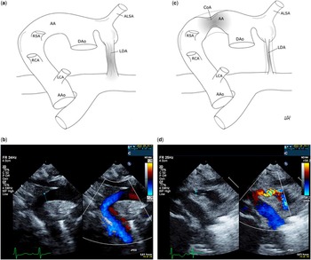
The large left arterial duct remained open, with significant left-to-right shunt. At 7 days of life, a 3-day course of indomethacin was started to close the arterial duct. Following the second dose of indomethacin, the patient developed weak femoral pulses. Blood pressure in the left arm and left leg were 73/56 and 60/43 mmHg, respectively. After the third dose, he developed respiratory distress, absent femoral pulses, and un-recordable lower-limb blood pressure. A stat echocardiogram revealed a closing left arterial duct and a coarctation of the right aortic arch (Fig 2c and d). There was also reduced left ventricular function. Prostaglandins were re-started. His respiratory distress improved, and there was improvement in his blood pressures: 59/45 mmHg in the left arm and 62/44 mmHg in the left leg. At this time, an echocardiogram showed relief of the aortic obstruction and mild improvement in ventricular function.
Figure 2: (a) Cartoon shows right aortic arch with aberrant left subclavian artery and left artery duct, AAo: ascending aorta, LCA: left carotid artery, RCA: right carotid artery, RSA: right subclavian artery, AA: aortic arch, DAo: descending aorta, ALSA: aberrant left subclavian artery, LDA: left arterial duct. (b) Echocardiogram: Colour compare showing right aortic arch and descending aorta with and without colour. (c) Cartoon shows coarctation in the right aortic arch and constriction of the left arterial duct, CoA: coarctation. (d) Echocardiogram colour compare showing the coarctation of the aorta on the right aortic arch. Cartoons by Stevi Golden-Potnik.

He was continued on prostaglandin infusion while being transferred to a tertiary paediatric cardiac centre for surgery, where he underwent patch augmentation of the aorta and ligation of the left arterial duct. His postoperative course was uneventful. A FISH assay showed 22q11 deletion, confirming the diagnosis of DiGeorge syndrome.
Follow-up assessment 2 months after the procedure showed no evidence of re-coarctation. His blood pressure was 94/42 mmHg in the right arm and 87/54 mmHg in the left leg. His echocardiogram showed that the aortic arch was wide open with a normal velocity in the descending aorta of 1.8 m/s.
Discussion
To our knowledge, this is the first reported case of a newborn with DiGeorge syndrome and a right aortic arch and left arterial duct, who developed a coarctation that was induced by indomethacin and relieved with prostaglandin. The clinical findings are consistent with coarctation secondary to constriction of ectopic ductal tissue present in the aorta. This constriction was induced by indomethacin. Prostaglandin was able to relieve the obstruction in the right aortic arch, irrespective of the opening effect in the left arterial duct.
Conclusions
This case demonstrates that the clinical presence of ductal tissue in a right aortic arch with no ipsilateral arterial duct can become constricted in response to indomethacin, causing coarctation. Even if an arterial duct is not visualised, prostaglandin can relieve aortic obstruction. In the acute setting, this allows for stabilisation before surgical correction or intervention is performed.
Acknowledgements
The authors would like to thank the Pediatric Cardiopulmonary laboratory, Children’s Hospital, London Health Science Center, London, Ontario, Canada for its support.
Financial Support
This research received no specific grant funding from any funding agency or from commercial or not-for-profit sectors.
Conflicts of Interest
None.