Introduction
Onchocerca flexuosa (Wedl, 1856) is a subcutaneous filariid nematode (Spirurida: Onchocercidae) whose primary host is the European red deer, Cervus elaphus (Bain & Schulz-Key, Reference Bain and Schulz-Key1974). It has also been reported in antelope (Dikmans, Reference Dikmans1933) and roe deer (Demiaszkiewicz, Reference Demiaszkiewicz1989; Demiaszkiewicz & Dróżdż, Reference Demiaszkiewicz and Dróżdż1990) but, based on the low number of animals affected, these appear to be atypical hosts.
Various species of Onchocerca mainly infect domestic animals (cattle and horses) and wildlife (red deer, roe deer and fallow deer), but they can also infect dogs and humans. Human onchocercosis can cause serious disease and is mainly localized in sub-Saharan Africa, Yemen, central and northern South America (World Health Organization, 2011). Although there are reported cases of zoonotic onchocercosis of ruminant origin, the first case of human infection with O. jakutensis from red deer was described in Europe (Koehsler et al., Reference Koehsler, Soleiman, Aspöck, Auer and Walochnik2007). Adult worms live in subcutaneous connective tissue of their hosts, free or leading to firm nodules where the worms are coiled. The microfilariae are located in the skin, near or far from adults, and their life cycle involves nematocerous dipterans of the families Simuliidae or Ceratopogonidae. The pre-patent period in deer is between 6 (Schulz-Key, Reference Schulz-Key1975a) and 9 months (Frank et al., Reference Frank, Wenk and Scherb1968).
Formation of nodules is the result of the reaction of the host against the parasite. Reproducing adults inside the onchocercoma are surrounded by fibrous tissue strongly infiltrated by inflammatory cells, the extent and identity of which depend on parasite-related factors, such as sex, vital status and mobility (Schulz-Key, Reference Schulz-Key1975b; Plenge-Bönig et al., 1995; Wildenburg et al., Reference Wildenburg, Plenge-Bönig, Renz, Fischer and Büttner1997). The patent period lasts several years (Rommel et al., Reference Rommel, Eckert, Kutzer, Körting and Schnieder2000).
Four species of Onchocerca have been described in European wild cervids: O. flexuosa, Wedl, 1856, adults of which are localized within nodules in the dorsal region and flanks of the red deer, while the microfilariae are found on the inside of the hind limbs; O. garmsi Bain & Schulz-Key, 1976, with adults free in subcutaneous tissue of the sternum and microfilariae concentrated in ventral subcutaneous regions, mainly along the sternum (Schulz-Key et al., Reference Schulz-Key, Bain and Wenk1976); O. jakutensis Gubanov, 1964 (syn. O. tubingensis Bain & Schulz-Key, Reference Bain and Schulz-Key1974), with adults found within nodules in the posterior dorsal region and thighs, and microfilariae in the ventral zone; and O. skrjabini Ruchjadev, 1964 (syn. O. tarsicola Bain & Schulz-Key, Reference Bain and Schulz-Key1974), adults of which are found in the tarsal and carpal regions, without creating nodules, and microfilariae in the skin of the ears and muzzle (Schulz-Key, Reference Schulz-Key1975a). Onchocerca flexuosa is the most prevalent species and is easily identified by its location and morphological characters (Bain & Schulz-Key, Reference Bain and Schulz-Key1974) as well as by molecular means using DNA sequences of the mitochondrial genes (Krueger et al., Reference Krueger, Fischer and Morales-Hojas2007).
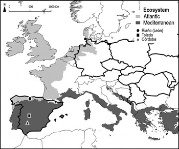
Onchocerca spp. in wild ruminants had previously appeared to be restricted to central and eastern Europe, perhaps because these were the only areas where they had been studied. Onchocerca flexuosa is the only species found in Spain, being reported in several regions of the country (Toledo: 39°25′N, 4°04′W, Córdoba: 38°02′N, 4°49′W and León: 43°03′N, 4°57′W) (fig.1).
European distribution of O. flexuosa in red deer (black-framed countries), previous reports of O. flexuosa in the Mediterranean (■, ▲) and in the present study in the Atlantic ecosystem (●) in Spain.

The aims of this study were to identify of O. flexuosa by morphology and mitochondrial 12S rDNA sequence analysis, to present the histopathological findings, to describe the epidemiology of the parasite and to analyse its potential importance in an Atlantic ecosystem of northern Spain.
Materials and methods
Study area
The study was carried out in the Riaño Regional Hunting Reserve, Province of León, north-western Spain, located in the Cantabrian Mountains. The reserve, where the red deer were shot, is made up of six municipalities (Acebedo: 43°2′N, 5°07′W; Boca de Huergano: 42°58′N, 4°55′W; Burón: 43°01′N, 5°03′W; Oseja de Sajambre: 43°08′N, 5°02′W; Posada de Valdeón: 43°09′N, 4°55′W and Riaño: 42°58′N, 5°00′W), covers an area of 78,995 ha and the greater part of its territory is in the Atlantic region, where the landscape is considerably mountainous. The region has a large and complex river network, as well as numerous tributaries in the form of mountain streams. The average monthly temperature is 10.2°C ( − 12°C/36°C). Winter is long and cold, and summer is short and temperate with an average temperature of 16–17°C. Rainfall is distributed throughout the year, with an average annual precipitation of 1088.6 mm, reaching its maximum value in October and its minimum in July (AEMET, State Metereology Station, 2009).
Sampling procedures
Between March 2005 and March 2007, 110 red deer (27 males and 83 females) were shot by the technical staff of the reserve in selective hunting, and their hides were collected. For management reasons, it was not possible to obtain red deer during the summer months. The deer sampled were classified into two age groups: young ≤ 2 years old (n= 24) and adults >2 years old (n= 86), as well as into three seasons (winter, spring and autumn). Subcutaneous nodules were observed. Afterwards, they were collected, measured, classified by size, according to their frequency distribution, into large (diameter >20 mm), medium (10–20 mm) and small ( < 10 mm), and preserved by freezing for later study. Some nodules of different sizes were prepared for histology, fixed in 10% neutral, buffered formalin, embedded in paraffin, sectioned, and stained with haematoxylin and eosin. Several female and male nematodes were removed intact from other nodules and preserved in 80% ethanol. Subsequently, they were cleared in lactophenol and identified by morphological characteristics from the description of this species by Bain & Schulz-Key (Reference Bain and Schulz-Key1974). The body length, width of the anterior end, length of the oesophagus and of its muscular part, and both spicule lengths were measured (in millimetres). For this study a Nikon Eclipse 80i microscope provided with differential interference contrast optics was used (Nikon, Tokyo, Japan).
Molecular analysis
Six adult males were used for molecular identification of the species, reserving the central part for DNA extraction. For molecular characterization, a portion of the mitochondrial 12S rDNA was amplified by polymerase chain reaction (PCR) using primers 12S OVC (5′-TCGGCTATGCGTTTTAATTTT-3′) as a forward primer and 12S OVB (5′-CAACTTACGCCCCTTTAGGC-3′) as a reverse primer, as described by Morales-Hojas et al. (Reference Morales-Hojas, Cheke and Post2006). PCR reactions of 25 μl consisted of each primer (0.5 μ m), 200 μ m deoxynucleoside triphosphates and 3 mm MgCl2. Proofreading polymerase (Finnzymes DyNAzyme EXT, New England Biolabs, Ipswich, Massachusetts, USA) was used for amplification. PCR cycling parameters for the rDNA reactions included denaturation at 94°C for 3 min, followed by 35 cycles of 94°C for 30 s, 50°C for 30 s, and 72°C for 1 min, followed by a post-amplification extension of 72°C for 7 min. Sequencing reactions were carried out using BigDye-terminator cycle sequencing chemistry (Applied Biosystems Inc., Foster City, California, USA), and reaction products were separated and detected using an ABI 3730 capillary DNA sequencer (Applied Biosystems) at the Plant-Microbe Genomics Facility, Ohio State University, Columbus, Ohio, USA. DNA templates were sequenced for both strands. The partial mitochondrial 12S rDNA sequences were aligned using Clustal X2 (Larkin et al., Reference Larkin, Blackshields, Brown, Chenna, McGettigan, McWilliam, Valentin, Wallace, Wilm, Lopez, Thompson, Gibson and Higgins2007), and similar sequences from GenBank corresponding to other Onchocerca species were compared.
Data analysis
The prevalence and mean intensity of the infection were determined, taking into account host sex, age and season, according to Bush et al. (Reference Bush, Lafferty, Lotz and Shostak1997). Data analysis was carried out using the chi-square non-parametric test in order to compare the prevalence, as well as the Mann–Whitney and Kruskal–Wallis tests to compare the intensity and length of the nodules. Statistical significance was determined at the P ≤ 0.05 level, and the statistical analysis was carried out using the SPSS 18.0 software package (SPSS Inc., Chicago, Illinois, USA). The 95% confidence intervals (CI) for prevalence were calculated according to Martin et al. (Reference Martin, Meek and Willeberg1988). The median intensity and the aggregation index (variance/mean ratio) were calculated in order to observe the distribution of the nodules of O. flexuosa in their hosts (according to Rózsa et al., Reference Rózsa, Reiczigel and Majoros2000).
Results
Gross lesions and distribution
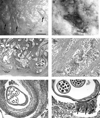
Nodules containing Onchocerca were observed from subcutaneous tissues of the back and flanks of the red deer. They were flat, round, ovoid or irregular in form; isolated nodules were frequently observed, but also laterally fused nodules, giving the appearance of lobulated nodules. They were solid and hard, and often surrounded by a congestive, sometimes haemorrhagic area (fig. 2B). The sections showed yellowish mucous material and whitish or yellowish interwoven nematodes, delimited by thick fibrous connective tissue.
Onchocerca flexuosa in the subcutaneous tissues of red deer: (A) a migrating male worm (arrows), scale bar = 2 mm; (B) numerous nodules, scale bar = 50 mm; (C) section of a nodule, with adult worms in compartments separated by infiltrated fibrous tissue, scale bar = 500 μm; (D) granuloma with worm remnants surrounded by macrophages, giant cells and a thick peripheral layer of fibrous tissue, scale bar = 150 μm; (E) granular tissue around a female with reduced somatic muscles and developing microfilariae in the uterus, scale bar = 100 μm; (F) Splendore–Hoeppli reaction around a female (arrows), scale bar = 50 μm.

Morphology and histology
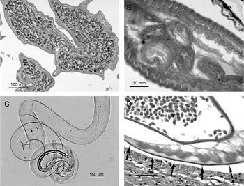
Adult worms are filiform, covered with a thin cuticle and measure 62.33 ± 6.59 (range: 52–71) in length with a width of the anterior end 0.18 ± 0.03 (0.14–0.20). The total length of the oesophagus is 7.88 ± 0.20 (7.66–8.04) and its muscular part 0.70 ± 0.14 (0.55–0.96). Males have a distinctive corkscrew-shaped end (fig. 3C), with caudal alae, post-cloacal caudal papillae and two unequal spicules, the left one being 0.63 ± 0.08 (0.54–0.77) in length, with narrow lateral alae, and the right one 0.20 ± 0.02 (0.16–0.23) in length. The spicule structure is further distinguished from that of other species by having a prominently recurved spicule hook.
Adult worms of Onchocerca flexuosa: (A) females with numerous microfilariae in the uterus, scale bar = 100 mm; (B) cross-section of a female, showing cuticular ridges with small protuberances and inner striations in the basal layer of cuticle, scale bar = 30 mm; (C) posterior region of a male to show the unequal spicules, scale bar = 150 μm; (D) cross-section of a male with cuticular ridges (arrows), large interspace between each one and the testis full of spermatocytes, scale bar = 50 μm.

In the nodules the worms are found in several compartments separated by an infiltrated fibrous tissue and leaving some space between them (fig. 2C). The majority of nodules are inhabited by several females, with or without microfilariae, and by one or two males lying in small cavities. Others house only male nematodes. There are inflammatory infiltrates of eosinophils, lymphocytes, plasma cells and macrophages, with strong fibroplasia giving rise to a granulation tissue around all females (fig. 2E). Amorphous eosinophilic material (Splendore–Hoeppli reaction) is observed on the surface of some worms (fig. 2F). Sometimes neutrophils and eosinophils are observed near the females, and many of them are surrounded by giant cells. Male worms are embedded in the infiltrate and no cellular inflammatory reaction is observed around them. Degenerate worms are surrounded by a granulomatous reaction consisting of remnants of worms, cell debris and eosinophils, surrounded by macrophages, giant cells and lymphocytes, and with a thick peripheral fibrous layer. In older nodules degeneration of the tissues and calcification of the worms frequently takes place (fig. 2D).
Histologically, male worms show a thin, three-layered cuticle; the layers distinguished by their varying density, with not too prominent ridges in the cortical layer of the cuticle and a large interspace between each ridge. The basal layer of the cuticle lacks inner striations. They show a well-developed musculature. In some specimens, the rounded testis is filled with spermatocytes (fig. 3D). Female O. flexuosa also have a thin cuticle with a three-layered structure. Cuticular ridges are generally inconspicuous or formed by small protuberances. The basal layer of the cuticle is undulating and the ripples are found between the inner striations which form thorn-like extensions (fig. 3B). The somatic musculature is much reduced and occasionally shows severe degeneration. The uterus of gravid females contains numerous microfilariae (fig. 3A).
Molecular and epidemiological analyses
Direct sequencing of PCR products yielded sequences ranging from 344 to 422 bases. Three of these sequences have been deposited in GenBank with accession numbers JQ733521, JQ733522 and JQ733523. Overlapping regions in each of the three sequences were identical. The sequences were also identical, in the same region, to those of other O. flexuosa sequences for this locus (GenBank accession numbers DQ523746, DQ523747 and NC016172, all from German isolates).
Due to the extreme difficulty of extracting entire parasites from the nodules and counting them, prevalence and intensity infection data are reported here as nodules/deer. Subcutaneous nodules were found in 94 of 110 red deer (85.45%, 95% CI: 78.86–92.04%) with a mean intensity of 9.53 ± 12.27 (range 1–74) nodules/infected host. Most deer had fewer than 25 nodules, with a median intensity of 10 nodules (CL: 5–10 for an actual (exact) confidence level of 96.1%). Data represent a typically aggregated/overdispersed distribution. The result obtained (14.05) for the variance/mean ratio indicates that the parasites are strongly aggregated among their hosts and not randomly or uniformly distributed.
Adult red deer had a higher prevalence (94.19%, 95% CI: 89.25–99.13%) than young ones (54.17%, 95% CI: 34.24–74.1%), with significant statistical variations (P< 0.001). No differences between males (88.89%, 95% CI: 77.06–100%) and females (84.34%, 95% CI: 76.52–92.16%) or between the 3 years of sampling were observed.
Significant variation was observed in seasons (P< 0.05), with the largest number of infected deer in winter (91.11%, 95% CI: 82.8–99.42%) and slightly lower in spring (89.47%, 95% CI: 79.71–99.23%), with a subsequent decrease until the autumn (70.37%, 95% CI: 53.14–87.6%).
With regard to mean intensity of the nodules, and also their prevalence, significant statistical differences in host age (P< 0.001) and seasons (P< 0.05) were observed, with higher values in winter (12.63 ± 16.73 nodules/red deer) (table 1).
Prevalence (%) and mean intensity (± SD) of infection of red deer with nodules of Onchocerca flexuosa from northern Spain, relative to host age, sex and season.

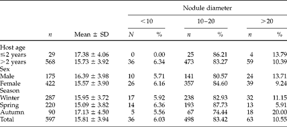
A total of 597 nodules, removed from 84 red deer, were measured. They had an average size of 15.81 ± 3.94 mm in diameter (range 6–34 mm). Most of the nodules (83.42%, n= 498) were medium sized, with lower proportions of large (10.55%, n= 63) and small ones (6.02%, n= 36). These data followed a normal distribution. All parameters examined showed the same pattern of distribution, except in young deer, where no small nodules were observed.
Statistically significant differences were only observed in seasons, with larger size of nodules (17.13 ± 4.50 mm, P< 0.05) and higher proportion of large nodules in autumn (20.00%, P≤ 0.005). Nevertheless, the medium-sized nodules were the most common, with the highest prevalence being in spring (87.73%, P< 0.05) (table 2).
The relationship between nodule diameter (mm) of Onchocerca flexuosa, relative to host age, sex and season; n, number of nodules measured; %, percentage of nodules according to their diameter.

Discussion
Male worms were found less frequently in the nodules than females, especially in those where females contained a large number of microfilariae. Males can leave the nodules and can be observed migrating subcutaneously (fig. 2A). Their mobility probably explains why they are less attacked by the host reaction. In contrast, female parasites, except for their anterior ends, are usually immobile due to an early degenerative alteration of the muscles, and were embedded in the nodular fibrous tissue. They lack a protective surface coat which, together with their immobility, makes them more susceptible to successful attack by the host's immune system (Franz et al., Reference Franz, Schulz-Key and Copeman1987; Plenge-Bönig et al., Reference Plenge-Bönig, Krömer and Büttner1995; Wildenburg et al., Reference Wildenburg, Plenge-Bönig, Renz, Fischer and Büttner1997). Granulomata observed around all females and degenerate worms indicate a strong immunological response against O. flexuosa.
The Splendore-Hoeppli reaction is typically characterized by intensely eosinophilic material around microorganisms (fungi, bacteria and parasites) or biologically inert substances. This reaction has been described surrounding adults of Schistosoma bovis in granulomata (Ferreras-Estrada et al., Reference Ferreras-Estrada, García-Iglesias, Pérez-Martínez, Manga-González, Ramajo-Martín, Escudero-Diez and García-Marín1998) but has also been observed in other Schistosoma spp., in strongyloidosis, filariae and cutaneous larva migrans (Rodig & Dorfman, Reference Rodig and Dorfman2001; Hussein, Reference Hussein2008). The eosinophilic material could represent the deposition of antigen–antibody complexes (immunoglobulins and major basic proteins) and debris from the host inflammatory cells (Madke et al., Reference Madke, Doshi, Pande and Khopkar2011).
The histology, morphological characteristics (form of spicules) and the measurements analysed, agree with those given in the redescription of O. flexuosa, carried out by Bain & Schulz-Key (Reference Bain and Schulz-Key1974) and Bain (Reference Bain1981). Similarly, the molecular identification of parasites supports the identification of O. flexuosa.
A comparison of the present results is difficult due to the paucity of epidemiological studies in this host–parasite system. In many European studies the data provided are isolated reports from morphological, faunistic, taxonomic or histopathological studies. The majority of them are based on the presence of microfilariae in the skin, few provide data on nodules and in none of them was the prevalence calculated from the number of adults removed from nodules. The prevalence of O. flexuosa in red deer varies considerably, ranging between 9.5% in the Czech Republic (Páv & Zajíček, Reference Páv and Zajíček1981) and 96% in Germany (Schulz-Key, Reference Schulz-Key1975a), with intermediate values in Romania (13%, Dulceanu & Ghitescu, Reference Dulceanu and Ghitescu1986), Poland (14.2–80.3%, Demiaszkiewicz, Reference Demiaszkiewicz1995; 23%, Dróżdż et al., 1993; 60%, Demiaszkiewicz, Reference Demiaszkiewicz1998), Ukraine (33.3%, Zhelizniak, Reference Zhelizniak2003) and Belarus (62.5%, Shimalov & Shimalov, Reference Shimalov and Shimalov2003). Our data on both prevalence and intensity of the infection were generally higher (exceeded only by those of Schulz-Key, Reference Schulz-Key1975a, in Germany), even when compared with the rest of Spain (33.6%, 3.93 ± 5.26, Santín-Durán et al., Reference Santín-Durán, Alunda, De la Fuente and Hoberg2001, and 24%, 3 ± 5, San Miguel et al., Reference San-Miguel, Álvarez, Rodríguez-Vigal and Luzón2003, in the central part of the country; 69.33%, Hernández-Rodríguez et al., Reference Hernández-Rodríguez, Acosta-García, Hernández-Redondo, Hernández-Rodríguez, Hidalgo-Argüello and De la Fuente-López2010, in the south; 87.01%, 7.57 ± 6.94, Díez-Baños et al., Reference Díez-Baños, Martínez-Delgado and Hidalgo-Argüello2007, and 92.73%, 7.71 ± 7.35, Hidalgo-Argüello et al., Reference Hidalgo-Argüello, Díez-Baños, Martínez-Delgado, Hernández-Rodríguez, Hidalgo-Argüello and De la Fuente-López2010, in previous studies in the north of Spain).
Temperature and moisture are the two most important environmental parameters for terrestrial organisms. Since the mid-twentieth century the Earth's surface and ocean temperatures have increased by an average of 0.75°C but there have also been changes in levels of precipitation and other forms of moisture (Lovejoy, Reference Lovejoy2008; Colwell et al., Reference Colwell, Dantas Torres and Otranto2011; Martin et al., Reference Martin, Pastoret, Brochier, Humblet and Saegerman2011). These changes, as well as other related factors with complex interactions, are affecting species and ecosystems, ecology and epidemiological cycles, especially those involving arthropod vectors, influencing their abundance and possibly also the development of the parasite.
Onchocerca flexuosa has been studied in different latitudes of Europe and Spain, each with their particular climatic patterns affecting the life cycle of the parasite. These patterns could explain the differences found in terms of prevalence, intensity and seasonality of infection.
Generally it is observed that adult red deer are more frequently infected (Santín-Durán et al., Reference Santín-Durán, Alunda, De la Fuente and Hoberg2001; San Miguel et al., Reference San-Miguel, Álvarez, Rodríguez-Vigal and Luzón2003). However, younger red deer are rarely investigated and this could lead to erroneous conclusions. Parasite transmission seems, at first, to be related to the lower time of contact with vectors for young deer, but the results indicate that red deer can be infected from birth. This takes place in May–June, which coincides with the beginning of the peak activity of the vectors. Thus, the first onchocercomata would appear in December and this could explain the appearance of parasitized deer younger than 1 year old in central and northern parts of Spain.
In contrast to the hypothesis of San Miguel et al. (Reference San-Miguel, Álvarez, Rodríguez-Vigal and Luzón2003), we believe that the size of the nodules is not linked to the time of infection (small nodules = recent infection, large nodules = mature infection). Under this assumption, small nodules would thus be more common in younger deer. Our results indicate the absence of small nodules, and a higher proportion of large ones, in younger deer than in adults. Neither could we relate the observed pattern of nodule presence and size to the period of peak activity of the vectors, or to the pre-patent period. The different sizes found are more likely due to other factors, which may include the number of the adults that they contain, whether they are inhabited by several females with or without microfilariae, whether males are together with females, or whether these nodules harbour only males. The male/female ratio of a single nodule, the sensitivity and reaction of each deer to the parasite, the reaction to dead adults and microfilariae, and the time during which the nodules remain at the predilection site of the red deer may be additional factors that influence the observed results in the studied C. elaphus population.
The role of particular vectors in the transmission cycles of many filariid species and their geographical distribution remain largely unknown. In the transmission of O. flexuosa to red deer in Europe, the simuliid species such as Prosimulium nigripes and Simulium ornatum are the only known vectors (Frank et al., Reference Frank, Wenk and Scherb1968; Dyková, Reference Dyková1972). However, their possible development in different species of Culicoides (Ceratopogonidae) has also been indicated (Schulz- Key & Wenk, Reference Schulz-Key and Wenk1981). Prosimulium nigripes has not been reported in our study area but other species, and/or species-complexes, within the genera Prosimulium and Simulium, besides S. ornatum, are present (Cordero del Campillo et al., Reference Cordero del Campillo, Castañón Ordóñez and Reguera Feo1994). The same species, and others within both genera, are also widely distributed in the locations where the presence of O. flexuosa has been cited in red deer (Adler & Crosskey, Reference Adler and Crosskey2012) but their competency as vectors for O. flexuosa is not known. Transmission of O. flexuosa has not been studied in Spain.
Considering the life cycle of this parasite, these results clearly indicate that in our study area (Atlantic Spain), there are favourable ecological factors for the development of O. flexuosa vectors. The abundance of rivers and mountain streams favours the survival of the vectors, thus allowing longer exposure to the definitive host. This could explain the high prevalence and intensity found throughout the entire study. In the central part of the country, the typical Mediterranean climate with summer drought would lead to lower survival of simuliid larvae, resulting in fewer adults and, therefore, a lower probability of infection. This may account for the lower prevalence and intensity as reported from these areas (Santín-Durán et al., Reference Santín-Durán, Alunda, De la Fuente and Hoberg2001; San-Miguel et al., Reference San-Miguel, Álvarez, Rodríguez-Vigal and Luzón2003).
The impact of O. flexuosa in deer is unknown. Infected red deer show no clinical signs except for the nodules under the skin found during the necropsy. Treatment is not required and control is difficult due to the ubiquity of the insect vectors.
The geographical distribution of O. flexuosa follows the Palaeartic pattern of its host and in this paper we document its most western geographical distribution in Europe.
Integrated studies, such as this manuscript, which combine classical morphology, histology and molecular biology, represent a practical, necessary and reliable tool for characterizing other Onchocercaspecies that infect cervids in the world, and determining their geographical distribution. These studies could provide the opportunity to observe host–parasite interactions and their response to environmental changes, contributing to knowledge for improved host management and conservation when pathogenic species are involved (Ferri et al., Reference Ferri, Barbuto, Bain, Galimberti, Uni, Guerrero, Ferté, Bandi, Martin and Casiraghi2009).
Acknowledgements
We are sincerely grateful to the Junta de Castilla y León for providing the red deer, and to all the forestry staff of the Riaño Regional Hunting Reserve for their co-operation, especially to Juan José Martínez Nistal, Chief of Wildlife Service in León Province. Our gratitude is extended to Coral Cordero Pérez, Rodrigo García Lastra and Telesforo Suárez Manga for their technical assistance, and also to Dr Juan Pablo Barrio Lera for his help in the production of the photographs. We also thank the Plant-Microbe Genomics Facility and staff at The Ohio State University for their contribution to this publication.
Financial support
This study was supported by a Research Project 2004-02580-C03-03/BOS from the Spanish Ministerio de Ciencia y Tecnología. A.M. holds a pre-doctoral fellowship from the University of León, Spain.
Conflict of interest
None.