Patients at risk for enteric vancomycin-resistant Enterococcus (VRE) colonization may be actively screened for VRE colonization to inform infection prevention practices and empiric antibiotic therapy.Reference Reyes, Bardossy and Zervos1 However, daptomycin nonsusceptible (DNS) and linezolid non-susceptible (LNS; including linezolid-intermediate or -resistant) VRE is of concern.Reference Kelesidis, Humphries, Uslan and Pegues2, Reference Bi, Qin, Fan, Ma and Gu3 At our hospital, surgical intensive care unit and liver transplant patients are actively screened for VRE colonization. We sought to (1) determine the daptomycin and linezolid minimum inhibitory concentration (MIC) distributions among enteric VRE from these patients and (2) identify modifiable risk factors for DNS and LNS enteric VRE.
We performed a retrospective analysis of prospectively collected VRE rectal swab surveillance cultures from patients ≥18 years of age admitted to a surgical intensive care unit and its associated step-down medical unit at Aurora St Luke’s Medical Center (Milwaukee, WI), a 650-bed tertiary referral center from June 1, 2017, to June 30, 2018. Only index isolates were included. VRE rectal swabs were collected using ESwab collection devices (Copan Diagnostics, Murrieta, CA) and cultured on chromID VRE agar plates (BioMèrieux, Durham, NC). Susceptibility testing was performed using daptomycin and linezolid ETESTs (BioMèrieux) using breakpoints of ≤4 mg/L and ≤2 mg/L, respectively.4 Relevant patient-, treatment-, and infection-level data were collected. Univariable analyses determined associations between independent variables and DNS (>4 mg/L) and LNS (>2 mg/L) VRE. All independent variables were included in a forward, stepwise multivariable logistic regression model to determine independent predictors of DNS and LNS VRE, with variables P ≤ .05 kept in the model. SAS version 9.4 software (SAS Institute, Cary, NC) was used for all calculations.
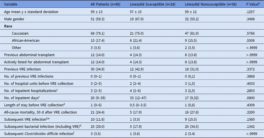
In total, 103 VRE surveillance cultures were performed during the study period; of these, 86 (83.5%) were index isolates and were included in the study. Patient demographics and subsequent infections are shown in Table 1; 75 patients (87.2%) had been hospitalized within 2 years before collection. The MIC50 and MIC90 were 2 mg/L and 3 mg/L for daptomycin and 3 mg/L and 6 mg/L for linezolid. Also, 81 (94.2%) isolates were E. faecium and 41 (68.6%) were LNS, including 11 linezolid-resistant isolates. Previous linezolid (17.4%) and/or daptomycin (16.3%) exposure was uncommon. Only 1 (1.2%) isolate was DNS precluding an analysis of DNS predictors. Previous cefazolin exposure (adjusted odds ratio [aOR], 0.30; 95% confidence interval [CI], 0.11–0.78) and previous tobramycin exposure (aOR, 0.20; 95% CI, 0.03–0.93) were inversely associated with LNS. Linezolid exposure was not associated with linezolid nonsusceptibility.
Patient Demographics and Subsequent Infectionsa

Note. VRE, vancomycin-resistant Enterococcus.
a All data presented as median (interquartile range) or no. (%) unless otherwise noted.
b Comparison between linezolid susceptible and linezolid nonsusceptible groups.
c To any hospital within the health system within 2 years before VRE isolate collection.
d Within 30 days after VRE collection.
e Two patients in the linezolid nonsusceptible group developed a bacterial infection after liver transplant.
f Defined as a positive C. difficile toxin PCR with antibiotic treatment for C. difficile within 30 days after VRE collection.
The high rate of linezolid nonsusceptibility observed among enteric VRE rectal surveillance isolates recovered from a vulnerable group of patients at risk for VRE infection was unanticipated. Previous linezolid exposure was infrequent and not associated with LNS. This finding is similar to a study of pediatric patients with LNS VRE, most lacked prior linezolid exposure.Reference Bagga, Buckingham, Arnold, Nesbitt, Guimera and Lee5 Daptomycin was active against all but 1 of our isolates; although most isolates had an MIC in the ‘susceptible dose dependent’ range.6 The MICs in this range require higher daptomycin doses to achieve optimal pharmacodynamic targets. Previous daptomycin exposure was also infrequent. We did not identify any strong, modifiable risk factors for LNS VRE.
Available antibiotic susceptibility data suggest that both daptomycin and linezolid retain activity against most clinical Enterococcus isolates.Reference Sader, Farrell, Flamm and Jones7, Reference Pfaller, Mendes, Streit, Hogan and Flamm8 Few studies have examined linezolid and daptomycin activity against enteric VRE from surveillance cultures. Cakirlar et alReference Cakirlar, Samasti and Baris9 found 40% linezolid resistance in a sample of enteric VRE surveillance cultures (n = 100) from Turkish hospitals, which is higher than the 11.6% linezolid resistance observed in our study. These authors used disk diffusion to determine linezolid susceptibility. Identification of linezolid nonsusceptibility is challenging because the wild-type MIC90 is near the susceptibility breakpoint.Reference Doern, Park, Gallegos, Alspaugh and Burnham10 Although we observed a high rate of LNS in our study, most LNS was not truly resistant but rather intermediate (MIC, 3 mg/L). Guidance from CLSI recommends linezolid resistance be confirmed by a secondary method; however, both ETEST and disk diffusion have previously demonstrated high specificity for linezolid resistance.4, Reference Doern, Park, Gallegos, Alspaugh and Burnham10 Given that the susceptibility testing was performed by experienced microbiology technologists using ETESTs, we are confident that the linezolid MIC data represent the true distribution and not an artifact of the testing methodology.
The clinical implications of our findings are important, and these data could influence empiric prescribing when VRE infections are suspected. However, a limitation of our work is that we did not further characterize each isolate in detail. In the absence of any identified predictors of LNS we will intensify our investigation to include a more robust analysis of these isolates beyond patients’ medication and environmental exposures before VRE colonization. Whole-genome sequencing of available isolates is underway to determine whether clonal spread of LNS VRE is occurring and whether or not these isolates harbor known linezolid-resistance determinants. The potential for horizontal transmission of a LNS clone(s) cannot be discounted; however, we have not appreciated horizontal VRE transmissions in our hospital. In the interim, linezolid and daptomycin susceptibility testing among enteric VRE isolates recovered from patients actively screened for VRE colonization may be warranted. These susceptibility data would allow clinicians to modify anti-VRE antibiotics, empirically for a presumed infection but also as perioperative or procedural prophylaxis. Without this information, empiric linezolid use in this population may provide inadequate VRE coverage.
In summation, we observed a high rate of LNS among enteric VRE surveillance cultures in a patient population at risk for VRE infection. We encourage clinicians and infection preventionists to consider the potential for LNS among enteric VRE in their institution(s). We recommend that additional studies be conducted to investigate linezolid and daptomycin susceptibility among enteric VRE recovered from surveillance cultures.
Financial support
This work was supported in part by a research grant from the Aurora Research Institute to Thomas J. Dilworth.
Conflicts of interest
All authors report no conflicts of interest relevant to this article.