Introduction
Halysites Fischer von Waldheim, Reference Fischer von Waldheim1828 is a genus of halysitid tabulate coral that is rare in the Upper Ordovician and widely distributed in the lower to upper Silurian (Hill, Reference Hill and Teichert1981; Wang and Zhan, Reference Wang and Zhan2015; Liang et al., Reference Liang, Elias and Lee2018). This cateniform (“chain”) coral was one of the early tabulates that appeared during the Great Ordovician Biodiversification Event (Webby et al., Reference Webby, Elias, Young, Neuman, Kaljo, Webby, Paris, Droser and Percival2004; Liang et al., Reference Liang, Elias and Lee2018). It is regarded as having evolved from Catenipora Lamarck, Reference de Lamarck1816 by the appearance of tubules containing tabulae between corallites (Flower, Reference Flower1961; Flower and Duncan, Reference Flower, Duncan, Pojeta and Pope1975; Scrutton, Reference Scrutton, Oliver, Sando, Cairns, Coates, Macintyre, Bayer and Sorauf1984). Many species of Halysites have been erected with detailed, traditional systematic descriptions since the type species Halysites catenularius (= Tubipora catenularia Linnaeus, Reference Linnaeus1767) was established. Yet, notwithstanding a few pioneering studies (Buehler, Reference Buehler1955; Hamada, Reference Hamada1959; Stasińska, Reference Stasińska1967, Reference Stasińska1981; Webby and Semeniuk, Reference Webby and Semeniuk1969; Webby, Reference Webby1975; Lee and Noble, Reference Lee and Noble1990), our knowledge about the fundamental growth characteristics of this genus remains strikingly poor.
The present study is based on specimens of Halysites from two localities in the Silurian of Estonia. They are identified to the species level through multivariate morphometric analysis. Although such methods have been applied successfully to Catenipora (Bae et al., Reference Bae, Elias and Lee2006a; Wang and Deng, Reference Wang and Deng2010; Liang et al., Reference Liang, Elias, Choh, Lee and Lee2016, Reference Liang, Elias and Lee2018), they have not previously been adapted for use on Halysites. Growth characteristics of the coralla are then examined, focusing on cyclomorphism, the formation of tubules, and modes of corallite increase. The findings are compared with those in previous studies involving species of Halysites and Catenipora. The results provide insight into the similarities and differences between these genera, and the phylogenetic implications are considered. This improves our knowledge of the paleobiology of ancient corals, as well as the evolutionary lineage and relationships of tabulate corals.
Materials and methods
Fifteen coralla forming the basis of this study were collected from two localities in the Silurian of western Estonia. One corallum was collected at Päri Quarry (Fig. 1.1), from argillaceous nodular limestone in the upper part of the Rumba Formation of early Telychian age (Fig. 1.2). Those strata represent shallow-water carbonate deposition in low-energy environments on the seaward or offshore shelf (Bassett et al., Reference Bassett, Kaljo, Teller, Holl and Bassett1989; Isakar et al., Reference Isakar, Ebbestad and Peel1999; Kiipli et al., Reference Kiipli, Kiipli and Kallaste2006; Mõtus and Hints, Reference Mõtus and Hints2007). The other 14 coralla are from Abula Cliff on the northern coast of Saaremaa Island (Fig. 1.1). The cliff exposes the topmost Vilsandi Beds (lagoonal dolomitic marlstones) and the basal part of the Maasi Beds, which form the middle part of the Jaagarahu Formation (Fig. 1.2). The coralla were collected from the pelletal limestone layers of the upper part of the section (Maasi Beds), which are interpreted as Sheinwoodian in age (Fig. 1.2). Those strata were deposited on a very shallow shelf in the zone of wave activity (Mõtus and Hints, Reference Mõtus and Hints2007; Vinn and Wilson, Reference Vinn and Wilson2012).
(1) Map of the study area in Europe showing the location of Abula Cliff on Saaremaa Island and Päri Quarry, Estonia; (2) Silurian stratigraphy in Estonia, with stars marking formations from which the studied coralla were collected (modified after Mõtus and Hints, Reference Mõtus and Hints2007).

Each of the 15 coralla was trimmed longitudinally to obtain the portion that includes the central growth axis and immediately surrounding area. The resulting sample was used in this study to avoid distortion of features resulting from divergence of corallites. Transverse and longitudinal thin sections of each sample were prepared. For the 11 best-preserved coralla, sets of transverse serial peels oriented perpendicular to the central growth axis and spaced as little as 0.05 mm apart were also prepared, adopting the method of Elias et al. (Reference Elias, Lee and Woo2008).
For a comprehensive morphometric evaluation of the coralla, 17 characters were measured or calculated (Fig. 2; Table 1). Compared with previous studies of Catenipora (e.g., Liang et al., Reference Liang, Elias and Lee2018), this represents a greater number of characters and includes measurements of tubules, which are present in Halysites but not in Catenipora. In the present study, quantitative linear or areal values were obtained for the following characters: tabularium area (V1), perimeter (V2), length (V3), and width (V4); corallite length (V5) and width (V6); outer wall thickness (V7); common wall thickness (V8) and width (V9); tubule length (V10) and width (V11); average size of lacunae (V16); and average spacing of tabulae (V17). In addition, four ratios were determined: corallite length to width (V12), tabularium length to width (V13), common wall width to corallite width (V14), and tabularium area to perimeter (V15). The majority of the coordinates used in the multivariate analysis are the average values of 20 randomly selected mature corallites characterized by relatively large size and normal shape, in transverse thin sections or peels located in the mature astogenetic stage at least 20 mm above the base of each corallum. To include a few coralla that do not possess many well-preserved corallites, 10 mature corallites were measured and the average values were used in the multivariate analysis. The average size of lacunae was calculated from transverse sections located at least 20 mm above the base of the corallum. The average spacing of tabulae was calculated based on measurements through at least one growth cycle including closely and widely spaced tabulae, made along the midline of 5 to 10 corallites in longitudinal sections. To account for intracorallum variation, three transverse sections spaced vertically 7 to 10 mm apart, all obtained at least 20 mm above the base of the corallum, were analyzed for V1–V16 in each of the 11 coralla that were processed by serial peels. Single transverse sections were analyzed for V1–V16 in each of the other four coralla. Thus, a total of 37 data sets were obtained from the 15 studied coralla. For each corallum, a single value for V17 was determined in longitudinal section. All of the measurements were obtained using image analysis software (IMT 5.0) and were processed using a statistical analysis system (PASW, version 17.0 for Windows) for multivariate analysis.
Schematic transverse section of Halysites, showing morphological characters measured in this study. V1, tabularium area; V2, tabularium perimeter; V3, tabularium length; V4, tabularium width; V5, corallite length; V6, corallite width; V7, outer wall thickness; V8, common wall thickness; V9, common wall width; V10, tubule length; V11, tubule width.

Morphological characters used in this study of Halysites Fischer von Waldheim, Reference Fischer von Waldheim1828.

For the determination of species, nonmetric multidimensional scaling (NMDS) and discriminant analysis were adopted to compare the 15 coralla with nine type specimens of morphologically similar species including H. catenularius (Linnaeus, Reference Linnaeus1767) (Thomas and Smith, Reference Thomas and Smith1954; Young and Noble, Reference Young and Noble1987; Mõtus and Klaamann, Reference Mõtus and Klaamann1999), H. junior Klaamann, Reference Klaamann1961 (Klaamann, Reference Klaamann1961, Reference Klaamann1966), H. senior Klaamann, Reference Klaamann1961 (Klaamann, Reference Klaamann1961), and H. priscus Klaamann, Reference Klaamann1966 (Klaamann, Reference Klaamann1966) (Table 2). Data for six of the type specimens were derived from images of transverse and longitudinal thin sections downloaded from Geoscience Collections of Estonia (http://geokogud.info/index.php?page=1) (Table 2, GIT specimens). The other three type specimens were measured from figures in the references cited (Table 2). Among them are two specimens from the same strata and locality on Gotland (Swedish Museum of Natural History specimens). They were identified as H. catenularius by Mõtus and Klaamann (Reference Mõtus and Klaamann1999), who illustrated one in transverse section and the other in longitudinal section. For the purpose of the present study, morphometric measurements from those sections were treated as if they came from one specimen to obtain the necessary transverse data (V1–V16) and longitudinal data (V17). Thus, in effect, nine type specimens were used for species determination in the multivariate analysis (Table 2).
Type specimens of Halysites used in the species determination of 15 coralla from Estonia.

To document growth characteristics in detail, the 11 best-preserved, serially sectioned coralla from our collection were selected for further study. The formation of tubules and modes of corallite increase were determined by careful examination of the transverse serial peels. Cyclic changes of corallite tabularial area during vertical growth have been shown to represent cyclomorphism in Catenipora (Bae et al., Reference Bae, Lee and Elias2006b, Reference Bae, Elias and Lee2013). For each of the 11 coralla in the present study, three mature corallites that remained undamaged during vertical growth were selected for the measurement of tabularial area and calculation of the average value in each serial section. Each vertical cycle of increasing and decreasing tabularial area, beginning and ending at minimum values of tabularial area, is considered to represent annual vertical growth, as in Catenipora (Bae et al., Reference Bae, Lee and Elias2006b, Reference Bae, Elias and Lee2013). The average size of lacunae was also determined in each serial section.
The transverse peels and thin sections illustrated herein are oriented as they appear looking down from the top of the corallum toward its base. Longitudinal thin sections are oriented with the growth direction upward.
Repositories and institutional abbreviations
Types, figured specimens, and other specimens examined in this study are deposited in the following institutions: Institute of Geology, Tallinn University of Technology, Estonia (GIT); Geological Survey of Canada, Ottawa (GSC); Palaeontological Museum, Uppsala University, Sweden (PMU); Swedish Museum of Natural History, Stockholm (RM).
Species determination
As a result of conventional morphological comparisons, it was found that Halysites catenularius, H. junior, H. senior, and H. priscus are morphologically similar to the examined coralla. For the determination of species of the examined coralla, multivariate analysis was adopted using the 17 morphological characters obtained from the 15 coralla and nine type specimens of the four species of Halysites (Table 2).
NMDS was conducted on the 15 coralla, including 11 for which three replicates were analyzed, as well as the nine type specimens of four species of Halysites. From the combinations of NMDS 1 and NMDS 2, four groups are distinguishable (Fig. 3.1). After linking the three replicates belonging to the same corallum, Groups 1 and 2 can be separated by grouping the coralla that are closest to each other in the morphospace. Groups 1 and 2 contain six and nine coralla, respectively. Among the 11 coralla for which three replicates were analyzed, it is noteworthy that intracolonial variation in the corallum from the Rumba Formation (arrow in Fig. 3.1) seems to be higher than that in the majority of the other coralla, which are from the Jaagarahu Formation. Group 3 represents H. junior, which is clearly separated from the other coralla and type specimens. Group 4 represents H. senior, which partially overlaps Group 2. The type specimens of H. catenularius and H. priscus are in the range of Group 2, indicating that they are similar to each other in morphology and are possibly conspecific.
(1) Nonmetric multidimensional scaling (NMDS) analysis of the 15 studied coralla, of which 11 coralla provided average values for three different levels in the mature astogenetic stage (shaded outlines indicate replicates belonging to the same corallum; arrow points to corallum from the Rumba Formation; the other coralla are from the Jaagarahu Formation), and nine type specimens belonging to four species of Halysites; (2) NMDS analysis of the 15 studied coralla, based on the average value for the highest level in the mature astogenetic stage (arrow points to corallum from the Rumba Formation; the other coralla are from the Jaagarahu Formation), and nine type specimens belonging to four species of Halysites.

A second NMDS was carried out on the 15 coralla, but including only the replicate obtained from the highest level at least 20 mm above the base of the corallum in the mature astogenetic stage, together with the nine type specimens of four species of Halysites (Fig. 3.2). The morphospace occupied by Group 1 is reduced substantially compared with the result of the first NMDS analysis. Type specimens of H. junior and H. senior are well separated from the other coralla and type specimens. Type specimens of H. catenularius and H. priscus remain in the range of Group 2. Comparisons of the average values of morphological characters between Group 1 and Group 2 (Table 3) indicate that they are very similar to each other without obvious overall differences (paired-samples Wilcoxon test, p = 0.4). Both of those groups are distinct from H. junior (Group 3) and H. senior (Group 4) (paired-samples Wilcoxon test, p < 0.05). It is obvious that the tabularium size (V1–V4) of Groups 1 and 2 is very different from those of H. junior and H. senior. Based on the morphological comparisons, coralla of Groups 1 and 2 are regarded as the same species. Type specimens of H. catenularius and H. priscus are included in Group 2, so all 15 coralla are identified as H. catenularius, which has taxonomic priority. Through multivariate analysis, H. priscus is considered to be conspecific with H. catenularius, which is consistent with a previous traditional systematic study (Mõtus and Klaamann, Reference Mõtus and Klaamann1999).

To examine the result of the classification, discriminant analysis was conducted on the 15 coralla, including those with three replicates, and nine type specimens of four species of Halysites. The morphospace of the discriminant analysis indicates clear separation of the three species of Halysites (Fig. 4). The cross-validation method of the discriminant analysis showed that two of the replicates of corallum GIT 806-13 (white arrows in Fig. 4) are closer to H. junior than to H. catenularius. This corallum has the largest corallite and tabularium sizes of the 15 coralla and is close to one type specimen of H. junior. However, corallum GIT 806-13 is retained within H. catenularius based on the results of the NMDS, together with its significantly smaller corallite and tabularium sizes than the other two type specimens of H. junior.
Discriminant analysis on the 15 studied coralla, including 11 with replicates representing three different levels in the mature astogenetic stage (shaded arrows point to replicates of corallum from the Rumba Formation; the other coralla are from the Jaagarahu Formation; white arrows point to two replicates of corallum GIT 806-13 referred to in the text), and nine type specimens belonging to four species of Halysites.

Through multivariate analysis based on 17 morphological characters, all 15 coralla are assigned to H. catenularius, and H. priscus is considered a junior synonym. Judging from the morphospaces of NMDS and discriminant analysis, it is found that H. catenularius, H. junior, and H. senior are closely related morphologically. As seen in Table 3, Halysites catenularius (Groups 1 and 2) is intermediate between H. junior (Group 3) and H. senior (Group 4) in terms of tabularium sizes (V1–V4) and corallite sizes (V5, V6), as well as tubule width (V11) and tabulae spacing (V17).
Systematic paleontology
Class Anthozoa Ehrenberg, Reference Ehrenberg1834 Subclass Tabulata Milne-Edwards and Haime, Reference Milne-Edwards and Haime1850 Order Halysitida Sokolov, Reference Sokolov1947 Family Halysitidae Milne-Edwards and Haime, Reference Milne-Edwards and Haime1849 Genus Halysites Fischer von Waldheim, Reference Fischer von Waldheim1828
Type species
Tubipora catenularia Linnaeus, Reference Linnaeus1767, from Silurian of Gotland (by monotypy).
Halysites catenularius (Linnaeus, Reference Linnaeus1767) Figures 5, 7–14
- 1767
Tubipora catenularia Linnaeus, p. 1270.
- 1854
Halysites catenularia (Linnaeus). Milne-Edwards and Haime, p. 270–272, pl. 64, fig. 1, la–c.
- 1954
Halysites catenularius (Linnaeus). Thomas and Smith, p. 766–768, pl. 20, fig. la–c; Buehler, Reference Buehler1955, p. 24–25, 28–29; Laub, Reference Laub1979, p. 274–281, pl. 32, figs. 2, 3, pl. 37, figs. 1, 2, pl. 41, fig. 2; Young and Noble, Reference Young and Noble1987, p. 1135–1137, figs. 5.10–11, 6.1; Mõtus and Klaamann, Reference Mõtus and Klaamann1999, p. 83, figs. 4A–B, 6A–B.
- 1966
Halysites priscus Klaamann, p. 60–61, pl. 22, figs. 5–7.
Neotype
PMU G 681, from unknown locality, Silurian of Gotland (selected by Thomas and Smith, Reference Thomas and Smith1954, p. 767).
Description
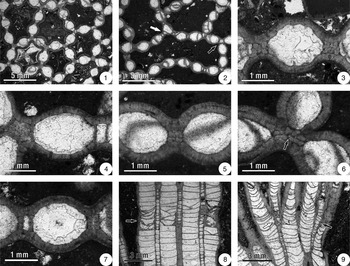
Coralla 60 × 30 to 130 × 80 mm across and 50–120 mm in height. Lacunae polygonal to elongated, size 23.59–89.08 mm2 (Fig. 5.1, 5.2). Rank junctions occur at tubules (black arrow in Fig. 5.2) or on lateral corallite walls (white arrow in Fig. 5.2). In transverse section, sporadic septa present in a few corallites (Fig. 5.3). Corallites subelliptical to subrounded (Fig. 5.1–5.7); may be distorted in direction of rank junction on lateral wall (white arrow in Fig. 5.2). Corallum average of transverse corallite length 1.92–2.43 mm, width 1.53–1.96 mm; tabularium area 1.28–2.09 mm2, perimeter 4.22–5.38 mm, length 1.50–1.93 mm, width 1.05–1.44 mm; outer wall thickness 0.23–0.32 mm; common wall thickness 0.37–0.59 mm, width 0.79–1.09 mm; tubule length 0.18–0.36 mm, width 0.30–0.45 mm. Coenenchymal tubules mostly rectangular, may be narrow, round, irregular, or absent in common wall (Fig. 5.4–5.7); irregular in rank junctions (Fig. 5.4). Balken structure may be present beside tubule (Fig. 5.3, 5.6) and in common wall (Fig. 5.5). In longitudinal section, corallite tabulae mostly complete, flat or concave, vertical spacing 0.38–0.54 mm, in some cases show cyclomorphic variation in spacing but relatively uniform thickness (Fig. 5.8, 5.9); abnormal spacing and appearance may accompany rejuvenation after damage to corallite (arrow in Fig. 5.8). Tubule tabulae mostly flat or slightly concave, generally more closely spaced than corallite tabulae, show cyclomorphic variation involving positive relation between spacing and thickness (Fig. 5.8). Tubules may develop into corallites (arrows in Fig. 5.9).
Transverse (1–7) and longitudinal thin sections (8, 9) of Halysites catenularius. (1) Polygonal lacunae; (2) elongated lacunae (white and black arrows point to rank junctions at lateral corallite wall and tubule, respectively); (3) corallite with low ratio of tabularium length to width (note the presence of septal spines, and balken structures in the common wall); (4) corallite with high ratio of tabularium length to width (note the irregular tubule in the junction area of three corallites and rectangular tubule on the right); (5) common wall of two corallites (note the absence of a tubule and presence of balken structures); (6) balken structures in the junction area of three corallites (arrow points to a tiny tubule in the center of the balken structures); (7) rectangular and narrow tubules on the left and right sides of a corallite; (8) growth pattern of tabulae in corallites and tubules (arrow points to an interval of abnormal spacing and appearance of tabulae in corallites related to rejuvenation after damage); (9) interstitial increase (arrows point to the development of juvenile corallites from tubules). (1, 3) GIT 806-8; (2, 4–7) GIT 806-4; (8) GIT 806-11; (9) GIT 806-9.

Materials
Fifteen coralla: GIT 806-1 from upper Rumba Formation, lower Telychian Stage, Päri Quarry, Estonia; GIT 806-2–15 from lower Maasi Beds, middle Jaagarahu Formation, middle Sheinwoodian Stage, Abula Cliff, Saaremaa Island, Estonia.
Remarks
Mõtus and Klaamann (Reference Mõtus and Klaamann1999) considered H. priscus and H. catenularius to be conspecific, which is supported by the present multivariate analysis. Laub (Reference Laub1979) and Young and Noble (Reference Young and Noble1987) synonymized H. junior with H. catenularius, but Mõtus and Klaamann (Reference Mõtus and Klaamann1999) noted that those species have very different diagnostic features. We find that the morphological characteristics in the coralla of H. catenularius are similar to, but distinct from the type specimens of both H. junior and H. senior. Halysites catenularius is intermediate between H. junior and H. senior in tabularium and corallite size, as well as tubule width and tabulae spacing. Balken structure is present in H. junior (Mõtus and Klaamann, Reference Mõtus and Klaamann1999) and H. senior as observed in the transverse sections in Geoscience Collections of Estonia. In the examined coralla of H. catenularius, this structure is commonly found next to the tubules. Based on the differences in corallite size, tabularium size, tubule width, and tabulae spacing, we conclude that H. catenularius, H. junior, and H. senior represent three different species that are closely related.
Growth characteristics
Cyclomorphism
Hamada (Reference Hamada1959) recognized periodic thickening of corallite tabulae and constriction of the outer wall in some species of Catenipora and Halysites, which he related to annual growth. Elias and Lee (Reference Elias and Lee1993) documented cyclomorphism involving high- and low-density bands in Catenipora rubra Sinclair and Bolton in Sinclair, Reference Sinclair1955, with high-density bands characterized by thicker and/or more closely spaced tabulae. In the same species, Young and Kershaw (Reference Young and Kershaw2005) noted banding represented by thickening of tabulae. They found banding to be indistinct in Catenipora escharoides Lamarck, Reference de Lamarck1816, but noted that septa may be slightly longer and more closely spaced in high-density bands. Gao (Reference Gao1992; Gao and Copper, Reference Gao, Copper, Lessios and Macintyre1997) identified external growth banding and internal density bands based on the spacing of tabulae in halysitid corals. Bae et al. (Reference Bae, Lee and Elias2006b) considered cyclic fluctuations of corallite tabularial area, bounded by minimum values, to record annual growth in Catenipora foerstei Nelson, Reference Nelson1963. In high-density cyclomorphic bands, they observed smaller tabularial areas, thicker tabulae, better-developed septal spines, and thicker corallite walls. Using the same methodology for recognition of growth cycles (i.e., tabularial area), Bae et al. (Reference Bae, Elias and Lee2013) documented cyclomorphism in C. rubra, C. foerstei, and two other species of Catenipora.
In the present study, longitudinal sections of H. catenularius show that corallite tabulae are relatively uniform in thickness, but there are some cycles involving the spacing of tabulae (Fig. 5.9). Tubule tabulae show cyclomorphism with a positive relation between thickness and spacing (Fig. 5.8). Following the quantitative approach developed by Bae et al. (Reference Bae, Lee and Elias2006b, Reference Bae, Elias and Lee2013), pairs of successive growth cycles bounded by minimum values of corallite tabularial area were detected from transverse serial peels of three coralla of H. catenularius (Fig. 6). Each cycle is considered to represent annual vertical growth of the colony. The annual vertical growth rate ranges from 4.9 to 9.1 mm (average 6.0 mm) as determined from the six cycles. A comparison of these cycles (see Fig. 6) shows that the growth rate is not related to the average tabularial area or the amount of variation in tabularial area. This differs from Catenipora foerstei, in which the growth rate tends to be positively correlated with variation of tabularial area (Bae et al., Reference Bae, Lee and Elias2006b). In H. catenularius, the average size of lacunae fluctuates during growth (Fig. 6). In some cases, there seems to be a weak negative correlation between changes of average lacuna area and changes of tabularial area.
Examples of vertical growth cycles, considered to be annual, detected from fluctuations of corallite tabularial area in transverse serial peels of three coralla representing Halysites catenularius (horizontal dotted lines mark cycle boundaries; vertical distance between boundaries is shown). (1) GIT 806-10; (2) GIT 806-14; (3) GIT 806-4. For each corallum, the left curve shows changes of tabularial area and the right curve shows changes of average lacuna size as determined from transverse serial peels. Vertical scale shown on right side of figure.

The inferred average annual growth rate of H. catenularius (6.0 mm) is higher than that reported for Halysites sp. (3.4 mm) from Ontario, Canada, but is about average for halysitid corals in general (Table 4). Scrutton (Reference Scrutton1998, fig. 12) summarized data for tabulate corals indicating that coenenchymal heliolitids had very low annual growth rates of about 2 to 6 mm (see also Nowiński, Reference Nowiński1991; Young and Kershaw, Reference Young and Kershaw2005, table 1; Liang et al., Reference Liang, Lee, Elias, Pärnaste and Mõtus2013). Favositids had annual growth rates ranging from 5 to 18 mm (Scrutton and Powell, Reference Scrutton and Powell1980; Scrutton, Reference Scrutton1998). The average growth rates for halysitid species range from 3.4 to 11.7 mm (Table 4), which is intermediate in comparison with heliolitids and favositids. It is noteworthy that average growth rates determined for the cateniform favositid Manipora amicarum Sinclair, Reference Sinclair1955, are 12.1 mm (Bae et al., Reference Bae, Lee and Elias2008) and 13.3 mm (Young and Kershaw, Reference Young and Kershaw2005, table 1). Nowiński (Reference Nowiński1991) reported a wide range of annual growth, from 2.0 to 20.0 mm, in species representing genera that were considered auloporids by Hill (Reference Hill and Teichert1981). Scrutton (Reference Scrutton1998) suggested that highly integrated colonies, such as coenenchymal tabulates, may have been physiologically most efficient at dealing with sedimentation, thereby compensating for their slow growth rates. Most cateniform tabulates are thought to have had an intermediate level of colony integration (Coates and Oliver, Reference Coates, Oliver, Boardman, Cheetham and Oliver1973). Strategies of halysitids for coping with sediment influxes may have included higher growth rates, as well as their cateniform growth pattern, which permitted trapping and shedding of sediment, and various modes of increase associated with rapid rejuvenation and regeneration of damaged and terminated polyps (Lee and Elias, Reference Lee and Elias1991; Bae et al., Reference Bae, Lee and Elias2006b, Reference Bae, Elias and Lee2013; present study).
Inferred annual growth rates based on internal characters in halysitid corals.

It has been proposed that some tabulate corals, possessing small corallites, moderate to high levels of colony integration, growth banding, relatively rapid growth rates, and growth forms similar to modern zooxanthellate scleractinians, were photosymbiotic (Coates and Jackson, Reference Coates and Jackson1987; Gao and Copper, Reference Gao, Copper, Lessios and Macintyre1997; Stanley and Lipps, Reference Stanley and Lipps2011; Zapalski, Reference Zapalski2014; Zapalski et al., Reference Zapalski, Wrzołek, Skompski and Berkowski2017). Other researchers, however, have considered such evidence to be equivocal (Scrutton, Reference Scrutton1998; Young and Kershaw, Reference Young and Kershaw2005). The suggestion that tabulates were zooxanthellate based on a comparison of their stable isotope signatures with those of scleractinians (Zapalski, Reference Zapalski2014) has also been questioned (Jakubowicz et al., Reference Jakubowicz, Berkowski, Correa, Jarochowska, Joachimski and Belka2015). The interpretation of halysitid tabulates is especially difficult, because cateniform growth forms are rare among the scleractinians (Coates and Oliver, Reference Coates, Oliver, Boardman, Cheetham and Oliver1973, table 2; Turnšek and Košir, Reference Turnšek and Košir2004). Distinct, annual, high- and low-density growth bands occur in a small proportion of zooxanthellate scleractinians, mostly having massive or hemispherical growth forms (Pratchett et al., Reference Pratchett, Anderson, Hoogenboom, Widman, Baird, Pandolfi, Edmunds and Lough2015). Such bands are comparable to those in some halysitids. However, growth bands are also present in some azooxanthellate scleractinians, including Lophelia pertusa (Linnaeus, Reference Linnaeus1758), which forms dendritic, hemispherical colonies. The banding in L. pertusa has been interpreted as annual (Mortensen and Rapp, Reference Mortensen and Rapp1998; Risk et al., Reference Risk, Hall-Spencer, Williams, Freiwald and Roberts2005), but may not be annual in all cases (Gass and Roberts, Reference Gass and Roberts2011). A plot of growth rates for modern scleractinians indicates a geometric mean of approximately 16 mm per year (Pratchett et al., Reference Pratchett, Anderson, Hoogenboom, Widman, Baird, Pandolfi, Edmunds and Lough2015, fig. 6). The lowest and highest annual rates, 0.8 and 333 mm (Pratchett et al., Reference Pratchett, Anderson, Hoogenboom, Widman, Baird, Pandolfi, Edmunds and Lough2015, table 4), were both obtained from zooxanthellate colonial species. Scleractinians with low and high growth rates tend to be massive and branching forms, respectively (Pratchett et al., Reference Pratchett, Anderson, Hoogenboom, Widman, Baird, Pandolfi, Edmunds and Lough2015). The range of reported annual growth rates for L. pertusa is 2.4–34.7 mm (Larcom et al., Reference Larcom, McKean, Brooks and Fisher2014). The growth rates of halysitids and all other tabulates (see previous paragraph) fall within the ranges of both zooxanthellate and azooxanthellate scleractinians. Definitive evidence that halysitids and other tabulate corals were photosymbiotic remains elusive.
Formation of tubules
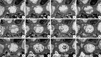
Tubules in H. catenularius were generated from small intramural openings between adjacent mature corallites (arrows in Fig. 7.3, 7.4, 7.7, 7.8), and in some cases beside juvenile corallites (arrows in Figs. 8.9, 9.6). Fission of an opening and fusion between adjacent openings were common (Fig. 7.3–7.12). Stasińska (Reference Stasińska1967, fig. 4) found that in some cases, tubules in Halysites arose from the fusion of two intramural openings. In H. catenularius, the size and shape of small intramural openings changed constantly with height in the corallum. Some openings seem to have disappeared in the common wall, but reappeared after a very short vertical distance. The walls around tubules thickened in some cases, resulting in balken structure (Fig. 8.1–8.5).
Transverse serial peels showing the formation of a tubule from small intramural openings in Halysites catenularius. (1–11) Development of small intramural openings in the common wall between corallites (e.g., arrows in 3, 4, 7, 8); (12) the resulting tubule in the common wall. GIT 806-8, figured peels spaced 0.05, 0.05, 0.05, 0.05, 0.05, 0.05, 0.05, 0.10, 0.25, 0.05, 0.15 mm apart, respectively. Scale bar shown in 1.

Transverse serial peels showing Type A interstitial increase in Halysites catenularius. (1–5) Balken structure in the thickened walls around a tubule; (6–8) tubule develops into a juvenile corallite; (9) appearance of small intramural openings (arrows) beside juvenile corallite; (10–12) juvenile corallite attains mature size. GIT 806-1, figured peels spaced 0.30, 0.35, 0.45, 0.50, 0.15, 0.25, 0.05, 0.05, 0.15, 0.20, 1.15 mm apart, respectively. Scale bar shown in 1.

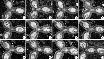
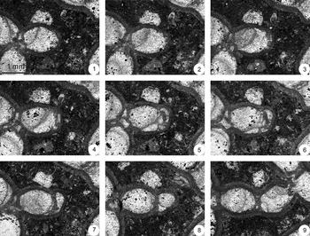
Transverse serial peels showing Type B interstitial increase in Halysites catenularius. (1–5) A tubule develops into a juvenile corallite between two mature corallites; (6–8) juvenile corallite attains mature size, while new tubules form on either side (arrows in 6, 8). GIT 806-10, figured peels spaced 0.65, 0.20, 0.15, 0.20, 0.05, 0.20, 1.35 mm apart, respectively. Scale bar shown in 1.

Stasińska (Reference Stasińska1967), Webby and Semeniuk (Reference Webby and Semeniuk1969, fig. 8), and Webby (Reference Webby1975, figs. 4, 5) reported that tubules associated with a new corallite were inserted interstitially at about the level where the corallite attained mature size. The present study reveals that tubules could appear earlier, beginning as intramural openings beside juvenile corallites when they reached approximately one-third of their mature corallite size (arrows in Figs. 8.9, 9.6). Young and Elias (Reference Young and Elias1995, p. 68, pl. 11, fig. 4) noted one example in Halysites of a tubule that was already present when an adjacent corallite was initiated. Juvenile corallites usually attained mature size over a short vertical distance, as demonstrated by Webby (Reference Webby1975) and the present study (Figs. 8.9–8.12, 9.5–9.8).
The small intramural openings that developed into tubules in H. catenularius (Fig. 7) are remarkably similar in appearance to intramural openings documented in the common wall between some corallites in four species of Catenipora (Bae et al., Reference Bae, Elias and Lee2013, fig. 1a–d). Some of the openings in Catenipora extended for a short or long vertical distance and then disappeared. Others developed into corallites with initial temporary connections to neighboring corallites, suggesting that they arose by a process of lateral increase (Bae et al., Reference Bae, Elias and Lee2013, fig. 1e, f). The intramural openings that disappeared were interpreted as aborted, incipient lateral offsets that originated from soft tissue on the common wall between corallites. The intramural openings in Catenipora were studied through transverse serial sections, so it is unknown whether they contain tabulae such as those in the tubules of Halysites (Fig. 5.8, 5.9).
Some authors have interpreted the tubules in Halysites as sites of polyps that differed from those occupying corallites, but most have considered them to represent intercorallite coenosteum (= coenenchyme; De Boer, Reference De Boer1973, pl. 3; Webby, Reference Webby1975; Hill, Reference Hill and Teichert1981; Scrutton, Reference Scrutton1998). Flower (Reference Flower1961, p. 42, 47, 48, pl. 8, fig. 2) suggested that in some “advanced” species of Catenipora, small depressions (“mesocorallite cavities” = calices) between corallites on the upper surface of the corallum were filled with solid balken structures during upward growth.
Interstitial increase
Interstitial increase in Halysites (= “coenenchymal increase” of Hill, Reference Hill and Teichert1981, p. F438) involved the formation of a new corallite through the expansion of a preexisting tubule (Webby and Semeniuk, Reference Webby and Semeniuk1969, fig. 8a; Webby, Reference Webby1975, fig. 5). In the coralla of H. catenularius, two types of interstitial increase are recognized depending on the location with respect to other corallites.
In Type A interstitial increase, the generation of a new corallite occurred in the junction area of three or four preexisting corallites. The new corallite developed by the expansion of a tubule that was usually irregular in shape, resulting in the extension of a rank and the associated lacunae (Fig. 8). The transformation of a tubule into a juvenile corallite is first recognizable at about half of its mature size. As the tubule developed into a corallite, a new tubule was initiated in its former place and on the other side of the new corallite (arrows in Fig. 8.9). Type A interstitial increase was the most frequent mode for the addition of corallites and occurred commonly in all the examined coralla of H. catenularius.
In Type B interstitial increase, the generation of a new corallite occurred between two preexisting corallites, which could be in the same rank (Fig. 5.9) or in two different ranks perpendicular to one another (Fig. 9). Similar to Type A interstitial increase, the new corallite developed by the expansion of a tubule (Figs. 5.9, 9). New tubules appeared on either side of the developing corallite (arrows in Fig. 9.6, 9.8). Although the frequency of Type B interstitial increase was much less than Type A interstitial increase, it also occurred in all the examined coralla of H. catenularius.
Lateral increase
Lateral increase in Halysites involved the formation of a new corallite through offsetting from a parent corallite (Webby and Semeniuk, Reference Webby and Semeniuk1969, fig. 8b; Webby, Reference Webby1975, fig. 4). In the coralla of H. catenularius, two types of lateral increase are recognized depending on the location with respect to a normal parent corallite. A third type, previously unreported in Halysites, was associated with regeneration of a damaged and terminated corallite or tubule. The processes of lateral increase in H. catenularius are similar to those in species of Catenipora (Dixon, Reference Dixon1976; Bae et al., Reference Bae, Lee and Elias2006b, Reference Bae, Elias and Lee2013).
In Type A lateral increase, the new offset initiated from the lateral wall of a juvenile or mature corallite through a lateral process and developed perpendicularly to the existing rank, thus forming a new rank (black arrows in Figs. 10.2–10.6, 11.6, 11.7). This type of corallite increase was rare and not observed in all the examined coralla of H. catenularius. Dixon (Reference Dixon1976, fig. 2b) recognized this type of increase in Catenipora. According to Stasińska (Reference Stasińska1967, p. 14), offsets rarely appeared on the lateral corallite wall of halysitids, and only in Catenipora. Clearly, however, such offsetting occurred in H. catenularius. Unlike Type A lateral increase in H. catenularius, the type C lateral increase recognized in species of Catenipora (Bae et al., Reference Bae, Lee and Elias2006b, Reference Bae, Elias and Lee2013) occurred at the junction of two corallites (see also Dixon, Reference Dixon1976, fig. 2d).
Transverse serial peels showing Type A lateral increase and Type B lateral increase in Halysites catenularius. (1–3) Initiation of Type A lateral increase by the appearance of a small protuberance (black arrows in 2, 3) perpendicular to the existing rank; (4–6) continuation of Type A lateral increase (black arrows), and initiation of Type B lateral increase by the appearance of a protuberance (white arrows) near the distal end of a corallite; (7–9) development of the juvenile corallites, followed by the joining of ranks. GIT 806-10, figured peels spaced 0.05, 0.10, 0.25, 0.05, 0.15, 0.15, 0.10, 1.2 mm apart, respectively. Scale bar shown in 1.

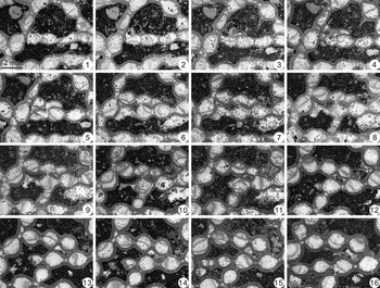
Transverse serial peels showing the appearance and development of an agglutinated patch of corallites in Halysites catenularius. (1–9) A temporary agglutinated patch of corallites formed by lateral increase along the outer wall of corallites (arrows in 2–4, 6, 7; note openings resulting in temporary fusion of irregularly shaped corallites in 5–9); (10–16) transformation into normal corallites and ranks, with further lateral increase (arrows in 12–14). GIT 806-13, figured peels spaced 0.20, 0.10, 0.15, 0.20, 1.00, 0.05, 0.30, 0.10, 0.35, 0.30, 0.80, 0.10, 0.05, 0.35, 0.75 mm apart, respectively. Scale bar shown in 1.

Type B lateral increase involved the development of an offset near the distal end of a single corallite (white arrows in Fig. 10.4–10.6; Fig. 12). Additional new corallites formed continuously one after another. Offsetting by this type of increase resulted in the rapid development of new ranks and lacunae during expansion of the corallum. Type B lateral increase was common in all the examined coralla of H. catenularius. This type of lateral increase in halysitids (termed “peripheral increase” by Buehler, Reference Buehler1955, and Hamada, Reference Hamada1959) is also well known from previous reports in Halysites (Stasińska, Reference Stasińska1967; Webby and Semeniuk, Reference Webby and Semeniuk1969; Webby, Reference Webby1975). It is comparable to the type D lateral increase documented in species of Catenipora (Bae et al., Reference Bae, Lee and Elias2006b, Reference Bae, Elias and Lee2013).
Transverse serial peels showing Type B lateral increase in Halysites catenularius. (1–4) Appearance of a lateral protuberance at the distal end of a corallite; (5-8) development of a juvenile corallite; (9) corallite attains mature size. GIT 806-10, figured peels spaced 0.05, 0.05, 0.05, 0.05, 0.05, 0.10, 0.25, 0.40 mm apart, respectively. Scale bar shown in 1.

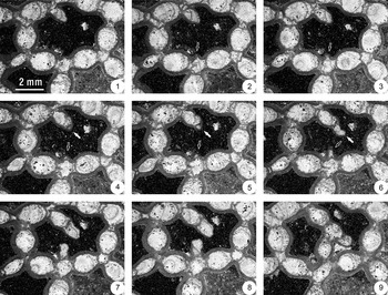
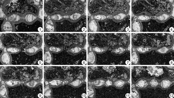
Unlike the four types of increase described above, the generation of a corallite by Type C lateral increase was associated with the regeneration of a terminated corallite or tubule. Corallites or tubules could be damaged and terminated by sediment influx (Fig. 13.1–13.6). During regeneration, a replacement corallite developed by lateral offsetting from an adjacent parent corallite (Fig. 13.7–13.11). When the new corallite was established, tubules appeared on either side of it (Fig. 13.12). This process allowed quick and successful recovery of damaged surface areas in colonies that were subjected to physical disturbances resulting in influxes of detritus. This type of lateral increase was very rare; damaged corallites usually recovered by rejuvenation without the introduction of an additional corallite (Fig. 5.8). Type C lateral increase in H. catenularius is comparable to type E lateral increase identified in species of Catenipora (Bae et al., Reference Bae, Lee and Elias2006b, Reference Bae, Elias and Lee2013).
Transverse serial peels showing Type C lateral increase and reconnection of a damaged rank in Halysites catenularius. (1–6) Damage between normal corallites and separation by sediment; (7–11) generation of an additional corallite by lateral offsetting and reconnection of the rank; (12) the new corallite attains mature size, with tubules on either side. GIT 806-13, figured peels spaced 0.70, 0.15, 0.05, 0.05, 0.15, 0.1, 0.15, 0.1, 0.1, 0.15, 1.00 mm apart, respectively. Scale bar shown in 1.

Temporary agglutinated patch of corallites
Temporary agglutinated patches of corallites are common in the examined coralla of H. catenularius. They formed by lateral increase along the outer wall of corallites, in areas of abnormal or damaged corallites (Fig. 11.1–11.8; note arrows in 11.2–11.4, 11.6, 11.7). During development of the agglutinated patch, walls between some irregularly shaped corallites opened, resulting in temporary fusion of corallites (Fig. 11.5–11.9). With subsequent growth of the colony and further lateral increase, the agglutinated patch of corallites was able to transform into normal ranks of corallites alternating with tubules (Fig. 11.9–11.16; note arrows in 11.12–11.14).
Agglutinated patches of corallites have not been reported previously in Halysites. Temporary agglutinated patches of side-by-side corallites, comparable to those in H. catenularius, were rare in some species of Catenipora (Lee and Elias, Reference Lee and Elias1991; Bae et al., Reference Bae, Lee and Elias2006b, Reference Bae, Elias and Lee2013). They formed by rejuvenation and/or regeneration during recovery from damaged and/or terminated corallites. In one species, C. foerstei, agglutinated patches could also form by increase involving normal, undamaged corallites (Bae et al., Reference Bae, Lee and Elias2006b, Reference Bae, Elias and Lee2013). In some cases, this resulted in a cluster of many corallites (Bae et al., Reference Bae, Elias and Lee2013, fig. 2).
Axial increase
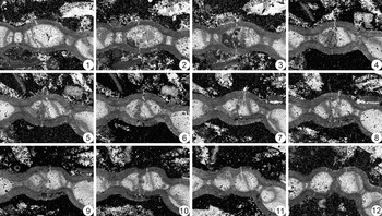
The axial mode of increase involved longitudinal fission of a corallite. In H. catenularius, this occurred in association with rejuvenation of a damaged corallite (Fig. 14). A new common wall appeared near the axial area of the damaged corallite (Fig. 14.3, 14.4). It subsequently extended from one lateral wall to the other, dividing the corallite in half (Fig. 14.5–14.8). The new wall was initially undulatory, and subsequently became straight. The resulting two juvenile corallites gradually enlarged into mature corallites of comparable size to the neighboring corallites (Fig. 14.9–14.12). Axial increase was extremely rare and not observed in all the examined coralla of H. catenularius.
Transverse serial peels showing axial increase in Halysites catenularius. (1–4) Damage of a normal corallite, followed by the entry of sediment; (5–8) rejuvenation of the damaged corallite, accompanied by the development of a new common wall dividing the corallite in half; (9–12) the resulting two juvenile corallites develop to mature size. GIT 806-8, figured peels spaced 0.25, 0.25, 1.15, 0.10, 0.05, 0.30, 0.10, 0.15, 0.15, 0.25, 1.00 mm apart, respectively. Scale bar shown in 1.

The process of axial increase has not been described previously in Halysites, although Young and Elias (Reference Young and Elias1995, p. 71) noted that connections between some corallite tabularia suggest axial increase may have occurred in a specimen of H. alexandricus Young and Elias, Reference Young and Elias1995. Lee et al. (Reference Lee, Jun, Bae, Elias, Hubmann and Piller2007) recognized four types of axial increase in Late Ordovician tabulate corals. Their type 2 axial increase associated with rejuvenation of a damage single corallite corresponds to that in H. catenularius. This type of axial increase is also known in some species of Catenipora (Lee and Elias, Reference Lee and Elias1991; Bae et al., Reference Bae, Lee and Elias2006b, Reference Bae, Elias and Lee2013; Lee et al., Reference Lee, Jun, Bae, Elias, Hubmann and Piller2007). It was an effective method for recovery and increasing the number of corallites after damage.
Phylogenetic implications
Regarding the evolutionary history of halysitids, it has generally been accepted that Quepora Sinclair, Reference Sinclair1955 (lacking septal spines) and/or Catenipora (with septal spines) gave rise to Halysites (Scrutton, Reference Scrutton, Oliver, Sando, Cairns, Coates, Macintyre, Bayer and Sorauf1984). Flower (Reference Flower1961, fig. 4) considered Quepora to be primitive, followed by Catenipora with septal spines and then “advanced” species of Catenipora with the addition of intercorallite balken structure. However, Quepora is now considered to be a junior synonym of Catenipora (see Laub, Reference Laub1979; Young and Elias, Reference Young and Elias1995; Liang et al., Reference Liang, Elias and Lee2018), and the earliest known halysitid has septa as well as balken (Catenipora tongchuanensis Liang et al., Reference Liang, Elias and Lee2018). According to Flower (Reference Flower1961; see also Flower and Duncan, Reference Flower, Duncan, Pojeta and Pope1975, fig. 1), Halysites arose from “advanced” Catenipora through the appearance of a tubule with tabulae instead of solid balken infillings between corallites (compare De Boer, Reference De Boer1973, pl. 3, with Flower, Reference Flower1961, pl. 8, fig. 2).
The mode of corallite increase is an important character in considering taxonomic assignments and phylogenetic relationships of tabulate corals (Lee and Elias, Reference Lee and Elias2000; Lee et al., Reference Lee, Jun, Bae, Elias, Hubmann and Piller2007; Liang et al., Reference Liang, Lee, Elias, Pärnaste and Mõtus2013). The present study shows that small intramural openings in some species of Catenipora are remarkably similar in appearance to those in Halysites catenularius. The ability to form intramural openings may reflect a relationship between the two genera. However, these structures were associated with lateral offsetting in Catenipora, whereas they gave rise to tubules that generated corallites by interstitial increase in H. catenularius. Corallite increase by the interstitial mode occurred in Halysites, but not in Catenipora. However, the three types of lateral increase identified in the present study of H. catenularius are comparable to types that have been recognized in species of Catenipora. Furthermore, the temporary agglutinated patches of corallites that formed by lateral increase in H. catenularius are like those in some species of Catenipora. In addition, the type of axial increase found in H. catenularius also occurred in some species of Catenipora. These similarities are consistent with a close phylogenetic relationship between Catenipora and Halysites.
Flower (Reference Flower1961, p. 42, 47, 48) suggested that species of Catenipora with and without balken may represent separate genera. He stated that the type species, C. escharoides Lamarck, Reference de Lamarck1816, apparently lacked balken (Flower, Reference Flower1961, p. 47). Young and Elias (Reference Young and Elias1995, p. 67, 73) noted the possibility that some species of Halysites having tubules with tabulae were ancestors of species in which tubules were partially or completely infilled with skeletal material, thereby reverting to a morphological state in which they would be classified as Catenipora. Both Catenipora and Halysites may be polyphyletic (Scrutton, Reference Scrutton, Oliver, Sando, Cairns, Coates, Macintyre, Bayer and Sorauf1984; Liang et al., Reference Liang, Elias and Lee2018).
Hill (Reference Hill and Teichert1981) placed the suborder Halysitina Sokolov, Reference Sokolov1947, within the order Heliolitida Frech, Reference Frech1897, in the subclass Tabulata. She considered the intercorallite tubules in Halysites to represent coenenchyme, a characteristic feature of heliolitids, but described Catenipora as lacking coenenchyme. As currently understood (Liang et al., Reference Liang, Elias and Lee2018), the earliest halysitid is Catenipora tongchuanensis of Late Ordovician, Sandbian age. Halysites is unknown until the Katian age (late Eastonian). The most primitive heliolitids are the coccoseridids, which have completely dilated septal and coenenchymal trabeculae or coenenchymal tubules with diaphragms in trabecular coenenchyme (Mõtus and Zaika, Reference Mõtus and Zaika2012). The earliest known coccoseridids are early Katian (early Eastonian), which is intermediate in age between the appearance of Catenipora and Halysites. Coenenchymal structures in halysitids and heliolitids are almost certainly not homologous (Young and Elias, Reference Young and Elias1995, p. 66, 67). Lateral increase is typical in Catenipora (e.g., Bae et al., Reference Bae, Lee and Elias2006b, Reference Bae, Elias and Lee2013), whereas coenenchymal increase is characteristic of heliolitids (Oliver, Reference Oliver and Macurda1968; for detailed examples, see Young and Elias, Reference Young and Elias1995, p. 48, 49, and Liang et al., Reference Liang, Lee, Elias, Pärnaste and Mõtus2013). A close phylogenetic relationship between Catenipora and coccoseridids, or between halysitids and heliolitids in general, seems improbable. We follow Scrutton (Reference Scrutton, Oliver, Sando, Cairns, Coates, Macintyre, Bayer and Sorauf1984, fig. 1, Reference Scrutton and Murray1985) and Young and Elias (Reference Young and Elias1995), who recognized the Halysitida and Heliolitida as separate orders with ancestors among the tabulate corals.
Conclusions
Multivariate morphometric analysis was successful in identifying the studied coralla from the Silurian Rumba Formation (Telychian) and Maasi Beds in the Jaagarahu Formation (Sheinwoodian) of Estonia. NMDS and discriminant analysis were based on 17 morphological characters obtained from 15 coralla and nine type specimens of four species of Halysites. As a result, the coralla were identified as H. catenularius, and H. priscus was confirmed as a junior synonym. Halysites catenularius, H. junior, and H. senior were closely related, with H. catenularius morphologically intermediate between the other two.
Growth cycles in H. catenularius, bounded by minimum values of corallite tabularial area, were presumably annual. The inferred average growth rate of 6.0 mm per year is typical for halysitids. Tubules in H. catenularius were generated from small intramural openings between adjacent corallites. The tubules appeared beside corallites that had reached at least one-third of their mature size. Two types of interstitial increase, in which a new corallite arose from expansion of a tubule, occurred in H. catenularius. This process took place at the junction of three or four preexisting corallites (Type A) or between two preexisting corallites (Type B). Three types of lateral increase, in which a new corallite arose through offsetting from a parent corallite, occurred in H. catenularius. An offset could be initiated from the lateral wall of a parent (Type A), from the distal end of a parent (Type B), or from a parent during regeneration of an adjacent terminated corallite or tubule (Type C). In areas of abnormal or damaged corallites, temporary agglutinated patches of corallites could form by lateral increase along the outer wall of corallites. Axial increase, involving longitudinal fission, occurred in association with rejuvenation of damaged corallites in H. catenularius.
The intramural openings, three types of lateral increase, temporary agglutinated patches of corallites, and axial increase in H. catenularius resemble features in some species of the halysitid genus Catenipora. These similarities are consistent with the generally accepted inference that Catenipora gave rise to Halysites. Evaluation of the possibility that both genera are polyphyletic will require further detailed analysis of additional species. A close phylogenetic relationship between halysitids and heliolitids seems improbable.
Acknowledgments
We are grateful to H. Pärnaste and B.-Y. Bae for their assistance in the field. This study was supported by grants from the Chinese Academy of Sciences (XDB26000000) and National Natural Science Foundation of China (grant no. 41402013 and J1210006) to KL, and the National Research Foundation of Korea (NRF) to D-JL (2018R1A2B2005578). We thank the reviewers, B. Hubmann and M. Zapalski, for their constructive comments.