Case report
Hybrid approach for postnatal treatment of hypoplastic left heart syndrome has been reported more than 10 years agoReference Roeleveld, Axelrod and Klugman1-Reference Hirata, Miyata and Hirahara3. Instead of continuous infusion of prostaglandin, duct stenting is usually performed for remaining the arterial duct widely open. At first, balloon-expandable stents have been used for this purpose; in the beginning of this decade, a specifically designed self-expandable stent (Sinus SuperFlex DS®; OptiMed Inc., Karlsruhe, Germany) received a CE-mark® for neonatal duct stenting of duct-dependent systemic circulation.Reference Goreczny, Qureshi and Rosenthal4,Reference Goreczny, Qureshi and Rosenthal5 .
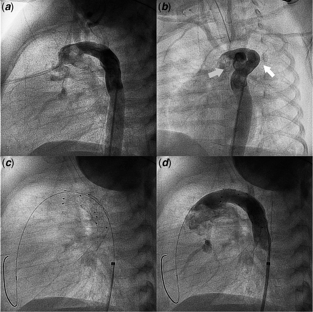
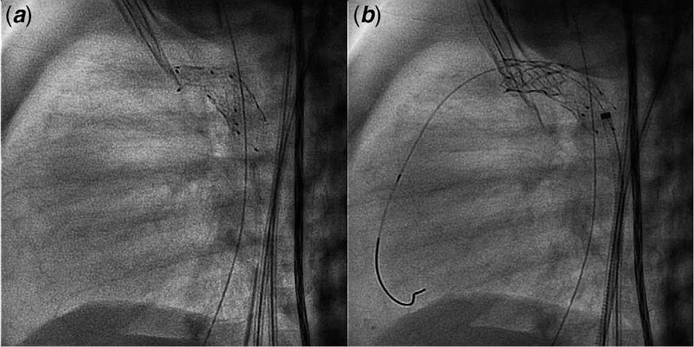
We report the case of a newborn with a hypoplastic left heart syndrome, following bilateral pulmonary artery bandings performed at the 5th day of life. He was referred to the cath lab at the 12th day of life (delay due to active infection) for ductal stenting. Prostaglandin infusion at 0.02 mcg/kg/min was discontinued in the cath lab. Arterial duct was wide, long, slightly tortuous, and with no significant stenosis (Fig 1a, 1b). Arterial duct minimum and maximum diameters were 6.5 and 7.5 mm, with an estimated length of 26–27 mm. From a 4F short sheath placed in the right femoral artery, a 9 mm × 20 mm sinus Superflex DS© stent was deployed in the arterial duct. A second 8 mm × 20 mm stent was placed in telescope technique, overlapping the first placed stent to cover the aortic arterial duct end (Fig 1c, 1d and Supplementary video S1). Enoxaparin twice a day, and aspirin at 5 mg/kg/day were administered during the first 2 days. Forty-eight hours after the procedure, signs of low cardiac output were detected, and a moderate gradient of 3.5 m/s in the stent flow was confirmed by echocardiography. The patient was referred to the cath lab, where an important collapse of the first sinus Superflex DS stent© at the pulmonary ductal end was detected (Fig 2a, Supplementary video S2). Angioplasty was discarded due to the high risk of an important recoil at a short-medium term. From a 5F long sheath placed in left femoral artery, a Palmaz Blue© stent (Cardinal Health Inc., Columbus, Ohio, United States of America) 7 mm × 15 mm was deployed at that location, with a good angiographic result (Fig 2b).
(a): 90° left lateral view. Angiography showing arterial duct and descending aorta. (b): Anteroposterior view. Angiography showing descending aorta and its bifurcation in arterial duct (white arrow), and aortic arch (grey arrow). (c): 90° left lateral view. Sinus SuperFlex stents covering arterial duct. (d): 90° left lateral view. Angiography showing a correct flow inside sinus stent and correct covering of the entire arterial duct.

(a): 90° left lateral view. Sinus SuperFlex stent collapse at the pulmonary ductal end. (b): 90° left lateral view. Palmaz Blue stent solving sinus SuperFlex stent collapse.

The sinus SuperFlex DS© stent has achieved an important expansion from its appearance and CE-mark for neonatal duct stenting. Its specific design for systemic ductal stenting in hypoplastic left heart syndrome provides specific sizes (7 to 9 mm) and lengths (12 to 20 mm), nitinol self-expandable design with an increased adaptability to ductal anatomy, and deliverance by a 4F short sheath, allowing not only venous, but arterial retrograde implantReference Goreczny, Qureshi and Rosenthal4. In the opposite side, its radial force is quite lower than force showed by balloon-expandable stentsReference Goreczny, Qureshi and Rosenthal5. Some groups use this stent in spite of some degree of arterial duct stenosis, performing some angioplasty with balloon after stent implantation, but its ideal setting is a wide non-stenotic arterial duct to keep patent. We describe the first report of an early collapse of a Superflex DS stent in a previous non-stenotic arterial duct. The reported complication raises questions about the risk factors and predictability, but slightly tourtous anatomy, telescoped stents, and aspirin administration have been present in other patients with no Superflex DS stent collapse. The possibility of an early collapse of the SuperFlex DS© stent should be kept in mind, and probably frequent echocardiographic studies should be carried out first days after prostaglandin discontinuation, to rule out this complication.
Acknowledgements
None.
Financial Support
This research received no specific grant from any funding agency, commercial or not-for-profit sectors.
Conflicts of Interest
None.
Ethical Standard
The research does not involve human and/or animal experimentation.
Supplementary material
To view supplementary material for this article, please visit https://doi.org/10.1017/S1047951120000189