Implications
This study demonstrates that the reproductive rabbit doe is in a very stressful condition from a metabolic and inflammatory point of view few days before parturition. This adverse condition seems well related with the number of offspring of the doe. Consequently, the new rabbit breeds require more care to prevent the adverse conditions in the peripartum period. In accordance with these results, feeding and managerial strategies for high prolificacy periparturient rabbit does should be revised.
Introduction
In modern intensive rabbit breeding, the reproductive management of the rabbit doe, the genetic programmes and the feeding systems have greatly increased the profitability of the system but, at the same time, several problems related to the health status and the welfare of does have also appeared, as reviewed by Pascual et al. (Reference Pascual, Xiccato and Fortun-Lamothe2006 and Reference Pascual, Savietto, Cervera and Baselga2013) and Castellini et al. (Reference Castellini, Dal Bosco, Arias-Álvarez, Lorenzo, Cardinali and Rebollar2010). One of these concerns the difficulties of the does during the peripartum. In fact, for the reproductive rabbit does, the periods of late pregnancy and early lactation, as for other livestock animal species (i.e. cow, pig, sheep, goat), represent critical phases (Xiccato, Reference Xiccato1996). In this period the doe is exposed to dramatic physiological and dietary challenges, including the abrupt transition from pregnancy to lactation that could be exacerbated if the number of foetuses in gestation is very high. The manner in which these changes occur and how they are managed are of great importance as they are closely linked with lactation success, the incidence of clinical and subclinical postpartum diseases and reproductive performance for the next gestation, all factors that affect significantly the profitability of the system (Castellini et al., Reference Castellini, Dal Bosco, Arias-Álvarez, Lorenzo, Cardinali and Rebollar2010).
The fast reproductive rhythm imposed to rabbit doe makes her unable to cover the energetic needs for lactation of multiple litters, final gestation stage and its own growth. Therefore, milk yield is decreased as the priority of restoring reserves for the future litter starts to increase (Pascual et al., Reference Pascual, Savietto, Cervera and Baselga2013) and this is accentuated in the last 3 days of gestation when the energy demands of the foetus are very high (Mocé et al., Reference Mocé, Santacreu, Climent and Blasco2004) and episodes of pregnancy toxaemia and liver disease may occur.
The genetic selection programs for reproductive traits have mainly focused on improving litter size (Pascual et al., Reference Pascual, Savietto, Cervera and Baselga2013), and the breeding of rabbits moved on from traditional production systems to more intensive ones using modern, prolific lines (Castellini et al., Reference Castellini, Dal Bosco, Arias-Álvarez, Lorenzo, Cardinali and Rebollar2010). The high number of foetuses of these breeds may trigger the pregnant does to respond to metabolic challenges happening in the last week of pregnancy. Blood metabolites as non-esterified fatty acids (NEFAs) and β-hydroxybutyrate (BHBA) could be useful indicators of the energy status of the animals (Pascual et al., Reference Pascual, Xiccato and Fortun-Lamothe2006). In addition, blood markers of inflammatory status can be useful to assess the health condition of the animals.
The aim of this study was to investigate the metabolic and inflammatory changes that occur in the reproductive rabbit doe close to parturition. Moreover, grouping the same does on the basis of the number of offspring, a retrospective study has been done focusing on the effect of the litter size on metabolism and health status.
Material and methods
This study complied with Italian laws on animal experimentation (DL n.116, 27/01/1992) and ethics. The experiment was carried out in the Università Cattolica del Sacro Cuore experimental barn (CERZOO) located in the Northern Italy (Piacenza).
Housing, animals and feeding
The study was carried out using nine rabbit does (Oryctolagus cuniculus) from commercial hybrid lines (HYPLUS PS 19; Groupe Grimaud, Roussay, France). During the study period (November to December), average daily temperatures ranged from 11.3°C to 18.4°C, and average relative humidity ranged from 65% to 75% maintained by a forced ventilation system. The does were 7 months old and were preparing for their third parturition. They were housed in individual flat-deck cages (0.4×0.6×0.35 m) equipped with drinker and a manual feeder. The does were bred under a constant photoperiod of 16 h light per day. The nest was prepared 2 days before parturition through the placement of a plastic floor and a metallic separator. After parturition, the litters were housed in the nest and within 3 days the nests were balanced at 10 puppies. The mothers were allowed to enter the nest and suckle once daily in the morning (0900 h) for a short time. The does were given ad libitum access to water and to a commercial pelleted diet for lactating does (Fertilap spec.7A; Purina – Cargill s.r.l., Milano, Italy) during the entire study period. Insemination was done 18 days after parturition, and weaning occurred at 34 days.
Blood sampling and analysis
At −3, 4 and 12 days from parturition (–D3, D4 and D12, respectively), blood was sampled and BW was measured immediately after the bleeding. The –D3 and D4 were selected because they were sampling times relatively close to parturition but immediately before and after the dramatic changes that are expected to happen around parturition. The D12 was included because at this time the does should have recovered from the negative energy balance. Blood samples were collected before the suckle at around 0830 h from the central auricular artery into 2 ml tubes containing sodium heparin as anticoagulant (5 units of heparin per ml of blood). The blood samples were immediately cooled in ice water and within 1 to 2 h after bleeding the plasma was obtained after centrifugation at 3500×g for 15 min at 4°C and stored at −20°C until analysed.
Samples were analysed for metabolic indicators (glucose (mmol/l), total cholesterol (mmol/l), triglycerides (mmol/l), creatinine (µmol/l), urea (mmol/l), aspartate aminotransferase or GOT (U/l), γ-glutamyl transpeptidase or GGT (U/l), alkaline phosphatase or ALP (U/L), NEFA (mmol/l), BHBA (mmol/l)), selected minerals (Ca (mmol/l), P (mmol/l), Mg (mmol/l), K (mmol/l), Cl (mmol/l), Zn (µmol/l)) and oxidative-inflammatory indicators (total protein (g/l), albumin (g/l), globulin (g/l), total bilirubin (µmol/l), haptoglobin (g/l), ceruloplasmin (µmol/l), reactive oxygen metabolites or ROMs (mgH202/100 ml), paraoxonase or PON (U/ml)).
Blood metabolites were analysed by an automated biochemistry analyser (ILAB 650; Instrumentation Laboratory, Lexington, MA, USA). Total protein, albumin, total cholesterol, total bilirubin, triglycerides, creatinine, urea, Ca, P, Mg, GOT, GGT, ALP were determined using kits purchased from Instrumentation Laboratory (IL Test). Globulin was calculated as the difference between total protein and albumin. Ions (K and Cl) were measured by the potentiometer method (ion-selective electrode connected to ILAB 650). Zn, NEFA and BHBA, were measured by the methods previously reported (Bionaz et al., Reference Bionaz, Trevisi, Calamari, Librandi, Ferrari and Bertoni2007). Haptoglobin and ceruloplasmin were analysed using methods described in Skinner et al. (Reference Skinner, Brown and Roberts1991) and Sunderman and Nomoto (Reference Sunderman and Nomoto1970), respectively, adapted to the ILAB 650 conditions. ROMs were measured by a commercial kit (Diacron International s.r.l., Grosseto, Italy). PON was measured by the method of Ferré et al. (Reference Ferré, Camps, Prats, Vilella, Paul, Figuera and Joven2002) adapted to the ILAB 650, as previously described (Bionaz et al., Reference Bionaz, Trevisi, Calamari, Librandi, Ferrari and Bertoni2007).
Grouping of animal and statistical analysis
The litter size characteristics of the rabbit does included in the study is provided in the Supplementary Table S1. Only one kit died at parturition and on average the nine does gave birth to 10.11±1.31 kits. In order to evaluate the effect of litter size on health-metabolic status of the does, the animals were retrospectively divided into two groups according to the number of offspring (including dead born kits). The threshold number of the kits to group the does was the average litter size. Consequently the two groups resulted: high litter size group (HI, does with >10.11 kits; n=5) and low litter size group (LO, does with ⩽10.11 kits; n=4).
The normal distribution of the data was checked by using Proc UNIVARIATE (SAS Inst. Inc., Cary, NC, USA; release 8.0) by NORMAL option. Data were not normally distributed and a log transformation was applied to satisfy normality and homogeneity of variance assumptions underlying linear models. Through the text, in tables and graphs the data are presented in the original scale (mean and s.e.m.).
Transformed data were subjected to ANOVA using the MIXED procedure of SAS. The statistical model applied included the fixed effect of days from parturition, litter size group and their interaction. The days from parturition within rabbit doe were considered as a repeated measure. The pair-wise comparison has been done using least significant difference (LSD) test.
The following model was applied:
 $$Y_{{ijk}} =\mu {\plus}{\rm T}_{i} {\plus}{\rm LS}_{j} {\plus}{\rm TLS}_{{ij}} {\plus}{\rm D}_{k} {\plus}{\varepsilon}_{{ijk}} $$
where Y ijk is the dependent variables (blood parameters and BW), µ the overall mean, T i the effect of days from parturition ( i =–D3, D4 and D12), LS j the effect of litter size ( j =HI and LO), TLS ij the interaction term, D k the random effect of the doe (k=1, … , 9), ε ijk the random error component.
Moreover, a principal components (PC) analysis was performed by the PRINCOMP procedure. All blood parameters were used for PC analysis excluding globulin (because of a high autocorrelation with total protein and albumin, from which it was mathematically calculated).
Results
Time effect
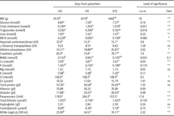
BW (Table 1) was higher before parturition (–D3), had the lowest values immediately after it (D4), then began to recover. A statistical difference was observed between –D3 and D4 (P<0.05). The effect of time around parturition on plasma parameters in reproductive rabbit does is shown in Table 1. Regarding metabolic indicators, glucose and cholesterol had the lowest levels before parturition (e.g. –D3 compared with D4 and D12; P<0.01). NEFA and GOT had almost two times concentrations at –D3 in comparison with D4 and D12 (P<0.05 for NEFA and P<0.01 for GOT); while creatinine and BHBA were higher at –D3 with respect to D4 (P<0.05). Triglyceride concentration was higher after parturition at D4 compared with –D3 and D12 (P<0.01), whereas urea had the highest concentration at D12 compared with –D3 (P<0.01) and D4 (P<0.05).
Effect of the days related to parturition (–D3=3 days before parturition; D4=4 days after parturition; D12=12 days after parturition) on BW and plasma parameters in reproductive rabbit does

NEFA=non-esterified fatty acids; BHBA=β-hydroxybutyrate; ROMs=reactive oxygen metabolites.
a,b,cMeans within each row, with no common superscript are significantly different (P<0.05). Statistical significance for the effect of time was established by using a conventional P-value lower of 0.05 (*) or 0.01 (**).
For mineral profile, plasma values of phosphorus were two times higher 3 days before parturition than during lactation (P<0.01). The chlorine was lower (P<0.05) before parturition (–D3) compared with D4 and D12. Plasma calcium had the highest values after parturition compared with –D3 and D12 (P<0.01), whereas potassium was lower at D12 compared with previous checks (P<0.05). Plasma levels of Mg did not change throughout the study period.
Regarding oxidative-inflammatory indicators, within the positive acute phase proteins, haptoglobin did not change around parturition, whereas at –D3 ceruloplasmin had the highest values (P<0.01) and, on the contrary, globulins had the lowest levels (P<0.05). Total bilirubin had a similar pattern of change like ceruloplasmin, with higher values at –D3 and a decrease after parturition (e.g. P<0.01 with D4 and P<0.05 with D12). The concentration of PON had an upward trend, from the lowest at –D3 to the highest values at D12 (P<0.01). ROMs reached the highest concentration at D4 and the lowest at D12 (P<0.01).
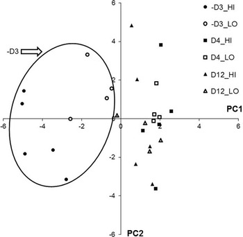
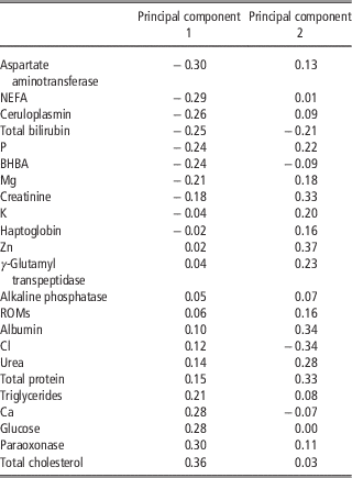
Figure 1 shows the result of PC analysis and Table 2 shows the eigenvector between all the variables considered. The first three PC explained 55% of total variation (25%, 19% and 11% for PC1, PC2 and PC3, respectively). Moreover, the PC1 was able to separate quite efficiently the samples according with the time relative to parturition (Figure 1). From the blood parameters submitted to PC analysis, the PC auto-vectors (Table 2) showed that GOT, NEFA, ceruloplasmin, total bilirubin, P and BHBA, were the predominant variables acting with a negative impact on PC1, while total cholesterol, PON, glucose, Ca and triglycerides were the predominant variables acting with a positive impact on PC1.
Scatter plot of the first two principal component scores. The samples were classified according with the time days from parturition (–D3=3 days before parturition; D4=4 days after parturition; D12=12 days after parturition) and the grouping based on litter size: high litter size group (HI) and low litter size group (LO). The x-axis represents component 1 (PC1) and the y-axis represents component 2 (PC2). In the oval are grouped samples from –D3.

Eigenvector coefficients between the blood variable submitted to principal component analysis and the Principal component 1 and 2

NEFA=non-esterified fatty acids; BHBA=β-hydroxybutyrate; ROMs= reactive oxygen metabolites.
Effect of litter size
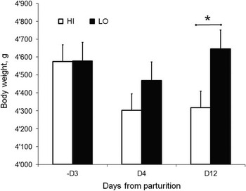
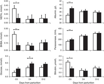
According with grouping based on litter size, the BW (Figure 2) was similar between HI and LO groups at –D3 and D4, but at D12 the BW was greater in LO than HI (P<0.05). The effect of litter size at different times during the transition period on plasma parameters in reproductive rabbit does is shown in Figure 3. According with the grouping, the HI does gave birth to 13.20±0.67 kits compared with 6.25±0.75 kits (P<0.01) for LO does.
Effect of litter size on BW changes at different days from parturition (–D3=3 days before parturition; D4=4 days after parturition; D12=12 days after parturition) of reproductive rabbit does in high litter size group (HI) and low litter size group (LO). Significance difference between groups at the same time is indicated by *(P<0.05).

Effect of litter size (white bars=high litter size group, HI; black bars=low litter size group, LO) at −3 (–D3), 4 (D4) and 12 (D12) days from parturition on plasma parameters in reproductive rabbit does: NEFA=non-esterified fatty acids (mmol/l); BHBA=β-hydroxybutyrate (mmol/l); albumin (g/l); paraoxonase (U/ml); glucose (mmol/l); bilirubin (µmol/l). Statistical significance between groups at each time point is indicated by using a conventional P-value lower of 0.1 (+) or 0.05 (*) or 0.01 (**).

The differences for blood profile parameters between the two groups were mostly evident before parturition (–D3). In particular, BHBA, NEFA and total bilirubin had the highest concentrations in HI compared with LO group (P<0.01 for BHBA; P<0.05 for NEFA and total bilirubin). The HI group at –D3 also had a lower concentration of albumin and PON (P<0.01). After parturition, the concentration of glucose at D4 was lower in HI compared with LO (P=0.09). Other blood parameters showed no or just marginal difference (Supplementary Table S2).
Discussion
In the last 2 decades, the breeding of rabbits moved on from traditional production systems to more intensive ones using modern prolific lines (see review of Castellini et al., Reference Castellini, Dal Bosco, Arias-Álvarez, Lorenzo, Cardinali and Rebollar2010). Consequently, the productivity of rabbit farms has increased but, at the same time, several problems related to the health status and the welfare of does also have appeared, for example excessive replacement rates, high mortality and culling rates and hypo-fertility (Rosell and de la Fuente, Reference Rosell and de la Fuente2009; Sánchez et al., Reference Sánchez, de la Fuente and Rosell2012). In addition, the nutritional requirements of the animals have had to be revised according with the new physiological demands (Xiccato, Reference Xiccato1996; Pascual et al., Reference Pascual, Savietto, Cervera and Baselga2013).
Effect of transition period in reproductive rabbit does
The nine rabbit does used in the present study did not have clinical symptoms of any disease during the experimental period. However, within the metabolic parameters analysed significant subclinical changes occurred during the peripartum period. A worse condition of the rabbit does was evident in late pregnancy (–D3) than in early (D4) and mid lactation (D12). The high concentrations of NEFA and BHBA before parturition confirmed that animals were in a catabolic condition (Bell, Reference Bell1995). Similar results were reported by Arias-Alvarez et al. (Reference Arias-Alvarez, García-García, Rebollar, Revuelta, Millán and Lorenzo2009), Theilgaard et al. (Reference Theilgaard, Baselga, Blas, Friggns, Cervera and Pascual2009) and Martínez-Paredes et al. (Reference Martínez-Paredes, Ródenas, Martínez-Vallespín, Cervera, Blas, Brecchia, Boiti and Pascual2012). The NEFA and BHBA increase during periods of body lipid mobilization, and this happens when energy from dry matter intake is not enough to cover the energy requirements of the animal (Brecchia et al., Reference Brecchia, Bonanno, Galeati, Federici, Maranesi, Gobbetti, Zerani and Boiti2006; Pascual et al., Reference Pascual, Xiccato and Fortun-Lamothe2006). In our study we confirmed that in the last days of pregnancy the reproductive rabbit does are at risk from the energetic point of view (Parigi-Bini et al., Reference Parigi-Bini, Xiccato and Cinetto1990).
In the last days of pregnancy the glucose demand by the foetuses is high and an imbalance occurs between the maternal ability to absorb or synthetize glucose and the foetal consumption (Gilbert et al., Reference Gilbert, Hay, Robert, Johnson and Battaglia1984; Jean-Blain and Durix, Reference Jean-Blain and Durix1985; Bruss, Reference Bruss1997), leading to a hypoglycaemic state. In fact, we observed a lower concentration of glucose during the last days of pregnancy that support the hypothesis of animals being in an energy deficient condition before parturition. Under these circumstances, hypoglycaemia leads to lipolysis in adipose tissue and to an increase of the blood NEFA (Jean-Blain and Durix, Reference Jean-Blain and Durix1985; Bruss, Reference Bruss1997). These observations are in agreement with research of Jean-Blain and Durix (Reference Jean-Blain and Durix1985) and with a recent study of Savietto et al. (Reference Savietto, Cervera, Blas, Baselga, Larsen, Friggens and Pascual2013), who observed high perirenal fat thickness losses already at 6 days before parturition, reaching minimal thickness at parturition and a recovery of perirenal fat thickness starting quickly in early lactation. A considerable degree of body fat mobilization before parturition was also reported by Brecchia et al. (Reference Brecchia, Bonanno, Galeati, Federici, Maranesi, Gobbetti, Zerani and Boiti2006) and Arias-Alvarez et al. (Reference Arias-Alvarez, García-García, Rebollar, Revuelta, Millán and Lorenzo2009) which observed that the blood concentration of NEFA reached the highest concentration at parturition in comparison to the first month of lactation. Pascual et al. (Reference Pascual, Savietto, Cervera and Baselga2013) hypothesized that this response is a result of the large degree of foetal growth during late pregnancy (almost doubled in the last 3 days; Mocé et al., Reference Mocé, Santacreu, Climent and Blasco2004), while the maternal body experiences to intense catabolism (Parigi-Bini et al., Reference Parigi-Bini, Xiccato and Cinetto1990). Moreover, the negative energy balance condition at the end of gestation could be worsened by the decrease of feed intake around the parturition (Xiccato, Reference Xiccato1996) which induces hypoinsulinaemia in does (Brecchia et al., Reference Brecchia, Bonanno, Galeati, Federici, Maranesi, Gobbetti, Zerani and Boiti2006), preventing the use of glucose as energy source and consequently the increase of lipid catabolism and NEFA concentrations (Rebollar et al., Reference Rebollar, Pereda, Schwarz, Millán, Lorenzo and Nicodemus2011). In our study, the plasma level of creatinine was higher before parturition and decreased after parturition likely due to muscle protein mobilization or catabolism as observed also in dairy cows (Bertoni et al., Reference Bertoni, Bani, Soressi and Molinari1994; Soriani et al., Reference Soriani, Trevisi and Calamari2012).
From the above results (higher concentration before parturition of NEFA, BHBA and creatinine) we can hypothesize that the condition of energy deficiency as well as the body fat and protein mobilization in the females (Parigi-Bini et al., Reference Parigi-Bini, Xiccato and Cinetto1990) can be excessively high and drive the liver in a partial condition of prepartum steatosis, as suggested from the very low prepartum concentration of plasma cholesterol (approximately six times higher at D4 and D12 compared with –D3) and triglycerides.
In women, the metabolic state described above is attributable to a disease that is known as acute fatty liver of pregnancy (AFLP) that occurs frequently in the last month of gestation (Ko and Yoshida, Reference Ko and Yoshida2006). The characteristic laboratory values of women during AFLP are an elevated concentration of aminotransferase, hyperbilirubinemia, hypoglycaemia (which is often profound), whereas albumin, blood cholesterol and triglycerides are low and blood creatinine may be elevated (Ko and Yoshida, Reference Ko and Yoshida2006). In domestic animals a similar situation is the pregnancy toxaemia syndrome. This syndrome, observed in ewes, goat as well as in rabbit (Greene, Reference Green1937), is associated with hypoglycaemia at the end of gestation, when the energetic requirements for foetal growth are very high and not completely covered by the feed intake. There is also a considerable fatty deposition in the liver so intense that it may interfere with the liver function (Bruss, Reference Bruss1997). Episodes of pregnancy toxaemia can be surmised in rabbit does, which lead females to higher mobilization of reserves in late pregnancy (Rommers et al., Reference Rommers, Meijerhof, Noordhuizen and Kemp2004; Martínez-Paredes et al., Reference Martínez-Paredes, Ródenas, Martínez-Vallespín, Cervera, Blas, Brecchia, Boiti and Pascual2012). Subsequently, body reserves are recovered during the first stage of lactation and reach a maximum around day 10 in lactation (Quevedo et al., Reference Quevedo, Cervera, Blas, Baselga and Pascual2007).
One of the events that can worsen the above mentioned negative energy balance is the inflammatory phenomenon (Elsasser et al., Reference Elsasser, Steele and Fayer1995; Bertoni and Trevisi, Reference Bertoni and Trevisi2013). Trevisi et al. (Reference Trevisi, D’Angelo, Gaviraghi and Bertoni2005) observed that, in goat, the inflammatory status before the parturition can affect the energetic deficit before and after parturition. During the acute phase response, the synthesis of liver proteins changes, resulting in an increase of these metabolites (i.e. haptoglobin, ceruloplasmin, serum amyloid A) while, at the same time, it starts the decrease of common liver proteins (i.e. albumin, lipoprotein, retinol binding protein, cortisol binding globulin, PON) which are referred to as negative acute phase proteins (Gruys et al., Reference Gruys, Toussaint, Niewold and Koopmans2005; Bionaz et al., Reference Bionaz, Trevisi, Calamari, Librandi, Ferrari and Bertoni2007). In our study we observed a higher concentration of ceruloplasmin and a lower concentration of PON and cholesterol before parturition, which seems to support the presence of inflammatory conditions in the prepartum rabbit females.
Mean concentration of minerals in does’ plasma before parturition is in agreement to normal values reported by Özkan et al. (Reference Özkan, Kaya and Akgül2012) in female, with exception of K that resulted higher. During the transition from late pregnancy to lactation a significant variation was observed for plasma level of Ca, K and Cl, but numeric differences are very low and consequently without important physiological implication. More interesting was the marked reduction observed for P after parturition. Nevertheless, this decrease is difficult to discuss due to scarcity of data in literature, but it is reasonable to assume a massive transfer into milk, as in rabbit milk P content is more than double than in other species (Maertens et al., Reference Maertens, Lebas and Szendro2006).
Multivariate analysis using PC analysis supports the above statements. The PC1 was able to characterize very well the samples collected before parturition from samples collected postpartum (Figure 1). Moreover, the blood parameters most associated to the PC1 (Table 2) confirm the observations made above of a condition of negative energy balance (NEFA, BHBA, glucose) and of a liver in inflammatory condition (total bilirubin, ceruloplasmin, total cholesterol, triglycerides, PON) 3 days before parturition.
Effect of litter size
In our study we also investigated the effect of litter size on metabolic and health status of the reproductive rabbit doe during the transition period. For this purpose we retrospectively grouped the does according to the number of offspring: high litter size group (HI, does with >10.11 kits; n=5) and low litter size group (LO, does with ⩽10.11 kits; n=4). The does were all from a commercial hybrid line (HYPLUS PS 19; Groupe Grimaud), received the same diet and the litter size of both groups were equalized after parturition (within 3 days from parturition); thus, we believe that the differences between two groups are related principally to the effect of the litter size. Interestingly, we observed differences from the comparison between HI and LO only before parturition (–D3).
The HI compared with LO females had higher concentrations of NEFA and BHBA 3 days before parturition. From this data we infer a greater body fat mobilization in late pregnancy for rabbit does of high litter size (HI group) when the foetus energy requirement increased dramatically (Mocé et al., Reference Mocé, Santacreu, Climent and Blasco2004). The lower concentration of albumin and PON and the higher concentration of bilirubin in HI compare to LO at 3 days before parturition support the idea that the last days of pregnancy are crucial from a metabolic and inflammatory point of view for the rabbit females. García et al. (Reference García, Argente, Muelas, Birlanga and Blasco2012) found an association between two acute phase serum proteins concentration (C protein reactive and serum amyloid A) and higher litter size variability, showing that does with higher litter size variability have poorer health status compared with does with lower litter size variability.
The BW drop after parturition was higher in HI compared with LO does, likely due to the higher numbers of kits at parturition. However, despite the litter equalization conducted after parturition (within 3 days from parturition), resulting in all does breastfeeding the same number of kits (10 kits per doe), only the LO were able to recover completely the pre-parturition live weigh in the first 12 days of lactation. On the contrary, the HI were not able to recover the pre-parturition live weight at 12 days from parturition. In support of this explanation we observed in HI rabbits a lower concentration of glucose for all the checked times of the study, suggesting that these animals were unable to fully recover the prepartum energy losses.
The different metabolic functions of a rabbit female (growth, gestation, milk production, health) must be covered by the available resources as feed intake and mobilization of body reserves (Xiccato, Reference Xiccato1996; Pascual et al., Reference Pascual, Xiccato and Fortun-Lamothe2006). It is well-accepted that resource allocation between metabolic functions is genetically controlled. Genetic selection for litter size at weaning has increased prolificacy, but also the ability to obtain resources without compromising the survival of rabbit females. However, it could have also increased the susceptibility of animals to the environment, focusing more on the maternal investment in the future litter rather than on the current one under restricted conditions to maximize their fitness. The genetic pressure to improve the litter size may have impaired the capacity of rabbits to react to the metabolic challenges taking place in the last week of pregnancy. Further studies are needed to increase the knowledge regarding this critical physiological phase of reproductive rabbit doe and to develop new nutritional and managerial strategies to overcome or attenuate these adverse conditions.
Conclusion
This study demonstrated that reproductive rabbit doe is in a very stressful condition from a metabolic and inflammatory point of view few days before parturition, and that this condition worsens with the increase of the number of offspring. The high number of offspring of the selected rabbit breeds requires more care to prevent or to attenuate these adverse conditions. Therefore, our results demonstrate that new nutritional and managerial strategies have to be taken into account to improve health, welfare and performance of the reproductive rabbit doe of the high prolificacy breeds.
Acknowledgements
The authors wishes to thank Prof. Giuseppe Bertoni (Istituto di Zootecnica, Scienze agrarie, alimentari e ambientali, Università Cattolica del Sacro Cuore) for his help in the experimental planning and Mr Pavia Dario, Minuti Giuseppe and Minuti Sergio.
Supplementary material
To view supplementary material for this article, please visit http://dx.doi.org/10.1017/S1751731114002675