Introduction
Aneurysms affecting the internal carotid artery (ICA) at the base of the skull are uncommon. Mycotic aneurysms affecting the extracranial carotid arteries are rare, with only 99 cases reported in the literature between 1950 and 2012.Reference Pirvu, Bouchet, Garibotti, Haupert and Sessa 1 , Reference Martens, Beelen and Degrieck 2 Internal carotid artery aneurysms are life-threatening lesions with a high risk of thromboembolic sequelae and devastating haemorrhage,Reference Reslan, Ebaugh and Raffetto 3 but the optimal management remains unclear.Reference Keshelava, Mikadze, Abzianidze and Kakabadze 4
The well-established open surgical approach requires optimal distal arterial access, which poses a risk to adjacent cranial nerves, and may lead to temporary or permanent dysfunction, particularly of the facial nerve. Endovascular repair is an attractive minimally invasive option.Reference Li, Chang, Yao, Guo, Liu and Wang 5 However, long-term results are lacking,Reference Li, Chang, Yao, Guo, Liu and Wang 5 , Reference Malikov, Thomassin, Margan, Keshalava, Bartoli and Branchereau 6 and anatomical limitations such as tortuosity may restrict stent placement. Mycotic aneurysms warrant excision of the affected segment, limiting endovascular options.
This study aimed to retrospectively review the outcome of three patients who underwent repair of a distal ICA aneurysm.
Materials and methods
Patient history
Patient one
A 50-year-old male presented with odynophagia and fever following extraction of an infected left lower molar tooth. Examination revealed a left peritonsillar swelling and left Horner's syndrome. Computed tomography (CT) and magnetic resonance imaging (MRI) scans revealed a 4 cm aneurysm of the left cervical internal carotid artery (ICA), extending from vertebrae C1 to C3, and two areas of cerebral infarction in the left ICA territory. The provisional diagnosis was a mycotic aneurysm, and the patient was commenced on intravenous clindamycin and ciprofloxacin.
Patient two
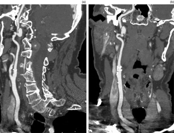
A 64-year-old male presented with a right occipital infarct. A CT angiogram revealed a right cervical ICA aneurysm at its transition with the petrous ICA (Figure 1).
(a) Lateral and (b) coronal views of pre-operative computed tomography angiograms of right cervical internal carotid artery aneurysm in patient two.

Patient three
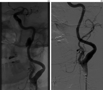
A 68-year-old male presented with headaches. A CT scan revealed a distal ICA aneurysm, which CT angiogram showed to be 1.8 cm in diameter and saccular (Figure 2). Medical history included prior abdominal aortic aneurysm repair, and bilateral popliteal and femoral artery aneurysms not requiring intervention.
Angiograms of left internal carotid artery aneurysm before (a) and after (b) stent deployment in patient three.

Intervention
Patients one and two both underwent pre-operative balloon occlusion testing, angiograms, CT and MRI. Both patients tolerated balloon occlusion testing. The distal limit of both aneurysmal sacs extended into the petrous segment of the artery. Endovascular stenting was deemed unsuitable in both patients because of vessel tortuosity.
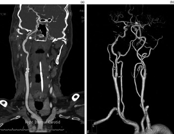
Patients one and two underwent open repair (Figure 3), involving the combined skills of a skull base otologist and a vascular surgeon. A Fisch type A approach,Reference Fisch, Fagan and Valavanis 7 including blind sac closure and facial nerve transposition, was employed for exposure. A reversed long saphenous vein graft was used in both patients, with proximal and distal anastomoses being constructed end to end.
Computed tomography angiograms of patient two following repair of right internal carotid artery aneurysm, showing: (a) coronal view of the repair and (b) a three-dimensional reconstruction.

In patient two, the distal arterial wall was friable, and, despite the exposure, suture placement and anastomotic repair was found to be technically very challenging. The facial nerve was thin and friable, and during retraction of the wound, the nerve was divided. It was repaired using a greater auricular nerve interposition graft. The patient underwent gold weight insertion and medial canthoplasty of his right eye for corneal protection during his admission.
The length of the procedure for patients one and two was 11 hours and 9 hours, and length of stay was 20 days and 19 days respectively.
Patient three underwent a stent graft repair of his aneurysm (Figure 2) using a Jostent® polytetrafluoroethylene-covered coronary stent graft measuring 5 × 26 mm over a 0.36 mm (0.014 inch) guide wire. Angiography performed immediately after stent deployment showed successful exclusion of the aneurysm sac. The length of the procedure was under 2 hours and the length of stay was 4 days.
Results
The aneurysms were successfully repaired in all patients, with no embolic or ischaemic sequelae. All patients had graft patency at follow up.
Patients one and two both experienced deficits of the facial and lower cranial nerves immediately following the operation, which recovered completely within eight and two months respectively, except the recovery of the facial nerve in patient two. At 12 months post-operation, patient two is starting to have movement in his face. Neither patient required the support of a tracheostomy or gastrostomy tube.
Patient one was found to be α1 antitrypsin deficient, and he required ongoing care by a neurologist and a vascular physician. Ultrasound examination showed graft patency 16 months post-operatively. He was implanted with a bone-anchored hearing aid (BAHA) to correct the conductive hearing loss following blind sac closure. His aided speech score was 99 per cent, compared to an unaided score of 46 per cent.
Patient two underwent a CT angiogram at six months; this revealed a 50–60 per cent distal anastomotic stenosis, which is being observed. Unfortunately, he continues to smoke.
Patient three had no neurological sequelae. Clopidogrel was added to his warfarin therapy. At six-months' follow up, CT angiogram revealed a patent stent, with a small endoleak possibly at the proximal fixation of the stent. The aneurysm sac is almost entirely excluded from flow and there is little embolic risk. Therefore, this is being monitored conservatively. An additional stent graft may be required in the future.
Discussion
Untreated internal carotid artery (ICA) aneurysms are associated with a high risk of thromboembolic sequelae,Reference Keshelava, Mikadze, Abzianidze and Kakabadze 4 with a reported mortality and stroke risk of 50 per cent.Reference Winslow 8 – Reference Windfuhr 10 These complex lesions are uncommon and require a multidisciplinary approach to management. The treatment options include occlusion (surgical or endovascular), surgical repair or bypass (including extracranial-intracranial), or endovascular repair.
Carotid occlusion carries a 24 per cent risk of ischaemic complications,Reference Landolt and Millikan 11 and pre-operative balloon occlusion testing may result in a false negative finding.Reference Liu and Couldwell 12 – Reference Sekhar and Patel 14 Carotid sacrifice also limits future treatment strategies should the patient develop pathology in the remaining extracranial arteries. This is particularly relevant as these patients may have an underlying predisposition to develop vascular disease, as seen in patients one and three. Therefore, carotid occlusion is not a preferred treatment option.
Surgical repair remains the most definitive strategy in the management of these lesions. Considerations include the anatomical exposure required, the potential for cerebral ischaemia during arterial clamping, conduit requirements and the pathology being treated.
The options for surgical repair of the aneurysm include: aneurysm resection and end-to-end anastomosis; aneurysmorrhaphy and patch angioplasty; or bypass grafting.Reference Tsai, Barot, Dalman and Mihm 15 Good results can be expected with surgical repair, with long-term stroke-free rates of 87 per cent reported at 20 years' follow up.Reference Attigah, Külkens, Zausig, Hansmann, Ringleb and Hakimi 16
The need to obtain adequate access to the ICA beyond the aneurysm can present a significant challenge (such as in patient two). For distal ICA aneurysms, a standard neck dissection will not allow adequate arterial exposure and alternative approaches are required. Since the description of infratemporal fossa exposure by Fisch et al., in 1984,Reference Fisch, Fagan and Valavanis 7 many approaches have been identified, each with various advantages and disadvantages.Reference Keshelava, Mikadze, Abzianidze and Kakabadze 4 Our exposure is based on Fisch and colleague's lateral skull base approach.Reference Fisch, Fagan and Valavanis 7
The disadvantages of the lateral skull base approach include the obligatory conductive hearing loss in the affected ear due to blind sac closure, which can be addressed by the use of a BAHA. More serious risks include temporary or permanent cranial nerve deficits. Malikov et al. reported on a series of 13 patients who all had a partial facial nerve palsy which was transient only.Reference Malikov, Thomassin, Margan, Keshalava, Bartoli and Branchereau 6 Lower cranial nerve palsy affecting speech and swallowing is often due to neuropraxia, as the lower cranial nerves may be adherent to the wall of the sac. Facial nerve palsy may occur if transposition is required, but this is often transient. If the nerve is divided, repair is required. In this case, the best expected outcome is House–Brackmann grade III palsy.Reference Tate and Tollefson 17 Trauma to the nerve is most likely to occur with the retraction required to facilitate distal arterial exposure.
In regards to defining the potential for intra-operative cerebral ischaemia, balloon occlusion testing has a role. Immediate severe cerebral ischaemia is uncommon with ICA clamping.Reference Kfoury, Patel and Mukherjee 18 If balloon occlusion of the ICA does cause significant cerebral ischaemia, this increases the complexity of the surgical repair. Shunt insertion may not be possible for distal ICA aneurysms. Use of cerebral protection strategies is a potential option. Additionally, extracranial-intracranial bypass combined with carotid occlusion can be considered.Reference Zhu, Tian, Zhou, Song, Xu and Mao 19
Management options may be limited by pathology, such as a mycotic aneurysm (patient one), requiring excision and surgical reconstruction with autogenous conduit. Mycotic aneurysmsReference Howell, Baburao and Graziano 20 – Reference Grossi, Onofrey and Tvetenstrand 22 treated non-surgically have high rates of rupture or thromboembolism, with mortality reaching 70–90 per cent.Reference Martens, Beelen and Degrieck 2 Prior to 1980, ligation was considered standard treatment,Reference Monsoon and Alexander 23 and had a mortality of 40 per cent and post-operative stroke incidence of 50–60 per cent.Reference Martens, Beelen and Degrieck 2 , Reference Monsoon and Alexander 23 , Reference Jebara, Acar, Dervanian, Chachques, Bischoff and Uva 24 More recent publications have advocated reconstruction using autogenous conduit, with non-septic mortality reported to have reduced to 4 per cent.Reference Martens, Beelen and Degrieck 2 , Reference Monsoon and Alexander 23 , Reference Jebara, Acar, Dervanian, Chachques, Bischoff and Uva 24 Mycotic aneurysms compound complexity and mortality in an already uncommon pathology. Prompt and aggressive surgical care is essential for optimal management of these lesions.
Autogenous conduit is the preferred option for carotid aneurysm reconstruction. Long saphenous vein is often well size-matched. For non-infected cases, prosthetic conduit can be used safely if autogenous conduit is not available.Reference Huilgol, Young, Lemech, Stephen, May and Harris 25
Endovascular repair results in less peri-operative morbidity than surgery.Reference Macina, Vidjak, Erdelez, Suknaic, Gašparov and Novacic 26 Furthermore, it avoids concerns regarding anatomical exposure, ICA clamping and conduit availability. Endovascular repair is not ideal for infected cases; however, it can be used as a temporising strategy, for example, to treat haemorrhage. In some cases, endovascular stents combined with antibiotic therapy can achieve successful long-term outcomes for infected aneurysms.
The options for endovascular repair include bare-metal stent placement with transstent coiling, or the use of stent grafts.Reference Heye, Maleux, Vandenberghe and Wilms 27 The limitations of endovascular repair include the requirement to obtain stable access to the target vessel and the availability of appropriate stents. Access to the target artery may be complicated by aortic arch and common carotid artery tortuosity and disease. In some cases, it may be possible to introduce the sheath through the common carotid artery in the neck rather than through the femoral artery, thus bypassing problems encountered in the aortic arch.
The ideal stent for distal ICA aneurysm repair would have a low profile, be compatible with 0.36 mm (0.014 inch) guide wire, have good trackability and have a monorail delivery system. Balloon expandable stent grafts can achieve good results; however, long and tortuous aneurysms may not respond well. Furthermore, stents may not be available to treat ICAs with large diameters or significant diameter changes between proximal and distal landing zones.
Future advances in stent design may overcome these problems. Despite these limitations, for suitable lesions, such as in patient three, endovascular repair is an attractive option and can yield good results.Reference Li, Chang, Yao, Guo, Liu and Wang 5 , Reference Zhou, Lin and Bush 28 Successful graft patency rates of 93.2 per cent at 15 months, stroke rates of 1.8 per cent and endoleak rates of 8.1 per cent have been reported.Reference Li, Chang, Yao, Guo, Liu and Wang 5
-
• Extracranial internal carotid artery aneurysms are rare, life-threatening lesions, with a high risk of thromboembolic sequelae and haemorrhage
-
• Treatment options include occlusion, surgical repair or bypass, or endovascular repair
-
• Surgical repair is a well-established, definitive management strategy for these lesions, despite some risk of long-term morbidity
-
• Endovascular stenting is a new, less invasive treatment modality for the appropriate patient, with good results
-
• Long-term results of endovascular stenting are lacking, and successes and failures should continue to be reported
In summary, the optimal treatment strategy for distal ICA aneurysms involves careful consideration of factors that are critical to each approach. In many cases, a preferred option will become evident. In equivocal cases, endovascular repair has short-term morbidity advantages; however, the long-term results are not known.
Conclusion
Future treatment of these rare and complex lesions will involve the complimentary use of surgical and endovascular techniques. To enable a better understanding of the strengths, limitations and outcomes of these techniques, it is essential that both successes and failures continue to be reported in the literature.
Acknowledgement
The authors would like to acknowledge Dr Richard Gallagher for his valuable role in managing the care of some of these patients.