Introduction
In 1978, Edwards and Henkel described a possible connection between the superior colliculus and the oculomotor nucleus (III) in the cat. They observed labeled terminals distributed in the periaqueductal gray (PAG) dorsal to III on both sides of the midline following tritiated amino acid injections into the superior colliculus. They termed this ventral portion of the PAG, the supraoculomotor area (SOA), based on its proximity to III, and suggested that this region was likely involved in oculomotor behavior considering the presence of the tectal terminal field. Their examination of retrograde labeling patterns following injections into SOA in the cat showed that this pathway derived from cells located primarily in the intermediate gray layer (SGI) of the rostral third of the superior colliculus. Based on the bilateral distribution of tectal terminals and the fact that the vertical meridian is represented in the rostral colliculus, they proposed that the likely targets of this projection are the dendrites of superior and inferior rectus motoneurons, which are bilaterally activated during vertical saccades. Intra-axonal staining in cats revealed that this terminal field actually represents a collateral projection of predorsal bundle axons that branches from the main axons as they run ventrally in the midbrain on the way to their decussation (Grantyn & Grantyn, Reference Grantyn and Grantyn1982). The predorsal bundle contains axons of crossed tectobulbospinal neurons that are the main output by which the superior colliculus directs gaze changes. A terminal field in the neuropil dorsal to III was also labeled in macaque monkeys following tritiated amino acid injections into the superior colliculus (Harting et al., Reference Harting, Huerta, Frankfurter, Strominger and Royce1980). Although the target of these terminals was described as the “visceral motor columns” of the Edinger–Westphal nucleus, the location was equivalent to that of the SOA. Axon collaterals of predorsal bundle fibers terminating in SOA were not noted following intracellular injections of collicular output cells in monkeys, although collaterals extending in this direction were observed (Moschovakis et al., Reference Moschovakis, Karabelas and Highstein1988a,Reference Moschovakis, Karabelas and Highsteinb).
Since this initial work, our understanding of the inputs and contents of the SOA has been extended. In monkeys, inputs to the SOA have been described from the deep cerebellar nuclei (May & Porter, Reference May and Porter1992; Bohlen et al., Reference Bohlen, Gamlin, Warren and May2021) central mesencephalic reticular formation (Bohlen et al., Reference Bohlen, Warren and May2016) and pretectum (Wasicky et al., Reference Wasicky, Horn and Büttner-Ennever2004). In cats, this region contains oculomotor internuclear cells projecting to the abducens nucleus (Maciewicz et al., Reference Maciewicz, Kaneko, Highstein and Baker1975; May et al., Reference May, Baker, Vidal, Spencer and Baker1987), but these cells are mainly found within III in monkeys (Langer et al., Reference Langer, Kaneko, Scudder and Fuchs1986). A population of peptidergic and nitridergic neurons that are believed to play a role in stress, and in eating and drinking behavior (Kozicz et al., Reference Kozicz, Yanaihara and Arimura1998; Ryabinin & Weitemier, Reference Ryabinin and Weitemier2006) is located in the monkey SOA (Horn et al., Reference Horn, Eberhorn, Härtig, Ardeleanu, Messoudi and Büttner-Ennever2008; May et al., Reference May, Reiner and Ryabinin2008; Erichsen & May, Reference Erichsen and May2012). This set of neurons, which project widely in the brain, has been designated the centrally projecting Edinger–Westphal population (EWcp) in contradistinction to the preganglionic Edinger–Westphal population (EWpg) that supplies the ciliary ganglion (Kozicz et al., Reference Kozicz, Bittencourt, May, Reiner, Gamlin, Palkovits, Horn, Toledo and Ryabinin2011). The EWpg is contained within SOA, and preganglionic motoneurons send dendrites out into SOA (May et al., Reference May, Warren, Gamlin and Billig2018). The ventral aspect of the SOA of monkeys has been shown to contain a specialized group of motoneurons located in the C-group, capping III (Büttner-Ennever & Akert, Reference Büttner-Ennever and Akert1981; Büttner-Ennever et al., Reference Büttner-Ennever, Horn, Scherberger and D’Ascanio2001). The C-group contains motoneurons supplying the medial and inferior rectus muscles. Similarly, the S-group, sandwiched between the two sides of III and located ventral to the SOA, contains motoneurons supplying the superior rectus and inferior oblique muscles. The S- and C-group motoneurons supply extraocular muscle fibers of a specific type, termed multiply innervated fibers (MIFs), due to the fact that the axons innervating them make numerous contacts along the length of the fiber (Spencer & Porter, Reference Spencer and Porter2006). In contrast, the motoneurons within III supply singly innervated fibers (SIFs) that are each contacted by a single, plate-like ending. The role of MIFs is not well understood. There is evidence that they may have graded activity, so they have sometimes been termed “nontwitch” muscle fibers (Chiarandini & Stefani, Reference Chiarandini and Stefani1979; Bondi & Chiarandini, Reference Bondi and Chiarandini1983; Nelson et al., Reference Nelson, Goldberg and McClung1986), although a portion of them also receives a plate-shaped ending near their midpoint, and these do display action potentials (Jacoby et al., Reference Jacoby, Chiarandini and Stefani1989). MIF motoneurons in cats have been shown to be active in all types of eye movements, but their firing rates differ from SIF motoneurons (Hernández et al., Reference Hernández, Calvo, Blumer, de la Cruz and Pastor2019). Finally, the dendrites of levator palpebrae superioris (levator) motoneurons, which control the position of the upper eyelid, and which reside in the caudal central subdivision of III (CC), extend their dendrites into SOA (Porter et al., Reference Porter, Burns and May1989; May et al., Reference May, Vidal, Baker and Baker2012).
Our conception of the functions of the rostral superior colliculus has also been modified since the work of Edwards and Henkel (Reference Edwards and Henkel1978). Collicular cells that fire tonically during fixation have been described in the rostral colliculus of the monkey and cat (Munoz & Wurtz, Reference Munoz and Wurtz1993a,Reference Munoz and Wurtzb; Peck & Baro, Reference Peck and Baro1997). Activity related to microsaccades has been found in this same region in monkeys (Hafed et al., Reference Hafed, Goffart and Krauzlis2009; Hafed & Krauzlis, Reference Hafed and Krauzlis2010). Buildup cells in this region are believed to play a role in pursuit initiation (Krauzlis, Reference Krauzlis2003). Furthermore, there is also considerable evidence from studies in cats for a role of the rostral colliculus in vergence and in lens accommodation (Sato & Ohtsuka, Reference Sato and Ohtsuka1996; Ohtsuka & Sato, Reference Ohtsuka and Sato1997; Ohtsuka & Nagasaka, Reference Ohtsuka and Nagasaka1999; Suzuki et al., Reference Suzuki, Suzuki and Ohtsuka2004). This has been reinforced by experiments in monkeys showing stimulation or inactivation of the rostral colliculus affects vergence (Chaturvedi & Van Gisbergen, Reference Chaturvedi and Van Gisbergen2000; Van Horn et al., Reference Van Horn, Waitzman and Cullen2013), and recording experiments that indicate it contains cells whose activity correlates with vergence angle (Cullen & Van Horn, Reference Cullen and Van Horn2011; Van Horn et al., Reference Van Horn, Waitzman and Cullen2013; Upadhyaya & Das, Reference Upadhyaya and Das2019; although see Walton & Mays, Reference Walton and Mays2003 for a different opinion). There is little evidence with respect to whether these newly described collicular populations have different downstream targets than saccade-related neurons (Büttner-Ennever et al., Reference Büttner-Ennever, Horn, Henn and Cohen1999).
In view of the several possible target populations within the SOA and the variety of possible sources of input resident in the rostral superior colliculus, we undertook a stepwise investigation of this tectal projection and its targets in macaques. The first step was to morphologically characterize the axonal arbors supplying the tectosupraoculomotor projection utilizing contemporary anterograde tracers. We then examined the relationship of the tectal terminal boutons to cells with descending outputs likely to belong to the EWcp population. In addition, we felt it was reasonable to reinvestigate the initial proposition that this projection to SOA targets vertical gaze motoneurons, by examining tectal inputs to superior rectus motoneurons and levator motoneurons. As a control for the vertical gaze hypothesis, we also tested for inputs to medial rectus motoneurons. Moreover, since medial rectus MIF motoneurons are located within the SOA, we examined the possibility of tectal input to these cells. In all these cases, we used dual tracer techniques in which an anterograde tracer was placed in the superior colliculus and a retrograde tracer was placed in another target. Finally, we investigated the morphology and topography of the cells of origin of this projection by use of retrograde labeling.
Materials and methods
Data from 18 Macaca fascicularis monkeys was used in this study. These included nine female and nine male adult and young adult monkeys. No sex-specific patterns were noted. The protocols were approved by the Institutional Animal Care and Use Committee of the University of Mississippi Medical Center. Surgeries took place in a dedicated surgical suite using sterile technique. Animals were sedated with ketamine HCL (10 mg/kg, IM), and they received a preemptive analgesic, Carprofen (3 mg/kg. IM). Following intubation, they were anesthetized with isoflurane (3%) for the surgical procedures, which took place with their heads in a stereotaxic frame (Kopf Instruments, Tajunga, CA). Atropine HCl (0.05 mg/kg, IM) was given to suppress respiratory tract secretion and dexamethasone (2.5 mg/kg, IV) was given to avoid edema. At the end of surgery, the wound edges were infiltrated with Sensorcaine, and the animals received Buprenex (0.001 mg/kg) as a postsurgical analgesic.
In order to examine tectal projections to the peptidergic EWcp population, we took advantage of the fact that these cells project widely within the brainstem and spinal cord (cat: Maciewicz et al., Reference Maciewicz, Phipps, Foote, Aronin and DiFiglia1983, Reference Maciewicz, Phipps, Grenier and Poletti1984; cebus monkey: Vasconcelos et al., Reference Vasconcelos, Donaldson, Sita, Casatti, Lotfi, Wang, Cadinouche, Frigo, Elias, Lovejoy and Bittencourt2003). We reasoned that SOA cells retrogradely labeled from the brainstem and spinal cord most likely belonged to the EWcp population. To this end we re-examined eight cases in our collection that had collicular injections of the anterograde tracers biotinylated dextran amine (BDA, n = 6) (Molecular Probes/Thermo Fisher, Waltham, MA), or Phaseoulus vulgaris leucoagglutinin (PhaL, n = 2) (Sigma Aldrich, St. Louis, MO), and injections of retrograde tracer, wheat germ agglutinin conjugated horseradish peroxidase (WGA-HRP) (Sigma Aldrich, St. Louis, MO) in the spinal cord (n = 2), medullary reticular formation (n = 4) or pontine reticular formation (n = 2). Details of these cases can be found elsewhere (Perkins et al., Reference Perkins, Warren and May2009; Wang et al., Reference Wang, Perkins, Zhou, Warren and May2013, Reference Wang, Perkins, Zhou, Warren and May2017). An additional case that just had a pontine reticular formation injection of WGA-HRP was also used.
The surgical approach for midbrain injections was as follows. After a midline scalp incision and craniotomy, the cerebral cortex overlying the midbrain was aspirated, revealing the surface of the superior colliculus and the caudal pole of the pulvinar. To inject the superior colliculus with 10,000 MW BDA (n = 11), a sharpened, 1.0 μl Hamilton syringe filled with a 10% BDA solution was driven to points 1.5–2.0 mm beneath the collicular surface in order to center its tip within the intermediate gray layer (SGI). The syringe was held in a micromanipulator at an angle of 20°, tip rostral in the sagittal plane, to compensate for the angle of the brainstem. An injection bolus of 0.1–0.2 μl was placed at 1–3 sites across the colliculus. To inject the superior colliculus with PhaL (n = 2), a solution of 2% PhaL in 0.1 m, pH 8.0 phosphate buffer (PB) was placed in a glass micropipette with a 15–25 μm diameter tip and injected into the colliculus by passing 7 μA of positive current for 7 minutes using a 50% duty cycle. To inject SOA, we widened the aspiration area medially to reveal the posterior commissure. Then a 1.0 μl Hamilton syringe, whose needle had been insulated with polyethylene varnish, was introduced at the midline, immediately rostral to the posterior commissure. It was held in a micromanipulator, angled 12° tip rostral in the sagittal plane and 2° tip medial in the frontal plane. The needle was driven through the third ventricle, so that it entered the PAG immediately above III. Electrical stimulation was used to produce eye movements and confirm proper localization (burst duration = 2.5–5.0 ms, pulse duration = 25–75 μs, current = 1–4 mA). Then 0.1 μl of 10% BDA (n = 1) or 0.02 μl of 2% WGA-HRP (n = 3) was injected. In all the midbrain injection cases, the aspiration defect was filled with gelfoam and the skin was reapproximated and stabilized with suture. Survival times varied with respect to the tracer used: BDA—17–21 days, PhaL—14 days, and WGA-HRP—2 days.
In the case of the dual tracer motoneuron experiments (n = 5), the collicular injection approach described above was followed by an injection of one or more extraocular muscles. An incision was made behind the brow and the eyelid skin was pulled forward. The orbicularis oculi muscle was disinserted from the supraorbital ridge. The target muscles were then isolated and stabilized with suture. A solution containing 2% choleratoxin subunit B conjugated to horseradish peroxidase (ChTB-HRP) (Sigma Aldrich, St. Louis, MO) (n = 4) or 2% WGA-HRP (n = 1) was injected using a 10 μl Hamilton syringe. A bolus of 5–10 μl of tracer was placed into one or more of the following: superior rectus muscle (n = 5; two on the left side and three on the right), left medial rectus muscle (n = 2), and levator muscle (n = 5; two on the left side and three on the right). The insertion of the orbicularis oculi muscle was re-established and the incision was closed with suture.
At the end of the survival period, animals were sedated with ketamine HCL (10 mg/kg, IM) and then euthanized following an IP injection of 50 mg/kg sodium pentobarbital. Once unresponsive, they were perfused with a buffered saline rinse, followed by a fixative containing 1–2% paraformaldehyde and 1.0–1.5% glutaraldehyde in 0.1 m, pH 7.2 PB. The brainstem was blocked in the frontal plane, postfixed for 1 h in the aldehyde solution, and stored at 4°C in 0.1 m, pH 7.2 PB.
Using a Leica VT100S vibratome (Leica Microsystems, Buffalo Grove, IL) the brainstem and spinal cord was sectioned at 100 μm for the BDA experiments and 50 μm for the PhaL experiments. The histochemistry used to reveal the tracers has been described previously (Chen & May, Reference Chen and May2007; Wang et al., Reference Wang, Perkins, Zhou, Warren and May2013). Briefly, to reveal the BDA, sections were exposed to a solution of avidin-HRP (0.2%) in 0.1 m, pH 7.2 PB at 4 °C overnight. After rinsing, they were reacted using diaminobenzidene (DAB) as the chromagen, with the reaction initiated by H2O2. Nickel ammonium chloride and cobalt chloride were added to intensify the reaction product by giving it a black color. To reveal the PhaL, sections were incubated in biotinylated anti-PhaL (Vector Labs) (1:2,000) in 0.1 m, pH 7.2 PB at 4°C overnight. They were then processed using an ABC kit (Vector Labs). Nickel/cobalt intensified DAB was again used as a chromagen. To reveal WGA-HRP or ChTB-HRP, sections were reacted with the chromagen, tetramethylbenzidene (TMB) in 0.1 m, pH 6.0 PB with 0.25% ammonium molybdate at 4°C overnight, with the reaction initiated by H2O2. In the dual tracer experiments, the sections were first reacted in TMB to display the retrogradely labeled cells. Then, the TMB reaction product was stabilized by incubating with the DAB solution without nickel/cobalt intensification. Finally, they were processed to reveal the BDA labeled axons, as described above. All sections were counterstained with cresyl violet before being coverslipped for light microscopy.
At a minimum, a series of one in three sections was processed and analyzed per case. The pattern of labeling was illustrated by use of an Olympus BH-2 microscope equipped with a drawing tube (Olympus, Center Valley, PA). A Wild M8 stereoscope with a drawing tube (Leica, Microsystems, Buffalo Grove, IL) was used to draw low magnification views. Illustrative examples were photographed using a Nikon Eclipse E-600 photomicroscope with a Nikon Ds-Ri1 digital camera (Nikon Instruments, Inc., Melville, NY). Images were obtained using Nikon Elements software, which allows multiple focal planes to be combined into a single image. They were adjusted for color and contrast to match the appearance in the microscope by use of Adobe Photoshop (Adobe, San Jose, CA).
Results
Collicular projections to SOA
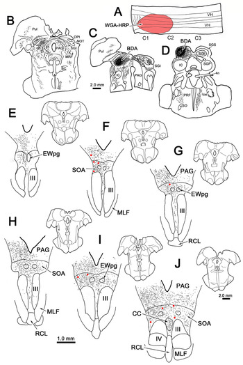
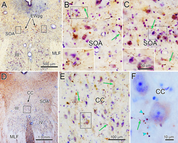
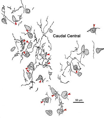
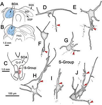
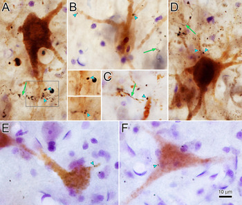
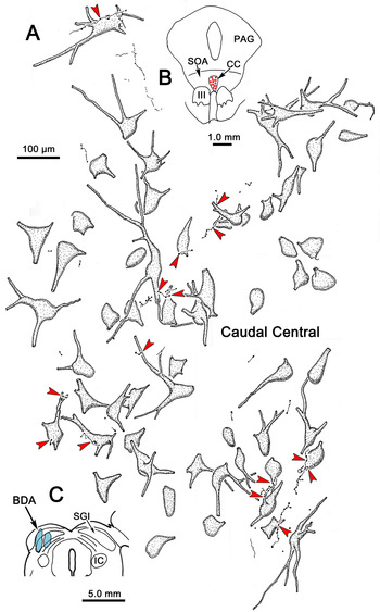
The first goal of the study was to characterize the distribution and morphology of the tectal axonal arbors within SOA. Fig. 1 shows a case with a large BDA injection of the left superior colliculus that was centered in the intermediate gray layer (SGI) and extended from the superficial to the deep gray layer (SGS & SGP) (Fig. 1C,D). It was well contained within the colliculus. Specifically, the injection site spread from the caudal to rostral pole of the target, but did not extend into the pretectum (Fig. 1B), and only spared the medial and rostralmost edge of the colliculus. Labeled axons ran ventrally through the midbrain reticular formation, and terminals were present ventrolaterally in the PAG, primarily on the ipsilateral side (Fig. 1E–J), and in the SOA, bilaterally (Fig. 1E–J). The density of the terminal field intensified at more caudal levels. Few terminals were observed within the confines of the oculomotor nucleus proper (III). However, relatively dense terminal label was encountered within the caudal central subdivision (CC), where levator motoneurons are found (Fig. 1J). Scattered terminals were also observed between the two sides of III, and within the boundaries of the preganglionic Edinger–Westphal nucleus (EWpg) (Fig. 1E–J). Photomicrographs in Fig. 2A–C provide a view of the terminal arbors in SOA from this case. Branch points were rarely observed on labeled axons (Fig. 2B,C). They primarily displayed en passant boutons. These boutons were mainly small, but some were larger. We were struck by the degree of terminal label in CC, and examined this structure in a more detailed fashion. As shown in Fig. 2D–F, this structure contained numerous BDA labeled tectal boutons. An example of a terminal arbor in which boutons (arrowheads) were located in the vicinity of a counterstained soma in CC is shown in Fig. 2F. The extensive nature of terminal arborization made in the CC by BDA labeled tectal axons is further demonstrated in Fig. 3. Note the numerous close associations (arrowheads) with counterstained cells, presumed to be levator motoneurons due to their size.
Distribution of superior colliculus projections to the macaque SOA. A BDA injection confined to the left superior colliculus (C,D) produced anterogradely labeled axonal arbors (stipple) bilaterally within SOA (E–J), and in EWpg (E–J) and CC (J). BDA labeled tectotectal neurons are indicated by black dots (B–D). A large injection of WGA-HRP placed in the upper cervical cord (A) produced a sparse distribution of retrogradely labeled cells (red dots) within SOA. Low magnification insets (E–J) are provided for reference to the progression of the chartings, which in this and other illustrated cases is rostral to caudal.

Morphology of BDA labeled tectal axons in SOA and CC (same case as Fig. 1). The left and right boxes in (A) indicate the areas of SOA shown in (B,C), respectively. Numerous axonal arbors (green arrows) are present on both sides. Boxes in (B,C) indicate regions shown at higher magnification in insets. The box in (D) indicates the region of CC shown at higher magnification in (E). Numerous axonal arbors (green arrows) are present in this subdivision. The boxed area in (E) is shown at higher magnification in (F), where the boutons (blue arrowheads) can be observed in the vicinity of a counterstained, presumptive motoneuron. Scale in (E) = (B,C). Scale in inset (C) = inset (B).

Tectal axonal arbors in CC. The relationship of BDA labeled axons from a collicular injection (same case as Fig. 1) to counterstained, presumptive levator motoneurons is illustrated. Many boutons displayed close associations (red arrowheads) with counterstained somata.

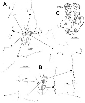
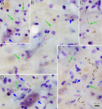
BDA can label axons of passage, so we confirmed the pattern of tectal termination using PhaL, a tracer with little propensity for axonal uptake (Gerfen & Sawchenko, Reference Gerfen and Sawchenko1984). Fig. 4C shows the location of a small PhaL injection site located in SGI, toward the caudal end of the superior colliculus. As might be expected for a much smaller injection site, far fewer labeled axons were present in the predorsal bundle (not illustrated). Nevertheless, PhaL labeled axonal arbors were found bilaterally within SOA (Fig. 4A1–7,B1 and 4). Some extended in from the PAG (Fig. 4B2). They were occasionally present in the region between III, as well (Fig. 4B3), but were not found within III, itself. As with the BDA labeling, the axons displayed few branches, and most of their boutons were en passant in nature. These features can also be observed in photomicrographs from this case (Fig. 5A–E). Note that while the labeled axons are generally of fine caliber, with small boutons (Fig. 5A–D), some were thicker and had larger boutons (Fig. 5E). This injection, and a similar PhAL case with a caudolateral injection, produced only a few labeled arbors in CC.
SOA axons labeled from the caudal superior colliculus. A small PhaL injection centered in SGI of the caudal left superior colliculus (C) labeled axons within the SOA. (A,B) The SOA region in two example sections is illustrated in the insets. The locations of individual numbered axonal arbors are indicated on the insets by arrows. Most were located in the SOA, but one example (B3) was located medial to III.

Morphology of PhaL labeled tectal axons in SOA (same case as Fig. 4). The labeled axons (thin green arrows) extended through the substance of the SOA, and were characterized by numerous en passant boutons. Most axons and boutons were quite fine (A–D), but some thicker examples (E) were observed. Branch points (pink arrowheads) were relatively rare (B,D,E). In most cases, the boutons displayed no organized relationship with counterstained somata (thick blue arrows) (A,B,D), including the large diameter presumed preganglionic motoneuron shown in (D). Scale in (E) = (A–D).

Comparison of the distribution of the tectosupraoculomotor axonal arbors across cases did not suggest a topographic projection; smaller injections involving just portions of the medial, lateral or caudal colliculus produced fewer labeled arbors, not a distribution limited to a subpart of SOA. Moreover, the morphology of the arbors does not suggest that individual axons concentrate their input on specific cells, but instead suggests that they distribute a few boutons to each of a number of neurons.
Relationship of tectal SOA projection to EWcp neurons
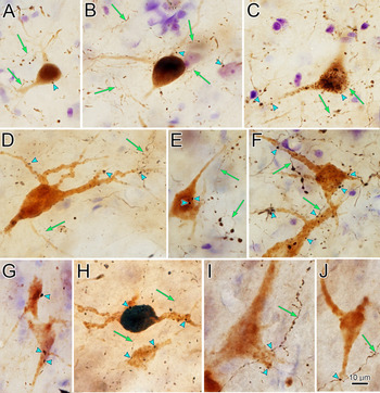
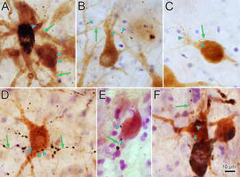
The case illustrated in Fig. 1 also had a large injection of WGA-HRP that encompassed much of the left cervical spinal cord between C1 and C2, and spread across the midline (Fig. 1A). It resulted in a sparse distribution of retrogradely labeled cells in the SOA (Fig. 1F,G,I,J, red dots). It is likely that these represent cells in the EWcp, a population found in the SOA, which is known to have widespread projections that include spinal cord (Kozicz et al., Reference Kozicz, Bittencourt, May, Reiner, Gamlin, Palkovits, Horn, Toledo and Ryabinin2011). As can be seen here, individual sections had only a few labeled cells, scattered on both sides of the midline. This suggests that while the projections of this peptidergic population as a whole may be widespread, only a few cells may be responsible for targeting any individual area. Overlap in the distributions of labeled tectal axonal arbors (stipple) and this set of cells is apparent in the illustration. This relationship is better appreciated in Fig. 6A–C. The neuropil contained numerous BDA labeled axons (arrows). In some cases, the boutons of these labeled axons lay in close association (arrowheads) with retrogradely labeled somata or dendrites, but most labeled boutons did not show any apparent relationship to the retrogradely labeled cells.
Close associations between BDA labeled tectal boutons and descending output neurons retrogradely labeled by WGA-HRP. These brown, retrogradely labeled cells displayed close associations (blue arrowheads) with boutons of BDA labeled tectal axons (green arrows). (A–C) Examples of retrogradely labeled neurons from a cervical spinal cord injection (case shown in Fig. 1). (D–F) Examples of retrogradely labeled neurons from two different medullary reticular formation injection cases. (G–J) Examples of retrogradely labeled neurons from two different pontine reticular formation injection cases. Scale in (J) = (A–I).

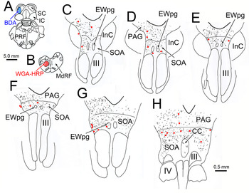
A similar pattern of relationships was apparent for cells retrogradely labeled from medullary reticular formation injections of WGA-HRP. An injection in the center of the left medullary reticular formation (Fig. 7B) that extended rostocaudally through a substantial portion of the medulla, only labeled a small number of cells in SOA, and these were scattered on either side of the midline (Fig. 7C–H). The labeled cells lay within the SOA terminal field (stipple) (Fig. 7C–H) produced by a BDA injection into the caudal end of the left superior colliculus (Fig. 7A). A few labeled terminals were also present in EWpg (Fig. 7C–G), and in CC (Fig. 7H). The labeled tectal terminals in SOA often showed close associations (arrowheads) with retrogradely labeled neurons in this (Fig. 6D,E) and other medullary reticular formation injection cases (Fig. 6F).
Overlapping distributions of tectal terminals and SOA cells projecting to the medullary reticular formation. An injection of WGA-HRP centered in the left medullary reticular formation (B) retrogradely labeled scattered cells (red dots) in SOA (C–H). Most were ipsilateral. An injection of BDA in the caudal left superior colliculus (A) labeled terminals (stipple) throughout SOA (C–H), within EWpg (C–G) and in CC (H).

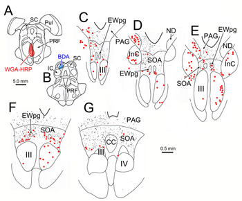
Once again, retrogradely labeled neurons (red dots) were present in the SOA (Fig 8C–G) following an injection of WGA-HRP into the left side of the pontine reticular formation that extended slightly across the midline (Fig. 8A). As this injection was centered in the paramedian pontine reticular formation and included the nucleus raphe interpositus, retrogradely labeled neurons were also present in gaze-related nuclei, including the interstitial nucleus of Cajal and the nucleus of Darkschewitsch (Fig. 8D,E). In this case, the BDA injection was located in the caudal pole of the left superior colliculus (Fig. 8B), and it produced a bilateral SOA terminal field (stipple) that overlapped with the distribution of the retrogradely labeled cells (Fig. 8C–G). BDA labeled axonal arbors were also present in CC (Fig. 8G) and within EWpg (Fig. 8C–F). The BDA labeled tectal terminals in SOA displayed a number of close associations with the cells projecting to the pontine reticular formation in this case (Fig. 6G) and another (Fig. 6I,J).
Tectal terminals overlap with SOA cells projecting to the pontine reticular formation. An injection of WGA-HRP centered near the midline in the left pontine reticular formation, and spreading slightly to the other side (A) retrogradely labeled scattered cells (red dots) in the SOA (C–G). There was a striking bilateral population of retrogradely labeled cells found just dorsal to the caudal pole of III (F). An injection of BDA in the caudal left superior colliculus (B) labeled terminals (stipple) throughout the SOA (C–G), and within EWpg (C–F) and CC (G).

This particular pontine injection produced one unexpected outcome, a dense bilateral distribution of retrogradely labeled neurons located just above the caudal end of III (Fig. 8F,G). We believe these caudal SOA cells were labeled because the needle track passed through the medial longitudinal fasciculus. In agreement with this supposition, this injection also labeled oculomotor internuclear neurons within III, mostly contralateral to the injection (Fig. 8C–F), a distribution matching that seen after abducens nuclei injections in the monkey (Langer et al., Reference Langer, Kaneko, Scudder and Fuchs1986). It is thought that oculomotor internuclear neurons project to the abducens nucleus via axons that run in or near the medial longitudinal fasciculus (cat: Maciewicz et al., Reference Maciewicz, Kaneko, Highstein and Baker1975), and it seems reasonable to propose that these SOA cells were also labeled by tracer spread along the injection needle track. Indeed, examination of a case in our collection (not illustrated) that did not have a collicular injection, but did have a medial pontine reticular formation injection that included the abducens nucleus, revealed numerous labeled cells in the caudal ventral SOA. It is reasonable to suggest that these are the cells in SOA that fire for divergent eye movements (Mays, Reference Mays1984; Judge & Cumming, Reference Judge and Cumming1986; Das, Reference Das2011; Pallus et al., Reference Pallus, Walton and Mustari2018) and project to the abducens nucleus (Ugolini et al., Reference Ugolini, Klam, Doldan Dans, Dubayle, Brandi, Büttner-Ennever and Graf2006). Furthermore, it is likely that divergence cells are among the premotor neurons controlling the lens accommodation component of the near triad, which are particularly numerous in the region containing labeled cells (Fig. 8F) (May et al., Reference May, Warren, Gamlin and Billig2018). Labeled collicular boutons were also observed in close association with these neurons found in caudal SOA (Fig. 6H).
Looking across the eight cases, the retrogradely labeled cells in SOA had relatively small somata (12–20 μm, mean—15 μm), similar to those of EWcp cells labeled with antibody to urocortin (Horn et al., Reference Horn, Eberhorn, Härtig, Ardeleanu, Messoudi and Büttner-Ennever2008; May et al., Reference May, Reiner and Ryabinin2008). The injections in all these cases labeled similar, bilaterally distributed, sparse cell populations even when the injections were unilateral. As shown here, only a few close associations were observed with any particular retrogradely labeled cell, either on its primary dendrites (Fig. 6B–D,F–I), secondary dendrites (Fig. 6D) or somata (Fig. 6A,C,E,G). A similar pattern of relationships was seen when tectal axons were labeled with PhaL and retrograde tracer was placed in the medullary reticular formation (not illustrated).
Relationship of tectal boutons and extraocular motoneurons
Fig. 9 shows the results from an example dual tracer case in which several injections of BDA were made within the superior colliculus (Fig. 9A,B) to label most of the tectal projection to the SOA. In addition, ChTB-HRP was placed in the right superior rectus muscle of this case. Retrogradely labeled motoneurons (red dots) were located in III and in S-group, which lies between the two oculomotor nuclei (Fig. 9C). Close associations were not observed between tectal terminals and SIF motoneurons in III. However, the superior rectus MIF motoneurons in S-group displayed very infrequent close associations (arrowheads) with anterogradely labeled tectal terminals (Fig. 9D–J). Rarely (Fig. 9J) were more than two close associations observed on an individual cell. In fact, the vast majority of S-Group motoneurons lacked close associations. Photomicrographs of these close associations (arrowheads, Fig. 10A–D) suggest the possibility of synaptic contact. However, in some cases (Figs. 9E and 10A), a through focus series (insets right of A) indicated the boutons and cell lay at different focal levels, suggesting the two were adjacent, but not in contact. In four of the five cases with superior rectus muscle injections, BDA labeled terminals were not observed in close association with retrogradely labeled superior rectus SIF motoneurons within III. In one case (not illustrated) a small region with close associations was observed. These appeared to all be emitted from collaterals of a thick axon extending from the MLF. Since the morphology of this axon did not match the others labeled in any of the other cases, we concluded that this terminal labeling in III probably represented staining of a nontectal axon.
Limited close associations between labeled tectal boutons and S-group motoneurons. (A,B) This case had a large BDA injection in the left superior colliculus with some spread into the periaqueductal gray. (C) A ChTB-HRP injection of the left superior rectus muscle labeled motoneurons (red dots) in III, and in the S-group, found on the midline between III. (D–J) Examples of retrogradely labeled superior rectus MIF motoneurons in S-group that displayed close associations (red arrowheads) with BDA labeled tectal axons. While S-group neurons with close associations are illustrated here, the vast majority of S-group cells lacked close associations with BDA labeled terminals.

Close associations (blue arrowheads) between tectal boutons and MIF motoneurons. (A–D) Examples of brown, retrogradely labeled superior rectus motoneurons in S-group (same case as Fig. 9). Green arrows indicate BDA labeled axons. Area of box in (A) is shown at two focal planes to the right, indicating that terminals in these presumed close associations actually lie above the dendrite. (E,F) Two rare examples of C-group medial rectus motoneurons (same case as Fig. 11) with possible close associations with BDA labeled tectal boutons. Scale in (F) = (A–E).

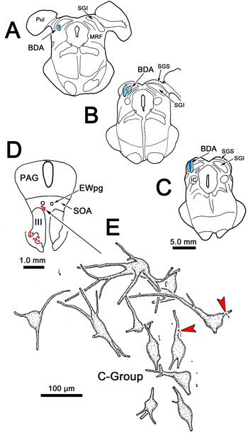
Since most of the tectal terminals lie in the SOA above the oculomotor nucleus, where the C-group is found, we also examined two cases in which the left medial rectus muscle was injected, producing retrogradely labeled motoneurons (red dots) in III and in C-group (Fig. 11D). In the example shown, the BDA injection sites occupied much of the lateral aspect of the left SGI, but did not spread outside the confines of the superior colliculus (Fig. 11A–C). This injection resulted in terminal labeling within SOA, but not within III (not illustrated). The SOA terminal field was actually sparser in the region containing the C-group motoneurons. In one of the two medial rectus injection cases, a very small number of terminals displayed close associations with a just handful of the retrogradely labeled MIF motoneurons in C-group (Fig. 11E). Two examples of these rare associations are shown in Fig. 10E,F. In view of the extremely limited evidence for close associations, we concluded that there is no substantive evidence that the collicular input targets medial rectus MIF motoneurons.
Relationship of labeled tectal boutons and C-group motoneurons. (A–C) Several BDA injections were made to include most of the left superior colliculus. (D) The location of the labeled medial rectus motoneurons (red dots) included MIF motoneurons found in the C-group following an injection of ChTB-HRP in the left medial rectus muscle. (E) Only a very small number of motoneurons displayed possible close associations (red arrowheads) with BDA labeled boutons.

Tectal projections onto levator motoneurons
More terminals were found in CC following this same injection (Fig. 12C). The levator muscle had also been injected with tracer in this case, resulting in retrogradely labeled motoneurons (red dots) throughout CC (Fig. 12B). The BDA labeled tectal terminals often displayed close associations (arrowheads) with the proximal dendrites and somata of the retrogradely labeled levator motoneurons (Fig. 12A). As shown in Fig. 13A–C, labeled axons (arrows) from this same case extended through CC providing multiple cells with a few associations, instead of being concentrated on individual cells. Similar densities of contact were present in other cases (Fig. 13D–F). These close associations (arrowheads) were located on both the somata (Fig. 13A,C–F) and proximal dendrites (Fig. 13A–C) of the retrogradely labeled levator motoneurons.
Close associations between tectal boutons and levator motoneurons. Retrogradely labeled levator motoneurons (red dots) are found in CC, located above the caudal pole of III (B). An injection of BDA into the left superior colliculus (C) (same case as Fig. 11) labeled axons within CC. (A) The boutons on these axons often showed close associations (red arrowheads) with retrogradely labeled levator motoneurons.

Relationship of BDA labeled boutons and ChTb-HRP labeled levator motoneurons. The axons labeled from tectal injections (green arrows) displayed numerous en passant boutons. Close associations (blue arrowheads) were present between these boutons and the dendrites (A–C) and somata (A,C–F) of the retrogradely labeled levator motoneurons. (A–C) Examples from the case illustrated in Fig. 12. (D–F) Examples from two other cases. Scale in (F) = (A–E).

Cells of origin of the tectosupraoculomotor projection
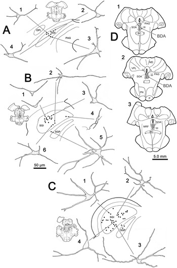
Subsequently, we turned our attention to the cells of origin of the tectal projection to the SOA. Fig. 14D shows an injection of BDA into III that spread up to the cerebral aqueduct to include SOA. It also extended a short ways beneath III. Within the superior colliculus, numerous retrogradely labeled cells (dots) were found in SGI, and a small number were located deep to SGI in the intermediate white (SAI) and the deep gray (SGP) layers (chartings, Fig. 14A–C). They were distributed throughout the mediolateral extent of SGI, and were, if anything, more numerous caudally. In three animals, we also attempted to retrogradely label cells following a WGA-HRP injection into III (not illustrated). In one case, we saw no labeled cells in the superior colliculus, and in the other two we observed just a handful of lightly labeled cells scattered throughout SGI, although other sources of input to III were well labeled. Perhaps the fact that the SOA projection is produced by thin collaterals of predorsal bundle axons (Grantyn & Grantyn, Reference Grantyn and Grantyn1982) makes it difficult to label the cells of origin of this projection with WGA-HRP. Also, it has been reported that not all axon terminals take up this tracer with equal avidity (Robertson & Grant, Reference Robertson and Grant1985).
Distribution and morphology of tectal neurons projecting to SOA. The BDA injection site, shown in (D), includes III, SOA and CC. Retrogradely labeled neurons (dots) were present throughout SGI along the rostrocaudal extent of the superior colliculus (Insets, A–C). (Labeled cells were present on both sides, but were just illustrated on the left side. Only labeled cells in the superior colliculus are plotted.) The labeled cells varied in somatic size, but were all multipolar cells with sparsely branched dendrites (numbered cells, A–C).

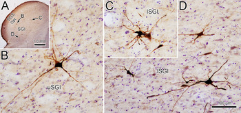
The BDA injection shown in Fig. 14D produced homogeneous staining of the dendrites of the retrogradely labeled cells (Fig. 14A–C). These neurons were generally multipolar in shape, and had long, gently tapering dendrites with very few branches. Their somata range in size from small (Fig. 14B1 and 4), to medium (Fig. 14B6 and C2), to large (Fig. 14B5 and C1 and 4). They had up to eight primary dendrites emerging from their somata. Examples of the homogeneous filled neurons found in a section through the superior colliculus (Fig. 15A) show the extensive labeling of their dendrites (Fig. 15B–D). These cells were present in both the upper sublamina of SGI (uSGI; Fig. 15B) and the lower sublamina of the layer (lSGI; Fig. 15C,D).
Multipolar tectal neurons supplying the SOA. Example cells from an individual section through the superior colliculus (A) show similar multipolar, isodendritc characteristics whether they occupy the upper (B) or lower (C,D) sublamina of the intermediate gray layer (SGI) (same case as Fig. 14). Scale in (D) = (B,C).

Discussion
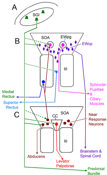
The results of this study support the presence of a bilateral tectosupraoculomotor projection in the macaque that is nontopographic, and likely produced by collaterals of predorsal bundle neurons. The findings present clear evidence that there are considerable species differences between the cat and macaque monkey with respect to the distribution of the cells of origin of the tectal projection to the SOA. Unlike the rostrally distributed cells of the cat, this pathway originates widely within the monkey’s intermediate gray layer. These findings also indicate that the original hypothesis that the projection to SOA primarily targets vertical gaze extraocular motoneurons is unlikely to be true. In fact, we observed only a few tectal terminals in close association with a handful of superior rectus MIF motoneurons (Fig. 16, green arrow to light blue S-group cell), no good evidence of input to medial rectus MIF motoneurons, and no close associations with the SIF motoneurons of either muscle. We did, however, observe close associations between tectal terminals and the levator motoneurons that control elevation of the upper eyelid (Fig. 16, green arrow to red cell). We also observed close associations between labeled tectal axonal boutons and SOA neurons projecting to the pontine and medullary reticular formation, as well as to the cervical spinal cord (Fig. 16, green arrow to purple cells). We believe these cells are most likely part of the peptidergic EWcp population. It is possible that collicular input modulates their activity, which is believed to play a role in stress, and in eating and drinking behaviors. Finally, we observed a projection to caudal SOA that may contact midbrain near response neurons (Fig. 16, green arrows to brown cells).
Summary diagram of collicular targets in the SOA. Collaterals of collicular axons projecting in the predorsal bundle (green arrows) target a variety of populations in the region of III at rostral (B) and caudal (C) levels. The cells of origin of this projection (green trapezoids) are scattered throughout SGI of the superior colliculus (A). In the SOA, these targets include peptidergic cells projecting to the brainstem and spinal cord (purple diamonds), and possible divergence cells projecting to the abducens nucleus (brown pentagons). Also targeted are levator motoneurons in CC (red hexagon). A small projection to superior rectus MIF motoneurons (smaller light blue hexagon) in S-group may be present (green arrow). Other targets that have yet to be examined (green dashed arrows) include the motoneurons in EWpg (pink hexagons) and the near response neurons in SOA (brown pentagons) that supply medial rectus motoneurons and EWpg motoneurons with input (Note that, although not shown, the tectal projection to all these targets is bilateral. To simplify presentation, not all laterality information is included in this schematic).

Technical considerations
The collicular injections were well confined to the target. The fact that terminals were labeled in the SOA following injections of both BDA and PhaL into the superior colliculus, in combination with the demonstration of the projection via retrograde tracing provides strong evidence that this pathway is indeed present in the monkey. This conclusion must be tempered by our difficulty in retrogradely labeling the collicular cells of origin of the SOA projection with WGA-HRP. Nevertheless, this finding corroborates earlier findings in monkey using transport of tritiated amino acids (Harting et al., Reference Harting, Huerta, Frankfurter, Strominger and Royce1980). That said, it is possible that a portion of the terminals labeled in the SOA following BDA injections might represent fiber-of-passage or axon collateral labeling of projections originating in the pretectum (Wasicky et al., Reference Wasicky, Horn and Büttner-Ennever2004). As always, it should be remembered that while close associations between labeled boutons and labeled cells are highly suggestive of synaptic contact, ultrastructural evidence is required to prove such connections.
The largely negative findings with regard to tectal terminals contacting MIF motoneurons must be considered with caution. Since the dendritic labeling in these cells was incomplete, it is possible that the tectal projection terminates primarily on distal dendrites, rendering this connection difficult to observe in the present material. However, the lack of a strong projection to the somata and proximal dendrites that were labeled would appear to suggest that this input, if present, is not a strong or driving influence on the activity of these cells.
With respect to the retrograde data from SOA injections, it should be noted that the BDA injection site extended above the SOA into the PAG (Fig. 12). This region did contain terminal label following collicular injections, so a portion of the retrograde label seen in the superior colliculus may be due to this spread. The BDA injection also extended ventral to III, presenting the possibility that decussating predorsal bundle axons may have picked up the tracer. We worried that this might explain the presence of more caudally distributed tectal neurons in the monkey than in the cat. However, injections of anterograde tracer limited to the caudal colliculus (e.g., Fig. 4) still labeled terminals in SOA, arguing against this interpretation.
Superior colliculus
The morphologic features and the heterogeneous size of the retrogradely labeled neurons observed following SOA injections is reminiscent of the characteristics of predorsal bundle neurons in the monkey (Moschovakis et al., Reference Moschovakis, Karabelas and Highstein1988a,Reference Moschovakis, Karabelas and Highsteinb; May & Porter, Reference May and Porter1992). This is in line with evidence from the cat that the SOA projection arises as collaterals of axons destined for the predorsal bundle (Grantyn & Grantyn, Reference Grantyn and Grantyn1982). It should be noted that far fewer cells were labeled following SOA injections than following injections of the entire predorsal bundle population (May & Porter, Reference May and Porter1992), so it may be that only a subpopulation of predorsal bundle axons exhibit collateral projections to the SOA. It appears that the distribution of the population providing SOA collaterals is fairly widespread, since there was no sublaminar specificity observed (Figs. 14 and 15). This is not true of other tectal targets where differential labeling of laminar outputs has suggested individual target regulation from uSGI and lSGI (May & Porter, Reference May and Porter1992; Zhou et al., Reference Zhou, Warren and May2008).
In the monkey, the cells of origin of the projection to the SOA extended throughout the rostrocaudal extent of SGI, in contradistinction to the rostrally restricted distribution in the cat (Edwards & Henkel, Reference Edwards and Henkel1978). This, and the fact that the projection is not specifically directed at vertical gaze motoneurons argues against the original theory that the projection to the SOA is related to control of vertical gaze. Other species differences in the rostrocaudal distribution of tectal projection populations have been reported. Most notably, tectotectal cells in the cat are restricted to a smaller rostral area than those of the monkey (Edwards, Reference Edwards1977; Behan & Kime, Reference Behan and Kime1996; Olivier et al., Reference Olivier, Porter and May1998). It has been suggested that this species difference in tectotectal projections may relate to the difference in normal oculomotor range of head-independent gaze changes in these two species (Olivier et al., Reference Olivier, Porter and May1998). Perhaps the greater extent of head-independent eye movements in the monkey may underlie the wider distribution of tectosupraoculomotor neurons as well. Arguing against this interpretation is the fact that many of the cells labeled in the present experiments lay in the lower sublamina of SGI (Fig. 15), which is also the source of the tectospinal projection (May & Porter, Reference May and Porter1992).
Oculomotor motoneurons
The present study does not provide evidence that the superior colliculus projects to SIF motoneurons in III. Despite extensive staining of the dendrites of these motoneurons in our material, we did not observe them reaching into the SOA. Superior rectus SIF motoneuron dendrites do enter the S-group region, but we did not observe them receiving close association from tectal boutons. While oculomotor motoneurons in nonprimate species can send dendrites into the overlying SOA (Edwards & Henkel, Reference Edwards and Henkel1978; Evinger et al., Reference Evinger, Graf and Baker1987), this has not been described for primates. Similarly, abducens motoneurons largely confine their dendrites to within the nucleus in primates (McCrea et al., Reference McCrea, Strassman and Highstein1986). In addition, the ventral location of superior rectus SIF motoneurons and medial rectus A-group motoneurons argues against contact from terminals in SOA. A better case might be made for the dorsally located, medial rectus B-group motoneurons. However, we observed no terminals on these cells, despite the fact we labeled motoneurons ipsilateral to the tectal injection, where the terminal field was densest. There is little physiological evidence for direct collicular input to extraocular motoneurons. A small, possibly monosynaptic, tectal input has been described for cat abducens motoneurons (Grantyn & Grantyn, Reference Grantyn and Grantyn1976), but no such input was seen with respect to cat medial rectus motoneurons (Grantyn & Berthoz, Reference Grantyn and Berthoz1977). This should perhaps be expected in that it is generally believed that the gaze target signal emanating from superior colliculus, which is encoded by the spatial coordinates of the activity in the tectum, must undergo a spatiotemporal transformation before it is appropriate to drive motoneurons and move the eyes (Hepp et al., Reference Hepp, Henn, Vilis and Cohen1989; Sparks & Hartwich-Young, Reference Sparks and Hartwich-Young1989; Moschovakis et al., Reference Moschovakis, Kitama, Dalezios, Petit, Brandi and Grantyn1998).
MIF motoneurons, due to their peripheral location vis-a-vis III, clearly lie within the tectosupraoculomotor terminal field. So it is perhaps surprising that few close associations between labeled boutons and labeled terminals were observed. Very sparse close associations were found on superior rectus MIF motoneurons in S-group (Fig. 16, green arrow, light blue cell) and no convincing evidence of input to medial rectus MIF motoneurons in C-group was found. In neither case is there evidence that collicular input is a major synaptic drive to these cells. This is not unexpected, if one considers that the main output of the superior colliculus is a burst of activity intended to drive premotor burst neurons and elicit saccadic eye movements. While MIF motoneurons are active during saccades, albeit with lower firing rates than SIF motoneurons (Hernández et al., Reference Hernández, Calvo, Blumer, de la Cruz and Pastor2019), the high frequency burst present in predorsal bundle axons before saccades does not seem appropriate to drive the primarily graded contraction and tonic action for which MIFs appear specialized (Hess & Pilar, Reference Hess and Pilar1963; Spencer & Porter, Reference Spencer and Porter2006).
Levator motoneurons
A considerable number of anterogradely labeled collicular axons arborized within CC in all our analyzed cases and a number of close associations were observed with retrogradely labeled levator motoneurons (Fig. 16, green arrow to red cell). Terminal fields in CC were not reported following predorsal bundle axon staining in cats (Grantyn & Grantyn, Reference Grantyn and Grantyn1982). However, data in the present monkey experiments strongly supports the possibility of direct input from the superior colliculus, although the number of contacts seen on individual levator motoneurons still suggests a modulatory, as opposed to driving, input. It has already been shown that levator motoneurons receive inputs from the vertical gaze centers: specifically, the M-group associated with the rostral interstitial nucleus of the medial longitudinal fasciculus (monkey: Horn et al., Reference Horn, Büttner-Ennever, Gayde and Messoudi2000; cat: Chen & May, Reference Chen and May2002) and the interstitial nucleus of Cajal (cat: Chen & May, Reference Chen and May2007; monkey: Horn & Büttner-Ennever, Reference Horn and Büttner-Ennever2008). This input is believed to regulate the vertical gaze-related movements of the eyelids (monkey: Becker & Fuchs, Reference Becker and Fuchs1988; Guitton et al., Reference Guitton, Simard and Codère1991; Fuchs et al., Reference Fuchs, Becker, Ling, Langer and Kaneko1992). These gaze pathways activate levator motoneurons during upgaze, and presumably decrease levator activity during down gaze. In this way, levator motoneuron activity is controlled by premotor neurons in a manner similar to that observed for superior rectus motoneurons. In addition, upgaze inputs to both populations are characterized by their use of the calcium regulatory protein, calretinin (Ahlfeld et al., Reference Ahlfeld, Mustari and Horn2011; Zeeh et al., Reference Zeeh, Hess and Horn2013; Che Ngwa et al., Reference Che Ngwa, Zeeh, Messoudi, Büttner-Ennever and Horn2014; Adamczyk et al., Reference Adamczyk, Strupp, Jahn and Horn2015). The present study suggests a difference between levator and superior rectus motoneurons, in that only the former receive this distinctive direct tectal input.
As noted above, the gaze-related signal produced by the superior colliculus is encoded topographically (Sparks & Hartwich-Young, Reference Sparks and Hartwich-Young1989), and this spatial code must be converted to a temporal code by the premotor gaze center neurons, before it is supplied to the extraocular motoneuron populations (Moschovakis et al., Reference Moschovakis, Kitama, Dalezios, Petit, Brandi and Grantyn1998; Grantyn et al., Reference Grantyn, Brandi, Dubayle, Graf, Ugolini, Hadjidimitrakis and Moschovakis2002). This leads to the question of why a direct tectal projection to levator motoneurons might be present. In this regard, the simplicity of levator actions may be of importance. Unlike extraocular muscles, the levator has no secondary actions. So the signal carried by upgaze collicular outputs may be useful in initiating upgaze lid movements. Moreover, it may be of value to ensure that the eyelid movement is not overtaken by the eye movement. The data with respect to this are conflicting. Although eye and lid saccades reach similar velocities, the initiation of the lid saccade actually lags the saccadic eye movement, (Becker & Fuchs, Reference Becker and Fuchs1988). This lag may be due to peripheral motor plant mechanics, including the fact that the levator muscle must counteract the passive downward force of the check ligaments (Evinger et al., Reference Evinger, Manning and Sibony1991; Sibony et al., Reference Sibony, Evinger and Manning1991). In fact, saccade-related levator motoneuron activity leads that of extraocular motoneurons by a few milliseconds (Fuchs et al., Reference Fuchs, Becker, Ling, Langer and Kaneko1992). Monosynaptic collicular input might help account for this small lead. However, if the tectal input initiates levator activity during upward saccades, the fact that the collicular output is not confined to the medial colliculus, which encodes upgaze, is surprising. Perhaps the lateral collicular projection to the levator motoneurons is from inhibitory neurons. Tectotectal inhibitory projections have been described (cat: Behan, Reference Behan1985; Takahashi et al., Reference Takahashi, Sugiuchi, Izawa and Shinoda2005; monkey: Munoz & Istvan, Reference Munoz and Istvan1998), so it is possible that inhibitory cells may also reach other midbrain targets. In light of these questions, it is clear that further physiological investigation will be required to confirm the presence and ascertain the function of this direct collicular projection onto levator motoneurons.
Centrally projecting Edinger–Westphal nucleus (EWcp)
We observed a number of close associations between BDA labeled tectal axonal boutons and small cells in the SOA retrogradely labeled from large injections into the upper levels of the cervical spinal cord, or the pontine and medullary reticular formation (Fig. 16, green arrows to purple cells). While we lack direct proof that these cells are peptidergic EWcp neurons, their scattered locations and relative small somatic diameters are in agreement with descriptions of this population, which has most recently been characterized by the use of urocortin 1 (macaque & human: Horn et al., Reference Horn, Eberhorn, Härtig, Ardeleanu, Messoudi and Büttner-Ennever2008; cat & macaque: May et al., Reference May, Reiner and Ryabinin2008; cebus monkey: Vasconcelos et al., Reference Vasconcelos, Donaldson, Sita, Casatti, Lotfi, Wang, Cadinouche, Frigo, Elias, Lovejoy and Bittencourt2003; review: Kozicz et al., Reference Kozicz, Bittencourt, May, Reiner, Gamlin, Palkovits, Horn, Toledo and Ryabinin2011). Furthermore, the EWcp neurons that use cholecystokinin, substance P and corticotropin releasing factor are known to have brainstem and spinal cord projections (cat: Maciewicz et al., Reference Maciewicz, Phipps, Foote, Aronin and DiFiglia1983, Reference Maciewicz, Phipps, Grenier and Poletti1984; Phipps et al., Reference Phipps, Maciewicz, Sandrew, Poletti and Foote1983; Chung et al., Reference Chung, Mason, Strassman and Maciewicz1987). SOA cells with descending projections have been noted previously in macaque studies (Castiglioni et al., Reference Castiglioni, Gallaway and Coulter1978; Langer & Kaneko, Reference Langer and Kaneko1990; Cowie et al., Reference Cowie, Smith and Robinson1994), but the present study is the first to document their sparse, scattered distribution in SOA based on a variety of injections. Given that only a small number of these cells were labeled from each of our injections, it seems possible that many of the other labeled collicular boutons observed in the SOA may be targeting EWcp cells projecting to other areas. In addition, our retrograde labeling in these cases rarely extended beyond primary dendrites. It is possible that more of the tectal input is directed at the distal dendritic tree of these cells.
Most functional studies of the urocortin-positive EWcp population have explored two possible roles for these cells: modulating responses to stress, and controlling the intake of food and fluids. The connection with stress is based on the fact that urocortin 1 binds and activates both receptor subtypes sensitive to corticotropin releasing factor (Wang et al., Reference Wang, Martínez, Vale and Taché2000). A number of rodent studies have revealed changes in the activity of urocortin-positive EWcp neurons due to stress (Weninger et al., Reference Weninger, Peters and Majzoub2000; Kozicz et al., Reference Kozicz, Li and Arimura2001; Vetter et al., Reference Vetter, Li, Zhao, Contarino, Liberman, Smith, Marchuk, Koob, Heinemann, Vale and Lee2002; Gaszner et al., Reference Gaszner, Csernus and Kozicz2004; Kozicz, Reference Kozicz2010). While the activity of the superior colliculus has primarily been studied with respect to precise targeting of saccades in trained animals, earlier studies noted the critical role of this structure in responding with swift gaze changes activated by the sudden appearance of stimuli in the periphery, the so called “visual grasp reflex” (Pasik et al., Reference Pasik, Pasik and Bender1966) and that the colliculus also plays a role in redirecting attention (Krauzlis et al., Reference Krauzlis, Lovejoy and Zénon2013). It might be reasonable that such an output would upregulate structures modulating the stress response levels of animals.
The role of EWcp urocortin-positive neurons in modulating eating and drinking behavior is also supported by a number of studies in rodents (Spina et al., Reference Spina, Merlo-Pich, Chan, Basso, Rivier, Vale and Koob1996; Weitemier & Ryabinin, Reference Weitemier and Ryabinin2005; Ryabinin & Weitemier, Reference Ryabinin and Weitemier2006). This population may also play a role in consumption of drugs of abuse (Zuniga & Ryabinin, Reference Zuniga and Ryabinin2020). An input from the superior colliculus may make sense for this function as well. An important role of the superior colliculus is directing the animal’s resources toward food or prey items (Comoli et al., Reference Comoli, Das Neves Favaro, Vautrelle, Leriche, Overton and Redgrave2012). Indeed, in rodents, the collicular area involved in prey capture sends axons into the SOA (Furigo et al., Reference Furigo, de Oliveira, de Oliveira, Comoli, Baldo, Mota-Ortiz and Canteras2010). So a projection to EWcp neurons may play a role in alerting a population of cells involved in decisions related to consumption to the fact that a palatable target is available.
Other targets
As noted above, the vast majority of labeled boutons observed in the SOA following collicular injections were not associated with any of the potential targets investigated in this study. So the main target of the SOA projection remains to be defined. Potential targets include the midbrain near response neurons (Fig. 16, dashed arrow to brown cell) and the motoneurons located in EWpg that control lens accommodation and pupillary diameter (Fig. 16, dashed arrow to pink cell). There is evidence that the rostral region of the superior colliculus may be involved in vergence and disjunctive saccades (macaque: Chaturvedi & van Gisbergen, Reference Chaturvedi and van Gisbergen1999, Reference Chaturvedi and Van Gisbergen2000; van Horn et al., Reference Van Horn, Waitzman and Cullen2013; Upadhyaya & Das, Reference Upadhyaya and Das2019). Furthermore, stimulation in the rostral colliculus in cats has also been reported to induce lens accommodation changes (Ohtsuka & Nagasaka, Reference Ohtsuka and Nagasaka1999), although terminals were not noted in the SOA following an injection of anterograde tracers into the area where stimulation produced lens accommodation (Sato & Ohtsuka, Reference Sato and Ohtsuka1996). In the present study, we did observe terminals contacting retrogradely labeled cells located dorsal to the caudal end of III in a pattern reminiscent of the premotor neurons controlling lens accommodation (May et al., Reference May, Warren, Gamlin and Billig2018), and hypothesized that these may be “near response” neurons projecting to the abducens nucleus (Ugolini et al., Reference Ugolini, Klam, Doldan Dans, Dubayle, Brandi, Büttner-Ennever and Graf2006) and controlling divergence (Mays, Reference Mays1984; Judge & Cumming, Reference Judge and Cumming1986) (Fig. 16, green arrow to brown cell). Additionally, stimulation of the superior colliculus has been shown to produce pupillary dilation (Wang et al., Reference Wang, Boehnke, White and Munoz2012; Wang & Munoz, Reference Wang and Munoz2018). Consequently, these other possible targets are worthy of further investigation.
Author contributions
The experiments were designed by P.J.M. and S.W., and were carried out by E.P., N.W., M.O.B., S.W., and P.J.M. Data analysis was done by M.O.B. and P.J.M. The manuscript was written by P.J.M. and edited by all authors.
Funding statement
This research was made possible by NIH grant EY014263 to P.J.M. and S.W.
Acknowledgments
We are beholden to Olga Golanov and Jinrong Wei for their excellent technical support for this work.
Data availability statement
The slide sets from the cases that support this study can be borrowed upon reasonable written request to the authors.
Competing interest
The authors have no competing interest to declare.