





Introduction
Schistosomiasis, caused by digenetic trematodes of the genus Schistosoma, remains one of the most significant parasitic neglected tropical diseases globally, with an estimated 250 million people infected, predominantly in sub-Saharan Africa (Lo et al., Reference Lo, Bezerra, Colley, Fleming, Homeida, Kabatereine, Kabole, King, Mafe, Midzi, Mutapi, Mwanga, Ramzy, Satrija, Stothard, Traoré, Webster, Utzinger, Zhou, Danso-appiah, Eusebi, Loker, Obonyo, Quansah, Liang, Vaillant, Murad, Hagan and Garba2022). Within the Lake Victoria basin, intestinal schistosomiasis, caused by Schistosoma mansoni, is hyper-endemic (Stothard et al., Reference Stothard, Webster, Weber, Nyakaana, Webster, Kazibwe, Kabatereine and Rollinson2009). This is largely driven by the extensive presence of Biomphalaria intermediate host snails along its shorelines and the high frequency of unsafe human-water contact in lakeshore communities (Magero et al., Reference Magero, Kisara, Suleman and Wade2024).
While public health control efforts in Uganda focus primarily on reducing morbidity in human populations through mass drug administration (MDA) of praziquantel (PZQ) (Oyene et al., Reference Oyene, Okoronkwo, Zoure, Amazigo, Nwoke, Katabarwa, Lakwo, Nwobi, Biritwum, Okeibunor, Isiyaku, Tekle, Diallo, Sitima, Elhassan and Boatin2025), considerably less attention has been paid to the long-term infection dynamics and disease consequences in sympatric non-human primate (NHP) hosts (Stothard et al., Reference Stothard, Mugisha and Standley2012). More broadly, the potential role of NHPs in maintaining transmission cycles has therefore emerged as a growing focus within One Health research frameworks (Standley et al., Reference Standley, Mugisha, Adriko, Arinaitwe, Rukundo, Ajarova, Mopya, Betson, Kabatereine and Stothard2013; Červená et al., Reference Červená, Brant, Fairet, Shirley, Petrželková and Modrý2016).
Although schistosomiasis-associated morbidity is well characterized in humans, long-term longitudinal data documenting the progression of natural infection and liver pathology in non-human great apes under continuous environmental exposure is very rare (Medkour et al., Reference Medkour, Amona, Laidoudi, Davoust, Bitam, Levasseur, Akiana, Diatta, Pacheco, Gorsane, Sokhna, Hernandez-aguilar, Barciela, Fenollar, Raoult and Mediannikov2020). Such a lack of evidence also constrains both wildlife health management and our understanding of the long-term dynamics of schistosomiasis within One Health transmission systems. Despite this well-established biological susceptibility, longitudinal observations of naturally acquired schistosomiasis in great apes have historically been scarce, largely because wild ape populations were assumed to have limited contact with snail-infested freshwater habitats (Červená et al., Reference Červená, Brant, Fairet, Shirley, Petrželková and Modrý2016; Richards et al., Reference Richards, Erko, Ponpetch, Ryan and Liang2019). However, systematic monitoring of threatened mammals has recently highlighted pathogens as significant drivers of population declines, necessitating closer scrutiny of parasitic infections (Leendertz et al., Reference Leendertz, Pauli, Maetz-rensing, Boardman, Nunn, Ellerbrok, Jensen, Junglen and Christophe2006).
On the Ngamba Island Chimpanzee Sanctuary (NICS), which is located in Mukono District and part of the Koome group of Islands within Lake Victoria, naturally acquired S. mansoni infection was first unequivocally documented by Standley et al. (Reference Standley, Mugisha, Verweij, Adriko, Arinaitwe, Rowell, Atuhaire, Betson, Hobbs, van Tulleken, Kane, van Lieshout, Ajarova, Kabatereine and Stothard2011). This semi-captive colony represents an uniquely informative cohort where the chimpanzees receive ongoing veterinary care but remain continuously exposed to a highly endemic lacustrine environment where S. mansoni transmission persists among local human and snail populations (Standley et al., Reference Standley, Mugisha, Adriko, Arinaitwe, Rukundo, Ajarova, Mopya, Betson, Kabatereine and Stothard2013). Baseline parasitological and ultrasonographical surveys conducted between 2010 and 2012 revealed persistent egg excretion and early hepatic pathology in several individuals despite repeated PZQ treatment at standard dosing (Standley et al., Reference Standley, Mugisha, Adriko, Arinaitwe, Rukundo, Ajarova, Mopya, Betson, Kabatereine and Stothard2013). Of special note here, the levels of liver fibrosis in animals can be adequately staged or graded (stages A [normal] to F [severe]), using the Niamey Ultrasound Protocol (WHO, Reference Richter, Hatz, Campagne and Jenkins2000), helping to differentiate early versus late levels of progressive hepatic disease (Masi et al., Reference Masi, Perles-barbacaru, Bernard and Viola2020).
Earlier observations on disease within chimpanzees raised critical unresolved questions regarding the long-term performance of repeated chemotherapy under conditions of sustained environmental exposure. In particular, it remains unclear whether periodic PZQ treatment alone would be sufficient to prevent progressive hepatic morbidity in such long-lived NHPs, or whether chronic low-intensity or intermittently patent infections continue to drive liver fibrosis over decadal timescales.
Critically, progression from early periportal thickening to irreversible pipe-stem fibrosis can occur slowly and remain clinically silent for many years, becoming apparent only at advanced stages (Sadun et al., Reference Sadun, von Lichtenberg, Hickman, Bruce, Smith and Schoenbechler1966; Richter et al., Reference Richter, Botelho, Holtfreter, Akpata, El Scheich, Neumayr, Brunetti, Hatz, Dong and Dietrich2016; Lara-Cano et al., Reference Lara-Cano, Castañeda-méndez, Uribe-esquivel and Barbero-becerra2024). Given the conservation status of chimpanzees and the potential for severe, life-limiting morbidity, long-term follow-up data are essential to inform both veterinary management and One Health surveillance strategies.
Here, we present a 12-year longitudinal re-evaluation of the Ngamba Island chimpanzee population, revisiting individuals previously examined in 2010–2012, as part of a more comprehensive annual animal health check, with some animals briefly sedated. Using a multi-modal diagnostic approach combining coprological, urine antigen-based and semi-invasive parasitological assays alongside standardized hepatic ultrasonography, we assess the persistence of S. mansoni infection and the progression of schistosomiasis-associated liver pathology under continued environmental exposure. At the same time, we reassessed the molecular DNA diversity of local S. mansoni, more than a decade later, to provide an insight into the long-term genetic dynamics. Although genotype–phenotype associations are not well established for S. mansoni, particularly in NHPs, our molecular analyses were included to confirm schistosome species identity and to provide exploratory insights into current schistosome diversity within this unique lacustrine transmission setting.
Materials and methods
Study site and animal population
The study was conducted at the NICS, a forested island located approximately 23 km southeast of Entebbe in Lake Victoria, Uganda (00°06′ S, 32°39′ E). The sanctuary encompasses approximately 40 ha of secondary rainforest and provides a semi-captive environment for 52 orphaned and rescued chimpanzees (Pan troglodytes), which receive daily care but retain access to natural habitats, inclusive of the lake shoreline. The geographical setting of NICS places the chimpanzee population within a region of Lake Victoria that is highly endemic for human intestinal schistosomiasis (S. mansoni) (Stothard et al., Reference Stothard, Webster, Weber, Nyakaana, Webster, Kazibwe, Kabatereine and Rollinson2009). Importantly, the chimpanzees have regular contact with to the lake’s shoreline, which is inhabited by Biomphalaria choanomphala, the intermediate hosts of S. mansoni, enabling sustained environmental exposure.
Of special note here, chimpanzees on Ngamba Island were initially averse to water contact; however, individual variation in water contact behaviours has been regularly observed. Notably, the chimpanzee ‘Sunday’ (SUN) displayed atypical tolerance of water, frequently approaching and entering shallow water areas (Stothard et al., Reference Stothard, Mugisha and Standley2012). This behaviour is likely associated with early-life human conditioning prior to arrival at the sanctuary and may have later influenced the behaviour of other group members through social learning. Such variation may affect localized exposure patterns at shoreline interfaces. In the studied chimpanzee population, no overt clinical signs attributable to intestinal schistosomiasis, e.g. abdominal ascites, have been observed, despite evidence of persistent and/or intermediate levels of fecal egg patency. Our study, conducted in 2024, represents a longitudinal follow-up to the baseline surveys performed in 2011–2012, revisiting the same semi-captive chimpanzee population again with portable ultrasonography.
Sample collection and preparation
A multi-modal diagnostic approach was employed over 3 consecutive days to maximize sensitivity for the detection of S. mansoni. Sample collection during the 2024 re-survey combined non-invasive parasitological surveillance with targeted clinical sampling during scheduled health checks. Non-invasive, passive sampling was conducted within the chimpanzees’ sleeping areas, where fresh fecal and urine samples were collected immediately after expression from 49 individually identified chimpanzees (see Online Appendix). Urine samples were collected non-invasively using falcon tubes during spontaneous urination. Sample collection was conducted under continuous direct observation by experienced sanctuary staff, allowing reliable identification of individual chimpanzees and accurate assignment of samples to each animal. For each subject, multiple samples were sought over a 3-day period to account for the known day-to-day variation in schistosome egg excretion. For molecular screening, 1 fecal sample per individual (n = 38) was preserved for subsequent DNA extraction and downstream polymerase chain reaction (PCR) analyses.
Additionally, targeted clinical sampling was performed on 8 chimpanzees during a scheduled health check in March 2024 (see Table 1). One individual from the original cohort (Mika) had died during the intervening period and was therefore unavailable for reassessment. This individual was replaced by a newly examined chimpanzee (Ruparelia) to maintain sample size, although this individual was not included in longitudinal comparisons. Eight chimpanzees underwent short-duration general anaesthesia administered by the sanctuary veterinarian as part of the annual health check, during which biological samples were collected and ultrasonographical examinations were performed. To facilitate invasive and imaging procedures, each chimpanzee was secured within a holding cage and anaesthetized via intramuscular injection. Anaesthesia was induced using ketamine (5 mg kg−1) and medetomidine (0.05 mg kg−1) administered intramuscularly under continuous veterinary supervision. Once sedation was achieved, animals were transferred to the sanctuary’s veterinary clinic, weighed and placed on an examination table being under monitored sedation for not more than 30 min.
Summary of the diagnostic test results performed on 9 Ngamba Island chimpanzees in June 2012 and then in the March 2024 follow-up

Table 1 Long description
The table lists nine Ngamba Island chimpanzees with age and sex, comparing diagnostic results from 2012 with a 2024 follow-up across KK, CCA, rectal snip biopsy, and ultrasound grading. In 2012, seven chimpanzees were KK positive; by 2024, all of those became KK negative, while Kalema remained KK negative and Ruperelia changed from not available in 2012 to KK positive in 2024. CCA results were positive for most animals in both years, including several that were KK negative in 2024; Katie, Kalema, and Ruperelia were CCA negative or not available in 2012 but CCA positive in 2024. Rectal snip biopsy shifted from positive in 2011 for most tested animals to negative in 2024 for all with reported follow-up, and Connie and Kalema were negative at both time points. Ultrasound findings commonly indicated ongoing pathology: Kidogo worsened from mild to moderate to severe in 2012 to severe in 2024, Rutoto remained severe, and several others stayed at moderate levels or moved from normal or mild to moderate. Ruperelia had no 2012 ultrasound data but showed mild pathology in 2024. Overall, parasitology and biopsy results improved or stayed negative by 2024, while antigen testing and ultrasound suggest persistent infection signals and chronic disease changes, noting that some 2012 values are missing for Ruperelia.
F, female; M, male; +, positive test result; −, negative test result. For the ultrasound results following the Niamey grading scale, ‘B’ indicates mild pathology, ‘C’ indicates presence of pipe-stems (moderate pathology) and ‘D’ indicates pipe-stems/occluding fibrosis (severe pathology).
Diagnostic assays for schistosomiasis
To assess the persistence of S. mansoni infection under conditions of long-term environmental exposure and repeated PZQ treatment, a multi-modal diagnostic framework was applied, integrating parasitological, antigen-detection and viability-based assays. This approach was specifically selected to address the known limitations of coprological diagnostics in chronic, low-intensity schistosomiasis, particularly in long-lived NHP hosts.
Conventional and antigen-based assays
Parasitological assessment of fecal egg excretion was performed using the Kato–Katz (KK) thick smear technique (Katz et al., Reference Katz, Chaves and Pellegrino1972). Duplicate smears were prepared from each stool sample using standardized 41.7 mg templates and examined under light microscopy at 100× magnification. Egg counts were expressed as eggs per gram of feces to provide a semi-quantitative estimate of infection intensity, categorized according to WHO guidelines developed for S. mansoni, acknowledging their extrapolative application to NHPs.
Given the reduced sensitivity of KK microscopy in chronic and post-treatment infections, a miracidia hatching test (MHT) was employed as a complementary qualitative assay to confirm egg viability and facilitate downstream molecular analyses (Weyher et al., Reference Weyher, Phillips-conroy, Fischer, Weil, Chansa and Fischer2010). Approximately 5 g of fresh stool were processed using a Pitchford funnel and exposed to indirect light to stimulate miracidial emergence. Following hatching, individual miracidia were visualized under a dissecting microscope, collected in 2.5 μL of water using a pipette and transferred onto Whatman FTA® cards (GE Healthcare Life Sciences; Amersham, UK) for downstream molecular analysis.
To detect active or recent infection independently of egg excretion, urine samples were screened using a point-of-care urine-circulating cathodic antigen (POC-CCA) assay (ICT International, South Africa) (Colley et al., Reference Colley, King, Kittur, Ramzy, Secor, Fredericks-james, Ortu, Clements, Ruberanziza, Umulisa, Wittmann and Campbell2020). The POC-CCA is an antigen-detection assay targeting schistosome gut-derived CCA and is recognized for its superior sensitivity in low-intensity and post-chemotherapy infections. Urine samples were tested immediately on-site following the manufacturer’s instructions in 49 individuals. Test cassettes were read visually, and all results were independently interpreted by 2 observers to minimize subjectivity, particularly in cases of trace reactivity.
Together, this integrated diagnostic strategy enabled robust detection of persistent S. mansoni infection while accounting for the limitations inherent to individual diagnostic modalities in chronic schistosomiasis.
Ultrasonography and morbidity assessment
To enable robust longitudinal comparison with baseline examinations conducted in 2011–2012, hepatic and portal pathology were assessed using standardized abdominal ultrasonography. Examinations were performed with a portable Chison Q5 ultrasound system equipped with a 3.5-MHz convex transducer (Mount International United Services Ltd., Gloucester, UK), consistent with equipment and settings used during earlier surveys.
Prior to scanning, abdominal hair was clipped to expose standardized intercostal windows on both left and right ventral aspects of the abdomen of the 8 chimpanzees, allowing reproducible visualization of the liver parenchyma, portal vein branches and periportal regions. Ultrasonographic assessment followed established scanning planes to ensure consistency across individuals and time points.
Liver pathology was recorded and graded according to the WHO–Niamey ultrasonographic classification system (WHO, Reference Richter, Hatz, Campagne and Jenkins2000; King et al., Reference King, Magak, Salam, Ouma, Kariuki and Blanton2003), which provides a standardized framework for assessing schistosomiasis-associated periportal fibrosis. This system has been widely applied in both human and NHP studies and enables direct comparison of disease severity across longitudinal surveys. Particular attention was paid to the presence and progression of periportal thickening and the development of Symmers’ pipe-stem fibrosis.
Rectal snip biopsy
As a qualitative method to detect the presence of calcified or retained schistosome eggs lodged within the rectal mucosa, rectal snip biopsies were performed according to Badran et al. (Reference Badran, El-alfi, Pfischner, Killough and Burns1955), as used before (Standley et al., Reference Standley, Mugisha, Adriko, Arinaitwe, Rukundo, Ajarova, Mopya, Betson, Kabatereine and Stothard2013). Rectal snip biopsy was included to increase diagnostic sensitivity of egg-patency in low-intensity infections, although its semi-invasive nature and limited diagnostic yield, in comparison to molecular methods, are acknowledged. Moreover, in 2011, previous surveys in the same chimpanzee population reported 62.5% positivity by this method, supporting its continued inclusion for comparative purposes. Using a metal proctoscope and metal biopsy spoon, 2- to 3-mm rectal mucosal tissue samples were collected under general anaesthesia. Biopsy specimens were fixed in 1% malachite green in 50% glycerol, compressed between glass slides and examined by light microscopy at 100× magnification (Khalil et al., Reference Khalil, Abdel-baki, Saleh, Abbas and Amir1993). This approach was included to supplement fecal egg-patent diagnostics in detecting tissue-retained eggs associated with chronic infection.
DNA extraction from fecal samples stored in ethanol
DNA was extracted from 38 individual chimpanzee fecal samples preserved in 70% ethanol using a modified QIAamp DNA Mini Kit (Qiagen, Germany) protocol as described by Cunningham et al. Reference Cunningham, Odoom, Pratt, Boatemaa, Asante-Ntim, Attiku, Banahene, Osei-Atweneboana, Verweij, Molyneux, Stothard and Adams2018 (GPLN paper). Briefly ∼0.1 g of preserved stool was placed in a 2-mL screw cap tube and underwent 3 rounds of washing with 1 mL of distilled water, during this stage a blank sample tube was included to act as a DNA negative extraction control. To the washed stool 0.9 g of 1.4-mm ceramic beads were added alongside 250 µL of 2% polyvinylpolypyrrolidone/phosphate-buffered saline solution. The sample then underwent mechanical lysis using the MagnaLyser (Roche) at 1509 ×g rpm for 30 s. Following mechanical lysis the rest of the DNA extraction followed the manufacturers protocol for the DNEasy blood and tissue kit (Qiagen, Manchester, UK) with the addition of Phocine herpes virus (PhHV) to the ATL buffer, to act as a DNA extraction, positive control. The extracted DNA was then screened using a Schistosoma spp. generic real-time (rt) PCR assay (Obeng et al., Reference Obeng, Aryeetey, de Dood, Amoah, Larbi, Deelder, Yazdanbakhsh, Hartgers, Boakye, Verweij, van Dam and van Lieshout2008) targeting the nuclear ribosomal internal transcribed spacer (ITS) gene. Positive samples were then further screened with a duplex S. mansoni and S. haematobium species-specific rtPCR using the primers and probes as described by Alzaylaee et al. (Reference Alzaylaee, Collins, Shechonge, Ngatunga, Morgan and Genner2020). The oligonucleotide sequences for the different primer and probe sets are detailed in Supplementary Table 2. With each rtPCR run, a PCR-negative and -positive control was used with well-characterized samples from prior research projects being used for the positive controls. For both the generic and species-specific schistosome rtPCR, the following volumes and concentrations were used: 2 µL of template, 400 nM of each primer, 100 nM of each probe and 6 µL of appPROBENo ROX super Mix (AppletonWoods, Birmingham, UK), with nuclease free water making up the rest of the reaction up to 12 µL. The PCR cycling conditions followed the super mix manufacturers recommendations, in brief it was as follows: initial denaturation at 95 °C for 3 min, followed by a 2 step-cycle for 40 cycles of 95 °C for 10 s and 60 °C for 30 s. The positive/negative status of each sample was recorded alongside the Ct values for each sample per assay.
FTA card DNA extraction and Schistosoma detection
Alongside fecal samples collected and preserved in ethanol, hatched miracidia from 2 individuals (SUN n = 49 and Natasha [NAT] n = 3) were spotted and preserved onto Whatman FTA cards. Miracidia preserved on FTA cards underwent alkaline DNA extraction following the methods described in the supplementary material of Webster et al. (Reference Webster, Alharbi, Kayuni, Makaula, Halstead, Christiansen, Juziwelo, Stanton, LaCourse, Rollinson, Kalua and Stothard2019). The addition of PhHV to the first extraction buffer served as an internal extraction control while the inclusion of a blank FTA card sample served as the extraction negative control. Following isolation of DNA, the samples underwent the same molecular screening previously described for the stool stored in 70% ethanol.
Schistosoma species identification and cox1 amplification
Of the 52 extracted miracidial FTA samples, 7 (SUN n = 4, NAT n = 3) had a 583bp region of the cox1 gene amplified and sequenced using the Asmit1 forward and Schisto reverse primers described in Huyse et al. (Reference Huyse, Webster, Geldof, Stothard, Diaw, Polman and Rollinson2009). The 25 µL PCR comprised 5 µL of template, 12.5 µL Q5® high-fidelity DNA polymerase (NEB, UK) and 400 nM of each primer, with the remaining volume being made up with nuclease free water. The manufacturers’ recommendations for PCR cycling were followed with an annealing temperature of 54 °C for 45 cycles. The PCR products were separated by electrophoresis on a 1% gel, stained with SYBR™ DNA gel stain (ThermoFisher Scientific, UK) for 45 min at 100 V. Alongside the PCR products, 100bp ladder from Bioline was run out on the gel to confirm band sizes. Following confirmation of target amplification, PCR products were cleaned using the HT ExoSAP-IT™ Fast High-Throughput PCR kit, after which samples were sent to Source Biosciences for Sanger sequencing. All cleaned 360bp cox1 sequences were submitted to the NCBI database and assigned the following accession numbers: PZ050358, PZ050359, PZ050360, PZ050361, PZ050362 and PZ050363
Schistosoma mansoni cox1 population genetics
All obtained cox1 sequences were aligned, cleaned and trimmed using Geneious Prime against reference sequence: JF508493. Of the 7 chimp miracidia samples, 6 (SUN n = 4 and NAT n = 2) were of sufficient enough quality to be used for further downstream analysis. In addition to the 2024 Ngamba chimp samples (SUN/NAT), an additional 4 cercarial samples (BIOM) and 3 miracidial sequences from human urine from Kimi Island (KK) were included. The sequences from the 2024 Ngamba and Kimi samples were aligned and trimmed against 26 reference sequences from the original 2010 Ngamba island study (H) (Standley et al., Reference Standley, Mugisha, Adriko, Arinaitwe, Rukundo, Ajarova, Mopya, Betson, Kabatereine and Stothard2013), plus 9 Kenyan reference sequences (KEN) from a 2007 study based in Nyanza (Hanelt et al., Reference Hanelt, Mwangi, Kinuthia, Maina, Agola, Mutuku, Steinauer, Agwanda, Kigo, Mungai, Loker and Mkoji2010) resulting in a 360bp consensus alignment (Supplementary Table 1). The aligned sequences were exported as a NEXUS file and a minimum spanning network tree was generated using PopART (Bandelt et al., Reference Bandelt, Forster and Röhl1999), with the following traits assigned to the different sequence types: original haplotype references (H) from the Ngamba 2010 study = ‘Ngamba2010,’ cox1 miracidial sequences derived from chimps SUN and NAT in 2024 = ‘Ngamba2024,’ the 2024 cox1 sequences from human urine (KK) and cercariae (BIOM) originating from Kimi island = ‘Kimi2024’ and the Kenyan reference sequences (KEN) originating from Nyanza = ‘Nyanza2007.’
Results
Parasitological findings from the 2024 follow-up survey
Non-invasive parasitological surveillance in 2024 yielded urine samples from 49 individually identified chimpanzees and fecal samples from 38 individuals, together representing 94.2% (49/52) of the resident sanctuary population at the time of sampling. Evidence of S. mansoni exposure or infection was assessed using a combination of urine antigen-based, copro-parasitological and invasive diagnostic assays (see Table 1), confirming the continued presence of transmission within the Ngamba Island ecosystem.
Urine-based POC-CCA testing identified circulating cathodic antigen positivity in 18 of 49 chimpanzees (36.7%; 95% CI: 24.7–50.7%), while 31 individuals remained consistently CCA-negative across all sampling days (see Online Appendix). In contrast, KK thick smear examinations detected patent infections in only 2 of 49 individuals (4.1%, 95% CI: 0.5–14.0%), with egg counts consistently within the WHO-defined light-intensity range where eggs were observed (see Online Appendix). This pronounced discordance between antigen detection and egg excretion closely mirrors patterns reported in the original Ngamba Island surveys and is characteristic of chronic, low-intensity schistosomiasis in long-lived NHP hosts (Standley et al., Reference Standley, Mugisha, Verweij, Adriko, Arinaitwe, Rowell, Atuhaire, Betson, Hobbs, van Tulleken, Kane, van Lieshout, Ajarova, Kabatereine and Stothard2011, Reference Standley, Mugisha, Adriko, Arinaitwe, Rukundo, Ajarova, Mopya, Betson, Kabatereine and Stothard2013).
MHTs conducted on selected stool samples confirmed the presence of viable eggs by the KK method in a limited number of cases (2/49), demonstrating that at least a proportion of infections remained biologically active. Successful hatching enabled downstream molecular processing, allowing continuity with the longitudinal parasite genetic monitoring framework established previously.
Ultrasonographic evidence of progressive hepatic morbidity
Longitudinal ultrasonographic comparison demonstrated progression of periportal pathology in multiple individuals relative to their 2011–2012 baseline classifications (Figure 1; Table 1). Using the WHO–Niamey grading system, several chimpanzees exhibited advancement from previously documented mild or moderate periportal thickening (grades B or C) to more severe patterns, including pipe-stem fibrosis consistent with grade D pathology (Table 1).
Examples of chimpanzee liver patterns as visualized by ultrasound. Image A shows the early stages of pipe-stem formation, consistent with a ‘C’ pattern of pathology, while Image B demonstrates pipe-stem formation with occluding fibrosis, indicative of a ‘D’ level of pathology.

No individuals demonstrated regression of established periportal fibrosis despite repeated annual PZQ treatment over the 12-year interval. In addition, hepatic abnormalities were detected in at least 1 individual who had previously been classified as ultrasonographically normal, indicating de novo development of schistosomiasis-associated morbidity during long-term environmental exposure.
Rectal snip biopsy findings
Rectal snip biopsies were successfully obtained from all 8 chimpanzees undergoing clinical examination in 2024 (8/8; 100%). No schistosome eggs were noted in any rectal mucosal samples (0/8; 0.0%), including individuals with concurrent urine POC-CCA positivity and/or ultrasonographical evidence of periportal fibrosis (see Table 1). These findings are consistent with previous observations at Ngamba Island, where rectal snip biopsy demonstrated limited sensitivity in semi-captive chimpanzees with predominantly low-intensity or chronic infections (Standley et al., Reference Standley, Mugisha, Verweij, Adriko, Arinaitwe, Rowell, Atuhaire, Betson, Hobbs, van Tulleken, Kane, van Lieshout, Ajarova, Kabatereine and Stothard2011, Reference Standley, Mugisha, Adriko, Arinaitwe, Rukundo, Ajarova, Mopya, Betson, Kabatereine and Stothard2013).
Molecular analysis of fecal and FTA samples and schistosome genetic diversity
The molecular analysis of the 38 chimpanzee fecal samples resulted in the internal positive control failing in 28.2% of the samples, indicating either failure to isolate DNA from the sample or the presence of inhibitors; these samples were therefore excluded from further analysis. Of the remaining samples (n = 28), 8 tested positive for the generic schistosome rtPCR, with a Ct range of 26.4–37.5. Of these 8 samples, 5 were identified as being S. mansoni using the species-specific rtPCR with the remaining 3 samples failing to amplify as either S. mansoni or S. haematobium. The average Ct value for the generic schistosome assay for the species-specific positives was 29.6, while the Ct value for the species-specific negatives was 36.6, indicating the 3 negative species-specific reactions is likely a sensitivity issue.
The internal extraction control was successfully amplified in all 52 of the FTA extractions, indicating 100% success with the DNA extraction process. When confirming the presence of schistosome DNA using the schistosome generic assay, 46 of the 52 samples tested positive (88.5%) with 44 of these 46 samples positive for S. mansoni using the species-specific assay, no samples were positive for S. haematobium. The average Ct value for the Schistosoma spp. assay was 24.8, while that of the species specific was 24.1. As with the fecal samples the 2 species-specific negatives had a higher average Schistosoma spp. generic Ct value of 33.9, indicating a lower yield of target DNA as an explanation for the negative results.
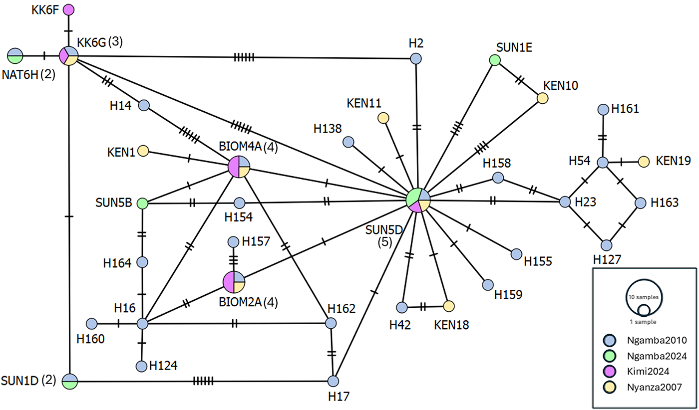
The 6, 360 bp, cox1 sequences from 2024 resulted in 5 separate haplotypes, 4 of which were grouped with haplotypes H1 (n = 2), H103 (n = 1) and H156 (n = 1) from the prior, 2010, Ngamba study (Standley et al., Reference Standley, Mugisha, Adriko, Arinaitwe, Rukundo, Ajarova, Mopya, Betson, Kabatereine and Stothard2013). The remaining 2 2024 barcode sequences (SUN1E and SUN5B) were novel as they differed from both the 2010 Ngamba study and the 2007 Kenyan study. These 2 novel sequences had a total of 6 unique base changes from haplotype H1, with SUN1E having a total of 4 base changes and SUN5B having 2 unique base changes when compared against H1 (Table 2). The network also identified 3 haplotypes that were previously found in Kenya (2007), Ngamba (2010) and Kimi (2024), indicating a wide distribution of these haplotypes, highlighting the putative gene flow across Lake Victoria. Haplotype H1 (SUN5D) from the 2010 Ngamba study was the only 1 observed across the different sites used in the haplotype network analysis.
Unique base changes found in the 2-novel barcode sequences from the Ngamba 2024 survey aligned against haplotype H1

Table 2 Long description
The table lists nucleotide bases at six mitochondrial genome positions for a reference haplotype H1 and two novel cox1 haplotypes, SUN1E and SUN5B. Positions are 1712, 1844, 1862, 1908, 1979, and 1991 from the start of the mitochondrial genome. H1 has A, T, A, A, A, and T at these sites. SUN1E matches H1 at 1844 and 1862 but differs at 1712 (G instead of A), 1908 (G instead of A), 1979 (G instead of A), and 1991 (C instead of T), giving four differences across the six positions. SUN5B matches H1 at 1712, 1908, 1979, and 1991 but differs at 1844 (C instead of T) and 1862 (G instead of A), giving two differences. The table reports only these selected sites, so it does not indicate whether additional differences exist elsewhere in the sequences.
Mt, mitochondrial.
Of note, the cox1 haplotype SUN5D was the most common haplotype, being present in the original Ngamba 2010 samples (H1) and the 2024 samples (SUN5D and NAT1G), as well as sequences from Kimi island (KK7D) and Kenya (KEN05) (Hanelt et al., Reference Hanelt, Mwangi, Kinuthia, Maina, Agola, Mutuku, Steinauer, Agwanda, Kigo, Mungai, Loker and Mkoji2010). The second most common haplotypes are jointly held by BIOM2A (BIOM3D, KEN16 and H36) and BIOM4A (BIOM4C, KEN09 and H10). The third most common haplotype is KK6G (KEN08 and H79) (Figure 2).
Minimum-spanning network of the cox1 DNA barcodes from the Ngamba 2024 study from S. mansoni miracidia isolated from chimps Sunday (SUN) and Natasha (NAT) as well as barcodes from the Ngamba 2010 survey, Kenya (2007) and the nearby Kimi Island. The number of barcodes per haplotype are given in brackets, where no brackets are shown, only a single barcode is present.

Figure 2 Long description
The diagram shows a genetic haplotype network of cox1 DNA barcodes from the Ngamba 2024 study, Ngamba 2010 survey, Kenya 2007 and Kimi Island. Nodes represent haplotypes, with sizes indicating the number of barcodes per haplotype. A central hub haplotype, SUN5D, connects to multiple other nodes, forming a star-like pattern. Several smaller clusters are visible around this central hub. Lines between nodes indicate genetic relationships. Different colors represent different studies: Ngamba 2024, Ngamba 2010, Kimi 2007 and Kenya 2007, as explained in the legend in the lower right corner.
Discussion
This 12-year longitudinal follow-up study provides compelling evidence that S. mansoni transmission persists within the semi-captive chimpanzee population of Ngamba Island and that long-term exposure is associated with progressive hepatic fibrosis, despite repeated PZQ treatment. By revisiting individuals previously examined in 2011–2012 using comparable parasitological, molecular and ultrasonographic methodologies, this study offers a rare insight into the natural history of chronic intestinal schistosomiasis in a long-lived NHP host.
Persistence of infection under repeated PZQ treatment
The continued detection of CCA in urine samples provides evidence of ongoing or recent S. mansoni infection in multiple chimpanzees more than a decade after the initial diagnosis (see Online Appendix; Table 1). As in the original Ngamba Island surveys, egg detection by KK was infrequent and restricted to low-intensity infections, highlighting the well-recognized limitations of coprological methods in chronic, low-burden schistosomiasis. This diagnostic discordance is consistent with human studies in endemic settings, where antigen detection outperforms microscopy in identifying residual or pre-patent infections, particularly following MDA (Lorenz et al., Reference Lorenz, Razafindrakoto, Rausche, Rasolojaona, Razafindralava, Zerbo, Höppner, von Thien, Rakotozandrindrainy, Doumbia, Klein, Kutz, Corstjens, de Dood, Hoekstra, van Dam, Jaeger, Schwarz, Tannich, Andrianarivelo, Rakotozandrindrainy, Rakotoarivelo, May, Rasamoelina and Fusco2025).
The persistence of CCA positivity despite periodic PZQ administration suggests that chemotherapy alone is insufficient to interrupt transmission in this lacustrine sanctuary environment. Rapid reinfection from contaminated shoreline habitats, where Biomphalaria snails are present and accessible to chimpanzees, remains the most parsimonious explanation. While reduced PZQ efficacy or altered pharmacokinetics in great apes cannot be entirely excluded, there is currently no evidence to support the emergence of drug resistance in this system. Instead, these findings reinforce earlier concerns that standard human dosing regimens may not be optimally effective for long-term control in semi-captive great apes repeatedly exposed to high-risk environments (Hoekstra et al., Reference Hoekstra, Casacuberta Partal, Amoah, van Lieshout, Corstjens, Tsonaka, Assaré, Silué, Meité, N’goran, N’gbesso, Roestenberg, Knopp, Utzinger, Coulibaly and van Dam2018). Given the high reinfection pressure and the limited impact on long-term infection prevalence, a strategy involving biannual PZQ treatment at adjusted doses may be warranted to achieve more effective parasite suppression and potentially reduce the risk of chronic morbidity (Olveda et al., Reference Olveda, Inobaya, McManus, Olveda, Vinluan, Ng, Harn, Li, Guevarra, Lam and Ross2017).
Progressive hepatic morbidity despite low-intensity infection
The most significant finding of this study is the clear progression of schistosomiasis-associated hepatic pathology in several individuals over the 12-year follow-up period (see Figure 1; Table 1). Comparative ultrasonography demonstrated advancement from mild or moderate periportal thickening to severe pipe-stem fibrosis in chimpanzees who had already exhibited pathology at baseline. Importantly, no regression of established fibrosis was observed, despite repeated PZQ treatment. Chimpanzees exhibit a high degree of susceptibility to S. mansoni infection (Richards et al., Reference Richards, Erko, Ponpetch, Ryan and Liang2019). Landmark experimental studies established that the clinicopathological progression of the disease in chimpanzees remarkably mirrors that observed in humans (Sadun et al., Reference Sadun, von Lichtenberg, Hickman, Bruce, Smith and Schoenbechler1966). These early investigations demonstrated that infected chimpanzees develop severe hepatic morbidity, including portal hypertension and the characteristic Symmers’ pipestem fibrosis (Sadun et al., Reference Sadun, von Lichtenberg, Hickman, Bruce, Smith and Schoenbechler1966). Unlike many other animal models, the chimpanzee’s long lifespan and physiological similarities to humans allow for the development of chronic lesions that are nearly identical to advanced human intestinal schistosomiasis (Cheever, Reference Cheever1987).
This pattern closely mirrors the natural history of chronic intestinal schistosomiasis in humans, where chemotherapy clears adult worms but does not reverse established periportal fibrosis (Cheever, Reference Cheever1987; King et al., Reference King, Magak, Salam, Ouma, Kariuki and Blanton2003). The chimpanzee thus once again proves to be an exceptionally faithful model for human hepatosplenic schistosomiasis, confirming earlier experimental and observational studies (Sadun et al., Reference Sadun, von Lichtenberg, Hickman, Bruce, Smith and Schoenbechler1966). The slow but relentless progression of fibrosis observed here underscores the cumulative impact of prolonged antigenic stimulation from trapped eggs, even in the context of low-level or intermittently patent infections.
The detection of new hepatic abnormalities in individuals previously classified as ultrasonographically normal further indicates that clinically silent infection can evolve into measurable morbidity over time. This finding has important welfare implications, as advanced periportal fibrosis may remain asymptomatic for extended periods before progressing to portal hypertension and its sequelae (Anjori et al., Reference Anjorin, Nabatte, Mpooya, Tinkitina, Opio, Kabatereine and Chami2024). Our findings demonstrate that even persistently low-intensity or intermittently patent S. mansoni infections are sufficient to drive progressive hepatic fibrosis over decadal timescales in chimpanzees, mirroring the natural history of human hepatosplenic schistosomiasis (Anjori et al., Reference Anjorin, Nabatte, Mpooya, Tinkitina, Opio, Kabatereine and Chami2024). This is consistent with previous findings in free-ranging primates, where Schistosoma infections are frequently subclinical and clinical disease manifestations are rarely documented under natural conditions (Standley et al., Reference Standley, Mugisha, Adriko, Arinaitwe, Rukundo, Ajarova, Mopya, Betson, Kabatereine and Stothard2013). Nevertheless, the presence of histopathological lesions by ultrasonography indicates that infection can occur without overt or clear external clinical expression, highlighting the importance of more rigorous pathological assessment(s) in understanding disease dynamics in wild great apes.
Molecular diversity of schistosome infections
Following DNA analysis, both the fecal and FTA samples we identified as being positive for S. mansoni only, no samples were positive for S. haematobium. The samples that were genus-positive but species-specific negative yielded lower quantities of DNA, which may have contributed to assay failure; however, the presence of non-S. mansoni schistosomes or potential hybrids cannot be excluded. The minimum spanning network tree identified 5 sperate haplotypes from the 6 partial-cox1 sequences derived from chimpanzees SUN and NAT with 3 of these matching previously identified haplotypes from the 2010 survey (H1, H10 and H36) and the remaining 2 being novel (SUN5B and SUN1E). While limited, this analysis demonstrates the persistence of schistosome haplotypes over a period of 10+ years as well as the possible introduction of new haplotypes. This demonstrates both the stability of schistosome populations but also the fact that parasite gene flow is occurring, even at this isolated location that has no permanent human population. Additionally, the haplotype analysis shows the broader picture of gene flow across Kimi island, Ngamba island and Kenya, with 4 haplotypes found across all these sites (KK6G, BIOM4A, BIOM2A and SUN5D). Possible routes of parasite gene flow, particularly the role that itinerant fishermen play across lake Victoria, including fishing just-offshore of Ngamba island, typically using human fecal baits, warrants greater surveillance.
Implications for chimpanzee health management and One Health perspectives
From a management perspective, this study confirms that intestinal schistosomiasis remains the dominant parasitic health threat to the Ngamba Island chimpanzees. Unlike transient or subclinical infections, schistosomiasis exerts a cumulative pathological burden with the potential to compromise long-term health and lifespan. The sanctuary setting, while providing protection from many threats faced by wild chimpanzees, paradoxically facilitates repeated exposure to infected freshwater habitats, creating conditions for sustained transmission.
These findings also have broader One Health relevance. The persistence of S. mansoni infection in chimpanzees living in close proximity to human communities highlights the permeability of transmission boundaries between human and NHP (Richards et al., Reference Richards, Erko, Ponpetch, Ryan and Liang2019). While the directionality and epidemiological significance of cross-species transmission remain unresolved, the presence of a long-term non-human reservoir complicates local control efforts and warrants continued integrated surveillance of humans, wildlife and intermediate host snails.
Conclusions
This 12-year longitudinal follow-up demonstrated that despite best efforts in regular administration PZQ with some tangible improvements, S. mansoni infection persists, however, within semi-captive chimpanzees on Ngamba Island. Of concern, there is an associated increase of progressive, irreversible hepatic fibrosis in certain animals. Together, analysis of these data provides rare longitudinal evidence that chronic S. mansoni infection in NHPs can result in progressive, irreversible hepatic morbidity, highlighting intestinal schistosomiasis as an underappreciated burden. The molecular analysis confirmed S. mansoni as the only schistosome present within the fecal samples noting that 3 historic cox1 haplotypes were still present, with an additional 2 novel cox1 haplotypes noted, not previously encountered. From both conservation and One Health perspectives, intestinal schistosomiasis represented here warrants more sustained surveillance and better integrated control strategies.
Supplementary material
The supplementary material for this article can be found at https://doi.org/10.1017/S003118202610242X.
Acknowledgements
We are grateful to the staff and management of the Ngamba Island Chimpanzee Sanctuary and the Chimpanzee Trust for their tireless dedication to the welfare of the chimpanzees. Permission for the research was granted by the Uganda Wildlife Authority. We thank Paula Cambero Guerra and Alfred Gadama for their assistance with English language editing. We also gratefully acknowledge the technical support provided by the Liverpool School of Tropical Medicine.
Author contributions
Author’s contribution. Inception and design of the study: J.A., L.J.C., J.R., E.J. LaC., and J.R.S.; sample collection, parasite dentification: J.A., T.M., D.O., S.M., A.R., F.B., E.J. LaC. and J.R.S.; analyses: A.J., C.G., and J.R.S.; molecular barcoding and analyses: A.J., L.J.C.,C.G.; first draft of the paper: A.J. and J.R.S. and all authors contributed to writing, editorial revision and final approval of the manuscript.
Financial support
This work was supported by Hungarian Eötvös State Scholarship [MAEÖ-00039-002/2023]. This work forms part of the MSc research dissertations of C.G. and A.J. receive salary support from the Wellcome Trust, these surveys were therefore supported, in part, by the National Institute for Health Research (NIHR) [using the UK’s Official Development Assistance (ODA) Funding] and Wellcome Trust [220818/Z/ 20/Z] under the NIHR–Wellcome Partnership for Global Health Research. The views expressed are those of the authors and not necessarily those of Wellcome, the NIHR or the Department of Health and Social Care.
Competing interests
The authors declare no competing interests. Professor J. Russell Stothard is the Editor-in-Chief of the journal but had no role in the peer review or editorial decision for this manuscript.
Ethical standards
All procedures were performed during routine annual veterinary health checks in 2024 under the supervision of the sanctuary’s veterinary team, without introducing any unduly cumbersome additional procedures beyond standard clinical care while animals were under brief sedation. Specimen collection and examinations adhered to the guidelines of the Uganda Wildlife Authority (UWA). While the majority of diagnostic procedures were non-invasive a semi-invasive rectal snip biopsy was obtained, as described before by Standley et al. (Reference Standley, Mugisha, Adriko, Arinaitwe, Rukundo, Ajarova, Mopya, Betson, Kabatereine and Stothard2013), to facilitate direct comparisons with prior studies, which was integrated into standard clinical monitoring.





 Open access

