INTRODUCTION
To date, approximately 20 extant species of the genus Scrippsiella Balech ex A.R. Loeblich III 1965, belonging to the subfamily Calciodinelloideae under the family Peridiniaceae (Fensome et al., Reference Fensome, Taylor, Norris, Sarjeant, Wharton and Williams1993), have been found in water from polar through tropical habitat and constitute a considerable fraction of the phytoplankton. Scrippsiella trochoidea (Stein) Loeblich III, 1976, the most common member of the genus, is a widespread and bloom-forming species (Honsell & Cabrini, Reference Honsell and Cabrini1991; Vink, Reference Vink2004; Wang et al., Reference Wang, Tang, He, Fukuyo and Azanza2008; Spatharis et al., Reference Spatharis, Dolapsakis, Economou-Amilli, Tsirtsis and Danielidis2009). This species is known to cause red tides in many aquatic environments like in Japan and China (Ishikawa & Taniguchi, Reference Ishikawa and Taniguchi1996; Qi et al., Reference Qi, Chen, Wang, Xu, Wang, Shen, Lu and Hodkiss2004), and is adapted to diverse environmental conditions due to its capability of encystment (e.g. Kim & Han, Reference Kim and Han2000).
Many ecological studies have been carried out in different aquatic environments in Iraq since the 1970s; however, only a few peridinioid species were recorded with no description or illustration of these taxa (Kell & Saad, Reference Kell and Saad1975; Maulood et al., Reference Maulood, Hinton, Whitton and Al-Saadi1981; Salman et al., Reference Salman, Jawad, Nassar and Hassan2013). Although S. trochoidea is reported from a wide variety of environmental conditions worldwide (e.g. Dodge, Reference Dodge1982; Steidinger & Tangen, Reference Steidinger, Tangen and Tomas1997) and it has been recorded in the Arabian Gulf (Al-Kandari et al., Reference Al-Kandari, Al-Yamani and Al-Rifaie2009; Attaran-Fariman & Bolch, Reference Attaran-Fariman and Bolch2012), this species has never been found in Iraqi waters. During survey of phytoplankton in the Shatt Al-Arab River, dense populations of S. trochoidea were observed in samples from different sites along the river. In this article we present a description of S. trochoidea recorded for the first time in the Iraqi aquatic ecosystem on the basis of light microscopic observations. New data on the distribution of this dinoflagellate along the Shatt Al-Arab River in relation to environmental variables are provided.
MATERIALS AND METHODS
Study area
The Shatt Al-Arab River passes through Basrah city and connects both the Tigris and Euphrates rivers with the Arabian Gulf at Fao city (Figure 1). Width and depth of the river vary from 400 to 1500 m and 7.5 to 12.5 m, respectively and it is affected by a diurnal tide (Huq et al., Reference Huq, Al-Saadi and Hameed1978). The river used to be a freshwater environment; however, due to dam constructions in Turkey and Iran, water discharge has reduced significantly over the last 20 years resulting in salt intrusion from the Gulf into the river. A water salinity of up to 8 was recorded at upstream stretches of the river during high tide (Hameed et al., Reference Hameed, Ali, Aljorany, Hassan and Al-Hello2013).
Map of the studied area.

Survey and data collection
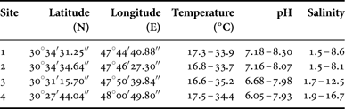
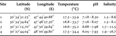
Monthly phytoplankton samples were collected from four sampling sites along the Shatt Al-Arab River (Figure 1; Table 1) between November 2009 and July 2010 (with the exception of February and June 2010) using Van Dorn water sampler (Wildco) and 20 µm mesh size plankton net. The samples were preserved with Lugol's iodine solution. Simultaneously, water temperature, pH and salinity were measured in situ using a multi-meter (WTW, 350i) (Table 1). In the laboratory, 1-L water samples for the phytoplankton quantitative analysis were concentrated to 30 ml. Cell concentrations were determined with Sedgwick Rafter counting cell under light microscopy.
List of the sampled sites in the Shatt Al-Arab River with ranges of the main environmental factors during the studied period.
Microscopic preparation
Cell morphology was examined with the Leica DM 2500 microscope (Leica, Wetzlar, Germany) equipped with epifluorescence (100 W short-arc mercury lamp) and Leica DFC 420C digital camera at 1000× magnification. LM observation of the thecal plate tabulation was performed on cells stained with Calcofluor White M2R (Sigma Chemical Co., St Louis, MO, USA) according to the method of Fritz & Triemer (Reference Fritz and Triemer1985).
Morphometric measurements were made from the calibrated digital LM images using Leica Application Suite v. 3.7 software (Leica Microsystems Ltd, Heerbrugg, Switzerland). Cell dimensions are given as the average ± SD. The nomenclature of Janofske (Reference Janofske2000) was applied for description of the sulcus composition. The general dinoflagellate classification scheme proposed by Fensome et al. (Reference Fensome, Taylor, Norris, Sarjeant, Wharton and Williams1993) was adopted.
The statistical analysis was conducted using the PRIMER v. 6.0 software package (Primer-E Ltd, Plymouth, UK; Clarke & Gorley, Reference Clarke and Gorley2006) and the figures were plotted using SigmaPlot v. 10.0.
RESULTS
During the phytoplankton survey, a dense population of Scrippsiella-like dinoflagellate was revealed in the Shatt Al-Arab River. Based on LM examination of the thecal plate arrangement, the observed dinoflagellate was assigned to Scrippsiella trochoidea.
SYSTEMATICS
Class Dinophyceae West & Fritch, 1927
Order Peridiniales Haeckel, 1894
Family Peridiniaceae Ehrenberg, 1830
Subfamily Calciodinelloideae Fensome et al., Reference Fensome, Taylor, Norris, Sarjeant, Wharton and Williams1993
Genus Scrippsiella Balech ex A.R. Loeblich III, 1965
Scrippsiella trochoidea (Stein) Loeblich III, 1976
(Figure 2)
Light and epifluorescent micrographs of Scrippsiella trochoidea isolated from the Shatt Al-Arab River: (A) Lugol's-preserved cell, centrally located nucleus (n) and peripherally arranged chloroplasts are visible; (B, C) squashed cell in ventral (B) and dorsal (C) view showing the thecal plate pattern; (D) squashed cell in ventral view; (E) Apical pore complex with surrounded plates, apical view; (F, G) detail of the sulcus area; (H) squashed cell in dorsal view showing the epithecal plate pattern; (I) antapical view showing the hypothecal plate pattern. Abbreviations: Po – apical pore plate; x – canal plate; 1–4′– apical plate series; 1–3a – anterior intercalary plates; 1–7″ – precingular plate series; 1–6c – cingular plate series; t – transitional plate; Sa – anterior sulcal plate; Sd – right sulcal plate; Ss – left sulcal plate; Sp – posterior sulcal plate; af – anterior flagellar pore plate; pf – posterior flagellar pore plate; 1–5″′ – postcingular plate series; 1–2″″– antapical plate series. Scale bars: A–D, F, H and I, 10 µm; E and G, 5 µm.
DESCRIPTION
The vegetative cells were tear-shaped, 16–27 µm long (20.80 ± 3.03 µm on average) and 13–23 µm wide (17.40 ± 2.89 µm on average) with conical to helmet-shaped epitheca and rounded hypotheca (Figure 2A). The epitheca was slightly longer than the hypotheca. The cingulum was wide, excavated, almost median and slightly descending. It is displaced by about half a cingular width (Figure 2B–D, H). A large, round nucleus was located in the centre of the cell (Figure 2A). The cells contained several disc-shaped chloroplasts arranged peripherally (Figure 2A).
The plate formula was Po, x, 4′, 3a, 7″, 6c (t + 5c), 6 s, 5″′, 2″″. The epitheca consisted of 16 plates (Figure 2B, C, H). The apical pore plate (Po) was round, surrounded by a raised rim to form a small distinct apical process (Figure 2A, E). The canal plate (x) was small, elongated and situated between Po, 1′, 2′ and 4′ on the ventral side of the cell (Figure 2B, E). The Po plate was located at the epithecal centre, encircled by four apical plates (1′–4′), of which plate 1′ was in contact with the sulcus and connected to the apical pore plate by the canal plate (Figure 2B, E, G). The first apical plate 1′ was narrow and had an ortho configuration (Figure 2B, D, E). Three anterior intercalary plates (1a–3a) were almost equal in size, contiguous and located dorsally. Plates 1a and 3a were pentagonal, whereas the plate 2a was hexagonal (Figure 2C, E, H). The precingular series consisted of seven unequal plates (1″–7″) (Figure 2B, C, H).
The cingulum consisted of six unequal plates (1c–6c), among which the first cingular plate (1c or transitional plate t) was much smaller (Figure 2B–D, F–H). In the sulcus, which consisted of six plates (Figure 2B, D–G), the hook-shaped anterior (Sa), left (Ss), right (Sd) and posterior (Sp) sulcal plates, and a small anterior (af) and posterior (pf) flagellar pore plates were discerned.
The hypotheca consisted of seven plates (Figure 2B–D, I). Postcingular series included five plates (1″′–5″″), among which plates 1″′, 2″′ and 4″′ were rectangular, whereas plates 3″′ and 5″′ were pentagonal. Two antapical plates (1″″–2″″) were nearly equal in size and pentagonal in shape (Figure 2B, C, I).
Scrippsiella trochoidea abundance and distribution in Iraqi waters
The dinoflagellate Scrippsiella trochoidea was occasionally observed in the composition of phytoplankton along the Shatt Al-Arab River. This species proliferated during a short time period, mainly November and December 2009, at all sampling sites and reached a maximum density of 0.44–0.51 × 10⁶ cells l−1 at the downstream stretch of the river (Figure 1, sites 3 and 4) in November 2009. Furthermore, lower densities were found only at the downstream sites in July 2010 (Figure 3).
Numerical abundance (106 cells l−1) of Scrippsiella trochoidea (olive green bars) and box plots for the ranges of salinity values (blue boxes) between November 2009 and July 2010 at four sampling sites along the Shatt Al-Arab River. The line through the middle of the box shows the median, the outer edges of the box correspond to the 25th and 75th percentiles and the crosses represent the extreme values.
Scrippsiella trochoidea was recorded in a wide range of water temperature (17.9°C–35.2°C). The pH value was above 7.0 during high population density. The density of the S. trochoidea population and its distribution were strongly correlated with a salinity pattern along the Shatt Al-Arab River. The development of this species in Iraqi waters was limited within salinities ranging from 2.3 to 16.7 (Figure 4). The Spearman rank correlation analysis revealed a significant positive relationship between the abundance of S. trochoidea and salinity (R = 0.816, P < 0.001); however, no significant correlations were found with water temperature and pH.
Relationship between the abundance of Scrippsiella trochoidea and salinity during November 2009–July 2010 in the Shatt Al-Arab River. The solid line represents the linear regression and the dashed lines correspond to the 95% confident limits.
Qualitatively, co-occurring phytoplankton was represented by rather abundant dinoflagellate Peridiniopsis sp., diatoms Cocconeis placentula, Entomoneis spp., Nitzschia spp., Pleurosigma sp., Thalassiosira spp., Synedra sp., Surirella sp., as well as by non-diatom algae Oocystis sp., Pediastrum sp., Actinastrum sp. and Scenedesmus spp. and cyanobacterium Oscillatoria sp.
DISCUSSION
Within Scrippsiella trochoidea, there are several cryptic species, representing strains of the same ribotype that are morphologically and genetically diverse but were initially considered as a single morphospecies (Montresor et al., Reference Montresor, Sgrosso, Procaccini and Kooistra2003; Gottschling et al., Reference Gottschling, Knop, Plotner, Kirsch, Willems and Keupp2005). This ambiguity in taxonomy of S. trochoidea is still ongoing among researchers. The thecal tabulation of the species has been reported as Po, x, 4′, 3a, 7″, 6c, 5–6 s, 5″′, 2″″ (Lewis, Reference Lewis1991; Janofske, Reference Janofske2000; Montresor et al., Reference Montresor, Sgrosso, Procaccini and Kooistra2003). Although the composition of sulcus is a conservative specific feature and a significant character for the taxonomy of armoured dinoflagellates (Balech, Reference Balech1980), the number of sulcal plates reported in S. trochoidea varies from five (e.g. Lewis, Reference Lewis1991; Montresor et al., Reference Montresor, Sgrosso, Procaccini and Kooistra2003) to six (Janofske, Reference Janofske2000). In the recent emended description of this taxon, Janofske (Reference Janofske2000) distinguished six sulcal plates, namely the anterior, left, right and posterior sulcal plates, and minor anterior and posterior flagellar pore plates, although the author did not give account to the difference in sulcal plate number with previous descriptions. However, in a study by Montresor et al. (Reference Montresor, Sgrosso, Procaccini and Kooistra2003), only five sulcal plates were described in 16 strains of S. trochoidea isolated from the Gulf of Naples. In addition to the anterior, left, right and posterior sulcal plates, a minor sulcal median plate was identified, which corresponded to the posterior flagellar pore plate in the plate notation proposed by Janofske (Reference Janofske2000), whereas the sixth anterior flagellar pore plate was not distinguished (Montresor et al., Reference Montresor, Sgrosso, Procaccini and Kooistra2003).
In the sulcus of the cells isolated from Iraqi waters, six sulcal plates were discerned in this study corroborating the observations of Janofske (Reference Janofske2000). Besides sulcus composition, the cells of S. trochoidea from the Shatt Al-Arab River were characterized by almost uniform-sized anterior intercalary plates that differed from the described pattern of anterior intercalary series with larger second intercalary plate compared with first and third ones (e.g. Lewis, Reference Lewis1991; Janofske, Reference Janofske2000; Montresor et al., Reference Montresor, Sgrosso, Procaccini and Kooistra2003). Some intrastrain variations in morphology within the S. trochoidea complex were observed previously by Montresor et al. (Reference Montresor, Sgrosso, Procaccini and Kooistra2003). Despite the minor differences in morphology, the vegetative cells of Scrippsiella isolated from the Iraqi waters were assigned to the S. trochoidea complex based on cell size, shape and characteristically narrow first apical plate.
In the present study, S. trochoidea was recorded along the Shatt Al-Arab River sites in a wide range of temperature and salinity, which is consistent with previous reports (e.g. Kim & Han, Reference Kim and Han2000). This reflects the capacity of the species to proliferate in different environmental conditions (Garate-Lizarraga et al., Reference Garate-Lizarraga, Hernández-Orozco, Band-Schmidt and Serrano-Casillas2001; Gomez, Reference Gomez2003; McQuoid, Reference McQuoid2005). The observations of S. trochoidea from this study can be considered as a new record for Iraqi flora. Seawater intrusion from the Arabian Gulf (Hameed et al., Reference Hameed, Ali, Aljorany, Hassan and Al-Hello2013) might have led to the introduction of this species into Iraqi inland ecosystems because S. trochoidea was reported in the adjacent Kuwaiti waters (Al-Kandari et al., Reference Al-Kandari, Al-Yamani and Al-Rifaie2009). The revealed positive relationship between abundance of S. trochoidea and salinity corroborates this assumption. Scrippsiella trochoidea is capable of persisting as coccoid cells for a long time and then germinating into a bloom of vegetative cells when conditions are favourable (Lundholm et al., Reference Lundholm, Ribeiro, Andersen, Koch, Godhe, Ekelund and Ellegaard2011). This ability might assist the species in spreading into adjacent aquatic systems, such as the Mesopotamian Marshes (Figure 1).
The dinoflagellate S. trochoidea is a potentially harmful bloom-forming species of unknown toxicity. This alga has been generally considered non-toxic (Hold et al., Reference Hold, Smith, Rappe, Maas, Moore, Stroempl, Stephen, Prosser, Birkbeck and Gallacher2001); however, the high biomass blooms of this species can negatively impact coastal ecosystems and cause mortality in marine biota. Hallegraeff (Reference Hallegraeff1993) indicated that a bloom of S. trochoidea can cause indiscriminate fish and invertebrate kills through the generation of anoxic conditions. Tang & Gobler (Reference Tang and Gobler2012) reported lethal effects of this species on Eastern oyster larva and Northern quahog larvae. Similarly, Shin et al. (Reference Shin, Jang, Jang, Ju, Lee and Chang2003) showed that egg production of the copepod Acartia omorii was diminished overtime with the consumption of S. trochoidea.
To date, diatoms were known to dominate the phytoplankton of the Shatt Al-Arab River (Huq et al., Reference Huq, Al-Saadi and Hameed1978; Al-Saadi et al., Reference Al-Saadi, Al-Lami and Kassim1996). The presence of bloom-forming and potentially toxigenic cryptic dinoflagellate species in Iraqi aquatic system underscores the need for further studies on ecology, toxicology and phylogeny of S. trochoidea in Iraq that will help in introducing a management programme for the waters of the Shatt Al-Arab River.
ACKNOWLEDGEMENTS
We are indebted to the Marine Science Centre (University of Basrah, Iraq) for financial support of sampling and for supplying facilities for analysis of samples. We are grateful to the anonymous referees for their constructive comments. The authors thank Dr Andrey Sazhin (P.P. Shirshov Institute of Oceanology, Moscow, Russia) for providing optical facilities.