Introduction
Mytilus coruscus is widely distributed along the eastern coastal regions of China, especially along the Zhoushan coastline. As a key component of the intertidal and subtidal ecosystems, it plays a crucial role in maintaining ecological balance by filtering water and providing habitat for other marine organisms. Moreover, M. coruscus is one of the most important economic shellfish species in China, contributing significantly to the aquaculture industry. Its high yield and market value makes it a vital resource for coastal communities, supporting both local economies and food security. Similarly to the other bivalves, the gonadal development in M. coruscus can be categorized into five stages: early gametogenesis, advanced gametogenesis, mature, spawning, and resting phases (Cheng et al. Reference Cheng, Xu, Liu and Qi2013). Due to the differential nutritional requirements during gametogenesis, the accumulation of nutrients in bivalves varies among gonadal development stages (Li et al. Reference Li, Zhao, Wang, Chen, Ma, Liu, Sun, Zhou, Ren, Dou and Wu2024). Carbon (C), nitrogen (N), and phosphorus (P) are fundamental elements in ecosystems, and essential for biological processes in organisms including M. coruscus (Sterner and Elser Reference Sterner and Elser2002). Tian et al. (Reference Tian, Diao and Zhang2024) found that seasonal variations in the accumulation of key elements, such as C, N, and P, in different tissues of male M. coruscus from Gouqi Island were primarily regulated by the stages of gonadal development. However, the dynamic changes in glycogen, amino acids, and fatty acids, which are crucial nutritional substances in the mussel (Grienke et al. Reference Grienke, Silke and Tasdemir2014), during different stages of gonadal development and their relationship with C, N, and P elements remain unclear.
Glycogen plays a central role in energy supply and metabolism during gametogenesis in many bivalves (e.g. Fearman et al. Reference Fearman, Bolch and Moltschaniwskyj2009; Qin et al. Reference Qin, Li, Li, Zhou, Xiang, Ma, Noor, Mo, Zhang and Yu2021). Indeed, glycogen has been suggested to serve as an energy source for bivalve growth and as an energy reserve stored in specific cells during gametogenesis (Qin et al. Reference Qin, Li, Li, Zhou, Xiang, Ma, Noor, Mo, Zhang and Yu2021). Gabbott and Bayne (Reference Gabbott and Bayne1973) also reported that gametogenesis depletes energy reserves, such as glycogen, in the mussel M. edulis. Moreover, the glycogen metabolism may also contribute to its conversion into lipids during gonadal development in mussels (Fearman et al. Reference Fearman, Bolch and Moltschaniwskyj2009). Cheng et al. (Reference Cheng, Xu, Liu and Qi2013) analysed the biochemical composition of the whole edible tissue of M. coruscus at various stages of gonadal development, finding that, although crude protein and fat content peaked during the mature stage, glycogen and ash contents were at their lowest. This suggests that, to meet the energy demands of gametogenesis, carbohydrates in the tissues are converted into more energy-efficient forms, such as lipids and proteins, for storage during gamete development (Napolitano et al. Reference Napolitano, Macdonald, Thompson and Ackman1992). However, the allocation of glycogen between different tissues during gonadal development in M. coruscus remains unclear.
Proteins are the primary constituents of mussel gametes (e.g. Berthelin et al. Reference Berthelin, Kellner and Mathieu2000; Ojea et al. Reference Ojea, Pazos, Martínez, Novoa, Sánchez and Abad2004). As the basic units of proteins, amino acids play key roles in gametogenesis and the development cycle of mussels. For example, glutamic acid and leucine levels increase significantly in the edible tissues of Ensis siliqua after the reproductive season (Napolitano et al. Reference Napolitano, Macdonald, Thompson and Ackman1992), while male M. edulis show higher levels of glycine, glutamic acid, and aspartic acid during the breeding season (Chen et al. Reference Chen, Huang, Zheng, Sun, Dong, Zhou, Zhu and Qin2021). These findings highlight the importance of specific amino acids in the gonadal development of bivalves.
Lipids are essential materials for gamete formation in mussels (Mao et al. Reference Mao, Tong, Xiao, Zhan, Xu and Wang2017). Fatty acids, as important components of lipids, can be metabolized into complex lipid compounds through various pathways and play a crucial role in the physiological functions of animals (Grabner et al. Reference Grabner, Xie, Schweiger, Zechner and Metabolism2021). Panayotova et al. (Reference Panayotova, Merdzhanova, Stancheva, Dobreva, Peycheva and Makedonski2021) analysed the fatty acid composition of Mediterranean mussels M. galloprovincialis and found that the content of saturated fatty acids (SFAs), which serve as energy reserves, was highest during the resting period. This could be because SFAs provide efficient energy storage (Adeva-Andany et al. Reference Adeva-Andany, Carneiro-Freire, Seco-Filgueira, Fernández-Fernández and Mouriño-Bayolo2019; Freites et al. Reference Freites, Fernández-Reiriz and Labarta2002), preparing for subsequent reproductive activities of mussels. Laudicella et al. (Reference Laudicella, Carboni, Whitfield, Doherty and Hughes2023) studied the lipid composition in the gonads of male mussels (Mytilus sp.) and found that long-chain fatty acids, such as eicosapentaenoic acid (EPA) and docosahexaenoic acid (DHA) accumulated in the testes as sperm developed. Martínez-Pita et al. (Reference Martínez-Pita, Sánchez-Lazo, Ruíz-Jarabo, Herrera and Mancera2012) found similar accumulations of EPA and DHA in the gonads of male Mediterranean mussels during sperm formation, as these fatty acids are crucial components of biological membranes (Bell and Sargent, Reference Bell and Sargent1985; Bell et al. Reference Bell, Henderson and Sargent1986; Soudant et al. Reference Soudant, Moal, Marty and Samain1997). This accumulation likely supports the formation of more membrane structures in mature sperm, aiding their motility and fertilization function during reproduction (Lubet et al. Reference Lubet, Brichon, Besnard and Zwingelstein1986).
Therefore, this study aims to investigate the dynamic changes in the key nutrients, such as glycogen, amino acids, and fatty acids, as well as their association with carbon, nitrogen, and phosphorus elements, during the gonadal development of male M. coruscus cultured on a mussel farm around Gouqi Island. Conducted under authentic mariculture conditions, this field investigation specifically examines nutrient flux patterns in relation to reproductive physiology. By measuring these nutritional components and analysing the changes in key nutrient elements, this study will explore the relationship between key nutrients and nutrient elements and clarify the nutrient accumulation process during gonadal development in bivalves. The findings provide operational benchmarks for optimizing feeding regimes and environmental management in bivalve cultivation systems. This mechanistic understanding of nutrient allocation processes during gametogenesis advances our knowledge of physiological energetics in commercially important mollusk species.
Materials and methods
Sample collection and tissue extraction
This experiment was conducted from October 2022 to September 2023. Each month, 20 healthy mussels (shell length 75–85 mm) were collected from a mussel farm (E 122°46′16″, N 30°43′) near Gouqi Island, Zhejiang Province. The mussels were dissected, and their gonads and somatic tissues (excluding gonads) were weighed separately using a microbalance (accuracy 0.001 g). Subsequently, the two types of tissues were immediately snap-frozen in liquid nitrogen and stored at −80℃ for further analyses.
After identifying the sex and gonadal developmental stages through tissue sections, nine male mussels from each gonadal developmental stage were randomly selected (Tian et al. Reference Tian, Diao and Zhang2024). The remaining gonadal and somatic tissues were divided into two groups. One group was weighed and stored at −80°C for amino acid, fatty acid, and glycogen analysis. The other group was placed in an oven and dried at 80°C to a constant weight for nutrient element analysis. For each set of three individuals, mixed samples for each of two types of tissues (gonads and somatic tissues) were used for nutrient element and nutrient content analysis.
Measurement of glycogen, amino acids, and fatty acids in different tissues
Glycogen analysis
The glycogen content in the gonadal and somatic tissues of the mussels was measured using a glycogen assay kit (Beijing Solarbio Technology Co., Ltd., Beijing, China). For each tissue, a 0.1 g sample was taken from the tissues stored at −80°C, placed in a 10 mL tube, and finely chopped. After adding 0.75 mL extraction solution (Glycogen content assay kit, Beijing Boxbio Science & Technology Co., Ltd), the mixture was boiled in a water bath for 20 mins (covered to prevent evaporation), with shaking every 5 mins for homogenization. Once the tissue was dissolved, the mixture was cooled and diluted to 5 mL with distilled water. After centrifuging at 2500 rpm for 10 min at 25°C, the supernatant was used to measure glycogen content at 620 nm using visible spectrophotometry.
Amino acid analysis
For amino acid composition analysis, 1.5 g samples from each tissue at each gonadal developmental stage were sent to Zhejiang Tiancheng Testing Technology Co., Ltd. for automated amino acid analysis (L-8900, Hitachi, Tokyo, Japan). The analysis measured 18 amino acids, including asparagine, serine, glutamate, proline, glycine, alanine, cystine, tyrosine, arginine, histidine, threonine, valine, methionine, isoleucine, leucine, phenylalanine, lysine, and tryptophan. The total amino acid (AAs) content and the total essential amino acid (EAAs) content were calculated based on the measured levels.
Fatty acid analysis
Fatty acids were measured according to the method of Folch et al. (Reference Folch, Lees and Stanley1957). The gonadal and somatic tissue samples were freeze-dried, and total lipids were extracted using chloroform/methanol (2:1, v/v), followed by methylation into fatty acid methyl esters (FAME). These were analysed using a gas chromatograph (Agilent 7890B) equipped with a detector (Flame Ionization Detector, FID) to determine the total fatty acid content and the percentage of each fatty acid. The gas chromatography conditions were set as follows: carrier gas (nitrogen) flow rate at 1 mL min−1, injection temperature at 250°C, pressure at 20.363 psi, total flow rate at 104 mL min−1, and split flow rate at 100:1. The column temperature was ramped from 70°C to 220°C and then held at 250°C. The detector temperature was set at 300°C. The column used is an Agilent J&W DB-23 from Agilent Technologies, with the exact model being 122-3232. The fatty acids analysed included saturated fatty acids (SFAs), unsaturated fatty acids (UFAs), monounsaturated fatty acids (MUFAs), and polyunsaturated fatty acids (PUFAs).
Measurement of carbon, nitrogen, and phosphorous in different tissues
Carbon and nitrogen analysis
For nutrient element analysis, approximately 0.001 g of dried and ground gonadal and somatic tissue samples were placed in tin capsules and analysed for carbon (C) and nitrogen (N) content using a C/N elemental analyser (FlashSmart, Thermo Fisher Scientific, USA). The C and N contents were calculated based on the weight of the samples.
Phosphorous analysis
Additionally, 0.01 g of each tissue sample was used for phosphorus (P) analysis. The samples were treated with potassium persulfate solution and autoclaved at 121°C for 45 min, then cooled to room temperature. After adding a color reagent, the mixture was centrifuged, and the absorbance at 880 nm was measured using a spectrophotometer (DR6000, Hach, USA). The P content was determined based on a standard curve created using a potassium dihydrogen phosphate solution.
Statistical analysis
According to Tian et al. (Reference Tian, Diao and Zhang2024), more than 85% of mussels cultured in Gouqi Island are males, in this study, we focus on the males in the following analysis. Then, data including the content of glycogen, amino acids, and fatty acids in the gonad and somatic tissues at different gonadal developmental stages were log-transformed to stabilize the variance. The Shapiro–Wilk and Levine tests were then used to test for normality and homogeneity, respectively. After confirming the transformed data met these assumptions, the effect of gonad development stages on these data was tested using a one-way ANOVA. Tukey’s multiple comparison test was applied to analyse significant differences at P < 0.05.
Furthermore, Pearson’s correlation analysis was performed on glycogen content and C content in the gonads and somatic tissues, respectively. Principal component analysis (PCA) was conducted to explore the relationships between the different amino acids and N content, as well as the fatty acids and P content in the gonads and somatic tissues. This analysis also aimed to analyse the dynamic changes of these nutrients during different gonadal developmental stages. Components with eigenvalues greater than 1.0 were extracted to explain the variability of these data. All the statistical tests were performed in R version 4.3.0 (R Core Team 2023).
Results
Changes in glycogen content in gonad and somatic tissues at different gonadal developmental stages
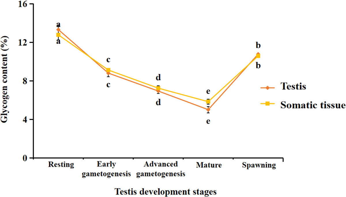
The one-way ANOVA results show that glycogen content in both gonadal and somatic tissues of male mussels significantly varied at different gonadal developmental stages (Table S1A). Glycogen content significantly decreased in both tissues from the resting stage to the maturity stages, reaching its lowest level at the maturity stage (Figure 1). Later, glycogen content began to significantly increase in both tissues during the spawning stage (Figure 1).
Changes of glycogen content among testis developmental stages in the gonads and somatic tissues of male M. coruscus. Error bars represent SD among the replications. Significant differences in glycogen content among different stages of testes development are indicated by different alphabets.

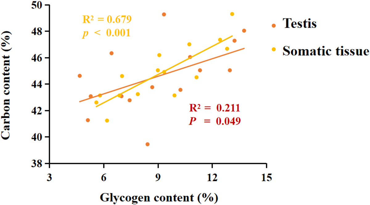
Pearson correlation analysis showed that glycogen content was positively correlated with C content in both gonads (R 2 = 0.211, P = 0.049) and somatic tissues (R 2 = 0.679, P < 0.001) (Figure 2).
Relationship between carbon content and glycogen in the testis and somatic tissues of male M. coruscus.

Changes in amino acid content in gonad and somatic tissues at different gonadal developmental stages
The results of one-way ANOVA indicate that the content of certain amino acids in the two tissues of male M. coruscus exhibits significant differences at different stages of gonadal development (Table S1B). In the testes, the contents of aspartic acid, glycine, tyrosine, isoleucine, EAAs, and AAs showed significant variation during gonadal development, with a trend of gradual increase as gonad development progressed. Notably, tyrosine, arginine, threonine, lysine, EAAs and AAs reached their highest levels during the mature stage (Table 1).
Variation of amino acid content among testis developmental stages in the testes and somatic tissues of male Mytilus coruscus.

Significant differences in glycogen content among testis development stages are indicated by different alphabets.
In the somatic tissues, significant differences were observed in the contents of aspartic acid, glutamic acid, proline, glycine, tyrosine, arginine, histidine, threonine, methionine, isoleucine, leucine, phenylalanine, lysine, EAAs, and AAs at different stages of gonadal development (Table S1B). Among these, aspartic acid, histidine, threonine, leucine, lysine, EAAs, and AAs peaked during the mature stage (Table 1). Additionally, with the exception of proline, and isoleucine, the contents of the most amino acids showed a general increasing trend during testes development.
To better understand the accumulation of amino acids and their relationship with N in the two tissues during different stages of gonadal development, PCA was performed. In the testes, the first two PCs explained 86.80% of the variation (Figure 3A, Table S2). PC1 was positively correlated with aspartic acid, serine, glutamic acid, glycine, tyrosine, threonine, lysine and AAs, while it was negatively correlated with proline, alanine, cysteine, histidine, valine, isoleucine, phenylalanine, tryptophan, EAAs and N content (Table S2). PC2 was positively correlated with serine levels and negatively correlated with aspartic acid, alanine, tyrosine, arginine, threonine, methionine, leucine, lysine, tryptophan, EAAs, AAs, and N content (Table S2).
Factor distribution of nitrogen and amino acids during testis developmental stages in testes (A) and somatic tissues (B) of male M. coruscus in PCA. Asp: Asparagine; Ser: Serine; Glu: Glutamate; Pro: Proline; Gly: Glycine; Ala: Alanine; Cys: Cystine; Tyr: Tyrosine; Arg: Arginine; His: Histidine; Thr: Threonine; Val: Valine; Met: Methionine; Ile: Isoleucine; Leu: Leucine; Phe: Phenylalanine; Lys: Lysine; Try: Tryptophan.

In the somatic tissues, the first two principal components explained 88.59% of the variation (Figure 3B, Table S2). PC1 was positively correlated with aspartic acid, glutamic acid, glycine, alanine, cysteine, histidine, threonine, valine, methionine, isoleucine, leucine, phenylalanine, lysine, EAAs, AAs, and N content, but negatively correlated with serine, tyrosine, and arginine (Table S2). PC2 showed positive correlations with proline and valine, and negative correlations with serine, alanine, cysteine, tyrosine, arginine, tryptophan, and N content (Table S2). Additionally, most of amino acids accumulated during the gonadal development stages in both the gonadal and somatic tissues, especially for aspartic acid, glycine, tyrosine, isoleucine, leucine, lysine, glutamic acid, glutamic acid, tyrosine and arginine, EAAs, and AAs (Figure 3).
Changes in fatty acid content in gonad and somatic tissues at different gonadal developmental stages
A total of 34 fatty acids and total fatty acids (FAs) were measured, and the contents of SFAs, UFAs, MUFAs, and PUFAs were calculated. The results of One-way ANOVA indicated that the content of most fatty acids in the two tissues of male M. coruscus varied significantly at different stages of gonadal development (Table S1C). Specifically, C8:0, C10:0, C11:0, C12:0, C14:0, C18:0, C20:0, C14:1, C16:1, C18:3 n-3, and FAs did not show significant changes across stages. Among the remaining fatty acids that exhibited significant differences, C4:0, C6:0, C11:0, C13:0, C14:1, C18:1cis, SFAs reached their highest levels at the resting stage and then decreased during the maturation period. Meanwhile C15:0, C16:0, C17:0, C21:0, C22:0, C23:0, C24:0, C15:1, C16:1, C17:1, C18:1trans, C24:1, C18:3 n-3, C20:2, C20:3 n-6, C20:3 n-3, C22:2, EPA, DHA, UFAs, MUFA, and PUFAs showed an increasing trend.
In the somatic tissues, significant differences in the content of C8:0, C10:0, C11:0, C14:0, C17:0, C21:0, C22:0, C23:0, C22:2, C24:0, C18:1cisC16:1, C20:3 n-3, and C20:3 n-6 were observed at different stages of gonadal development (Table S1C). Among these, C11:0, C12:0, C14:0, C15:0, C16:0, C17:0, C18:0, C20:0, C21:0, C22:0, C23:0, C24:0, C14:1, C18:1cis, C18:1trans,C18:3 n-6,C18:3 n-3, C20:3 n-3C20:3 n-3 and C22:2 showed an increasing trend, while C13:0 and C17:1 reached their highest levels during the resting stage and then decreased at the maturation period. No significant differences were observed in the content of the other fatty acids in the somatic tissues across different stages of gonadal development (Figure 4, Table 2).
Factor distribution of phosphorus and fatty acids during testis developmental stages in testes (A) and somatic tissues (B) of male M. coruscus in PCA.

Variation of fatty acid content among testis developmental stages in the testes (%) and somatic tissues (%) of male Mytilus coruscus. Significant differences in glycogen content among testis development stages are indicated by different alphabets

To clarify the accumulation of fatty acids in the two tissues of male M. coruscus during different stages of gonadal development and the relationship between P and amino acids, PCA was conducted. The PCA results indicated that the first PCs explained 79.49% of the variation in the testes (Figure 4A, Table S3). PC1 was positively correlated with C15:0, C16:0, C17:0, C18:0, C21:0, C22:0, C23:0, C15:1, C16:1, C20:1 n-9, C24:1, C18:2trans, C18:3 n-3, C20:2, C22:2, DHA, UFAs, and MUFAs, while negatively correlated with C4:0, C6:0, C8:0, C10:0, C11:0, C12:0, C13:0, C20:0, C14:1, C20:3 n-6, EPA, and SFAs. PC2 was positively correlated with C4:0, C16:0, C17:0, C21:0, C22:0, C23:0, C16:1, C18:1cis, and SFAs, while it was negatively correlated with C15:0, C24:0, C18:1trans, C18:2trans, C24:1, C18:3 n-6, C20:3 n-3, C20:3 n-6, C22:2, EPA, DHA, UFAs, PUFAs, FAs, and P content.
In the somatic tissues, the first two PCs explained 67.93% of the variation (Figure 4B, Table S3). PC1 was positively correlated with C12:0, C14:0, C16:0, C20:0, C23:0, C18:1cis, C18:1trans, C20:1 n-9, C18:3 n-6, C20:3 n-6, C22:2, EPA, SFAs, UFAs, MUFAs, and P content, while negatively correlated with C4:0, C6:0, C8:0, C13:0, C17:0, C18:0, C21:0, C17:1, C24:1, C18:3 n-3, C20:2, C20:3 n-3. PC2 was positively correlated with C6:0, C8:0, C11:0, C12:0, C13:0, C17:1, C18:2trans, EPA, MUFAs, and P, while negatively correlated with C10:0, C15:0, C16:0, C17:0, C18:0, C21:0, C22:0, C24:0, C14:1, C15:1, C20:3 n-3, C22:2, DHA, PUFAs, FAs (Table S3). Additionally, although the fatty acid contents in the somatic tissues did not cluster together at different stages of gonadal development, most fatty acids accumulated during the gonadal development stages in the testis, especially for FAs, UFAs, PUFAs, MUFAs, EPA and DHA (Figure 4).
Discussion
Relationship between glycogen content and gonadal development in male Mytilus coruscus
The glycogen content in different tissues of male M. coruscus varies across different stages of gonadal development. In this study, glycogen levels were highest in both tissues at the resting period, with a gradual decrease during the proliferation and maturation phases. Previous studies have confirmed that changes in glycogen levels in bivalves are closely associated with reproductive activity, with the highest glycogen levels typically observed in the non-reproductive phases (Dridi et al. Reference Dridi, Romdhane and Elcafsi2007; Ojea et al. Reference Ojea, Pazos, Martínez, Novoa, Sánchez and Abad2004; van Houcke et al. Reference van Houcke, Medina, Linssen and Luten2016). This is likely due to the rapid breakdown of glycogen to provide energy for the body’s activities, making glycogen the preferred energy form for supporting gametogenesis and growth in bivalves, including mussels (Li et al. Reference Li, Li, Wang, Li and Zhang2020, Reference Li, Song, Meng, Li and Zhang2017). Moreover, glycogen metabolism also plays a crucial role in lipid biosynthesis, which is essential for the formation of gametes. During gametogenesis, the breakdown of glycogen provides glycerol-3-phosphate, a key precursor for the synthesis of lipids such as triglycerides and phospholipids (Scanes Reference Scanes, Scanes and Dridi2022). These lipids are critical for the structural integrity of gametes and for providing energy reserves necessary for early embryonic development (Li et al. Reference Li, Zhao, Wang, Chen, Ma, Liu, Sun, Zhou, Ren, Dou and Wu2024). Therefore, the utilization of glycogen-derived glycerol-3-phosphate for lipid biosynthesis is an integral part of the reproductive process in bivalves.
In bivalves, glycogen is primarily stored in the mantle and gonads, with nearly all tissues undergo glycogen hydrolysis and glucose formation to provide ATP for gametogenesis (Cumplido et al. Reference Cumplido, Marinho and Bigatti2020; Murata et al. Reference Murata, Touhata and Miwa2020). Glycogen accumulation in both tissues begins during the spawning phase and peaks during the resting phase. This may be because these stages represent periods of low gonadal activity, allowing glycogen to accumulate in preparation for the upcoming gametogenesis. During the reproductive and growth phases, which have high energy demands due to gametogenesis and proliferation, glycogen levels in both tissues decline, reaching their lowest point during gonadal maturation. This suggests that the glycogen stored in both the gonads and somatic tissues is gradually consumed during gonadal development (Bacca et al. Reference Bacca, Huvet, Fabioux, Daniel, Delaporte, Pouvreau, Van Wormhoudt and Moal2005; Cumplido et al. Reference Cumplido, Marinho and Bigatti2020).
The positive correlation between C content and glycogen accumulation was also observed in our study. This correlation can be explained by the fact that glycogen is a polysaccharide composed of glucose, which contains carbon, hydrogen, and oxygen atoms (Adeva-Andany et al. Reference Adeva-Andany, Gonzalez-Lucan, Donapetry-Garcia, Fernández-Fernández and Ameneiros-Rodríguez2016). Carbon, as a major element in glucose, is metabolized into glucose and stored as glycogen. Thus, the accumulation of C in these tissues is closely related to glycogen storage (Burke et al. Reference Burke, van Loon and Hawley2017).
Relationship between amino acids and gonadal development in male Mytilus coruscus
Significant differences were observed in the amino acid content across different stages of gonadal development in male M. coruscus tissues. Among the amino acids, aspartic acid, glycine, tyrosine, isoleucine, EAAs, and AAs exhibited significant changes, with their levels gradually increasing during gonadal development. Dridi et al. (Reference Dridi, Romdhane and Elcafsi2007) found similar results in Pacific oysters (Crassostrea gigas), where the total amino acid content in the gonads and visceral mass increased with gonadal development. This may be due to the increased demand for amino acids during gonadal development, as cellular metabolism and protein synthesis intensify (Cheng, Reference Cheng2014). While amino acids are crucial for protein synthesis (Dong et al. Reference Dong, Qin, Xue, Du, Lin, Xu and Zhu2018), they may also contribute to energy production, supporting gametogenesis and embryonic development (Izquierdo et al. Reference Izquierdo, Fernández-Palacios and Tacon2001; Li et al. Reference Li, Han, Zheng and Wu2021; Lovenberg et al. Reference Lovenberg, Weissbach and Udenfriend1962).
According to the PCA results, both the gonadal and somatic tissues in male M. coruscus accumulate essential amino acids during gonadal development. This suggests that essential amino acids play a crucial role in gonadal development in male M. coruscus (Wu, Reference Wu2013). Isoleucine, leucine, and lysine exhibited trends similar to those of EAAs. This may be due to branched-chain amino acids (BCAAs) such as isoleucine can activate protein synthesis and other vital processes as signaling molecules (Neinast et al. Reference Neinast, Murashige and Arany2019). Among them, leucine, a ketogenic amino acid, not only generates energy through oxidative metabolism but also stimulates protein synthesis, inhibits protein degradation, and enhances protein activity (Bandyopadhyay et al. Reference Bandyopadhyay, Todorova, Pavlova and Overholtzer2022; Posey and Davis Reference Posey and Davis2023). Lysine is an essential amino acid that plays important roles in regulating metabolic balance, enhancing the absorption of other proteins, and promoting growth and development (Bibhas et al. Reference Bibhas, Anoy, Mahesh, Gayen, Mandal, Sardar and Tarafdar2022).
In addition to EAAs, aspartic acid, glutamic acid, and glycine also showed a trend of gradual accumulation during gonadal development. These non-EAAs are critical for cellular structure and function. Among them, aspartic acid acts as a neurotransmitter with multiple physiological functions and plays an important role in reproductive regulation (Li et al. Reference Li, Han, Yin, Li and Yin2018). Glutamic acid, a precursor for synthesizing glutathione and glutamine, is involved in protein and amino acid synthesis and influences cellular energy metabolism (Kunio et al. Reference Kunio, Hisayuki and Eiji2013; Brosnan and Margaret, Reference Brosnan and Margaret2013; He et al. Reference He, Furukawa, Toyomizu, Nochi, Bailey and Wu2021). Glycine, as a neurotransmitter, participates in signal transmission and the synthesis of various nitrogen-containing compounds like glutathione (Aragón and López-Corcuera Reference Aragón and López-Corcuera2003), thus facilitating gonadal development. These amino acids play critical roles in protein synthesis and cellular metabolism during the growth phase, contributing to growth and development.
In the gonads, AAs were found in lower quantities than in somatic tissues during all stages of gonadal development. This may be due to the mantle being the primary storage tissue in Mytilus species (Guo, Reference Guo2017), while the gonads focus on gametogenesis, requiring dynamic nutrient allocation rather than long-term storage (Tabakaeva and Tabakaev Reference Tabakaeva and Tabakaev2016). Tyrosine and arginine exhibited stage-specific accumulation patterns: while abundant in somatic tissues during the growth phase, they peaked in gonads during maturation. This shift aligns with their distinct metabolic roles. Tyrosine serves as a precursor for L-DOPA, which is enzymatically converted to dopamine – a neurotransmitter regulating gonadotropin release and reproductive behavior in mollusks (Schenck and Maeda Reference Schenck and Maeda2018). Elevated tyrosine levels during maturation may thus support neuroendocrine signaling essential for spawning synchronization. Arginine accumulation correlates mechanistically with its dual roles in gonadal maturation. First, as a substrate for nitric oxide synthase (NOS), arginine drives nitric oxide (NO) production, a signaling molecule modulating apoptosis during oocyte maturation and spermatid differentiation (Gupta and Uversky Reference Gupta and Uversky2024). Second, arginine-derived polyamines (e.g., spermidine) are critical for DNA stabilization in developing gametes and redox homeostasis during rapid cell proliferation (Halloran et al. Reference Halloran, Stenhouse, Wu and Bazer2021). These interconnected pathways underscore arginine’s importance in meeting the metabolic demands of gamete formation.
Relationship between fatty acids and gonadal development in male Mytilus coruscus
The content of various fatty acids in two tissues of male M. coruscus showed significant differences at different stages of testicular development. In this study, no significant differences were found in the fatty acid content in the somatic tissue of male M. coruscus at different gonadal developmental stages, but fatty acids in the testes accumulated progressively during gonadal development. A similar trend was observed by Dridi et al. (Reference Dridi, Romdhane and Elcafsi2007) in Pacific oysters (C. gigas). The increase in fatty acids during gonadal development is likely to meet the energy and structural demands of gonadal development and function maintenance. During testicular development, male M. coruscus requires a large amount of energy to support the formation and development of reproductive cells. Lipids are the primary energy reserve for gametogenesis and larval development in most marine bivalves (Holland, Reference Holland1978), and fatty acids are important components of lipids and a key energy source for organisms. Therefore, during gonadal development, male M. coruscus may synthesize more fatty acids to meet energy demands. Studies by Pazos et al. (Reference Pazos, Román, Acosta, Abad and SaíNchez1997) have shown that fatty acid levels are good indicators of gonadal maturation. Additionally, fatty acids are essential components of cell membranes (Grabner et al. Reference Grabner, Xie, Schweiger, Zechner and Metabolism2021), playing an important role in the structure and function of cells. Therefore, during gonadal development, male M. coruscus may synthesize more fatty acids to maintain both, the normal structure and function of the testes.
In the testes, UFAs also accumulated progressively with gonadal development. The accumulation trend of PUFAs was similar to that of MUFAs, with the accumulation of PUFAs being particularly prominent. This could be because UFAs, as efficient energy sources, support sperm production and maturation. PUFAs, as the main component of fatty acids in mussels, are closely related to growth and reproduction (Alkanani et al. Reference Alkanani, Parrish, Thompson and McKenzie2007). For example, Leonardos and Lucas (Reference Leonardos and Lucas2000) found a positive correlation between PUFAs and ω3 fatty acids and juvenile mussel growth. Levental et al. (Reference Levental, Malmberg, Symons, Fan, Chapkin, Ernst and Levental2020) demonstrated that higher levels of PUFAs influence gamete maturation and maintain membrane fluidity. Furthermore, the accumulation of EPA and DHA in PUFAs during gonadal development reached higher levels during the testicular maturation phase, consistent with the findings of Dridi et al. (Reference Dridi, Romdhane and Elcafsi2007). High levels of EPA and DHA play a significant role in the structure and function of cell membranes during gametogenesis (Soudant et al. Reference Soudant, Moal, Marty and Samain1996, Reference Soudant, Van Ryckeghem, Marty, Moal and Sorgeloos1999).
In contrast to UFAs, SFAs in the testes primarily accumulated during the resting phase, likely because the resting phase of M. coruscus from Gouqi Island occurs mainly in summer (Tian et al. Reference Tian, Diao and Zhang2024). On one hand, mussels may store energy for gonadal development in the fall, and on the other hand, they may require significant energy to cope with the high-temperature environment during summer. SFAs are generally more stable than UFAs and are better suited for energy storage (Chen et al. Reference Chen, Ju and Chen2008). Moreover, SFAs are the most abundant fatty acids in the testes during different stages of gonadal development, which may be because marine environments provide food sources with a higher proportion of SFAs (Liu et al. Reference Liu, Ke, Li, Liao and Huang2014; Lv et al. Reference Lv, Han, Sun and Wang2009).
In contrast to UFAs, SFAs in the testes primarily accumulated during the resting phase, likely because the resting phase of M. coruscus from Gouqi Island occurs mainly in summer (Tian et al. Reference Tian, Diao and Zhang2024). On one hand, mussels may store energy for gonadal development in the fall, and on the other hand, they may require significant energy to cope with the high-temperature environment during summer. SFAs are generally more stable than UFAs and are better suited for energy storage (Chen et al. Reference Chen, Ju and Chen2008). Moreover, SFAs are the most abundant fatty acids in the testes during different stages of gonadal development, which may be because marine environments provide food sources with a higher proportion of SFAs (Liu et al. Reference Liu, Ke, Li, Liao and Huang2014; Lv et al. Reference Lv, Han, Sun and Wang2009).
Phosphorus accumulation and its role in gonadal development
Furthermore, P gradually accumulated in both tissues with testicular development. P is an important component of DNA and RNA and plays a role in gene expression and genetic material replication (Bird and Eskin, Reference Bird and Eskin2021). As testicular development progresses, M. coruscus may require more P to support the synthesis of genetic material and promote the development and division of reproductive cells. P is a component of ATP, which serves as the energy reserve molecule within cells, providing the energy required for cellular activities (Nicholls et al. Reference Nicholls, Chin, Williams, Lenton, Flaherty and McGrath2023). Gonadal development and reproductive processes typically require substantial energy, and thus, the accumulation of P in mussels may be related to ATP synthesis and energy metabolism. P is also a key component of phospholipids, which are vital for the synthesis and function of cell membranes (Sterner and Elser Reference Sterner and Elser2002). As testicular development progresses, more P may be needed to maintain membrane integrity and function to support the growth and differentiation of reproductive cells. Additionally, P is involved in cell signaling (Sabbagh, Reference Sabbagh2013; Michigami, Reference Michigami2021; Michigami et al. Reference Michigami, Kawai, Yamazaki and Ozono2018; Kritmetapak and Kumar, Reference Kritmetapak and Kumar2021), potentially influencing hormone regulation and intercellular communication during gonadal development. Therefore, P gradually accumulates in both tissues during gametogenesis.
The accumulation of P is consistent with that of various fatty acids: in the testes, P accumulates in parallel with FAs, UFAs, and PUFAs; in the somatic tissue, P accumulates alongside FAs, SFAs, UFAs, PUFAs, and MUFAs. This may be because P is a major component of cell and organelle membranes (Sterner and Elser Reference Sterner and Elser2002), and fatty acids are key components of lipids such as phospholipids (Grabner et al. Reference Grabner, Xie, Schweiger, Zechner and Metabolism2021). P may be required for the synthesis of these fatty acids, which could explain the synchronized accumulation of P and fatty acids in the mussel’s tissues.
Conclusion
This study systematically examined the accumulation patterns of glycogen, amino acids, fatty acids, and phosphorus in male M. coruscus at different stages of gonadal development and explored their relationship with reproductive development. The results demonstrated that glycogen levels were highest during the resting phase and gradually decreased during the proliferation and maturation phases, which is consistent with glycogen’s role as a primary energy source supporting gametogenesis and growth in bivalves. The accumulation of glycogen was positively correlated with carbon content, indicating a consistent relationship between glycogen and carbon storage in the tissues of male mussels.
Additionally, amino acids, especially EAAs, showed a gradual increase during gonadal development, reflecting the heightened demand for protein synthesis and cellular metabolism. Branched-chain amino acids (BCAAs) like isoleucine, leucine, and lysine play significant roles in activating protein synthesis and supporting reproductive cell development. Non-essential amino acids, such as aspartic acid, glutamic acid, and glycine, also accumulate progressively, highlighting their importance in cellular function and reproductive regulation.
In terms of fatty acid accumulation, no significant differences were found in somatic tissues across developmental stages, but fatty acids progressively accumulated in the testes. This trend, particularly for PUFAs, is likely associated with the energy and structural demands required for gametogenesis. P also showed a gradual accumulation in both tissues, paralleling the accumulation of fatty acids, and plays a vital role in supporting genetic material synthesis, energy metabolism, and cell membrane function during gonadal development.
In conclusion, the study highlights that the accumulation of glycogen, amino acids, fatty acids, and phosphorus follows distinct temporal patterns during gonadal development in male M. coruscus. These nutrients are essential for supporting reproductive development, cellular functions, and energy metabolism. The observed temporal patterns of nutrient accumulation highlight the complex interplay between energy storage and reproductive demands, which has not been comprehensively documented in previous studies. Our findings have practical implications for mussel breeding and aquaculture management. Understanding the specific nutrient requirements during different stages of gonadal development can inform targeted feeding strategies to optimize reproductive success and overall health of cultured mussels.
Future studies should further investigate the molecular mechanisms underlying these accumulations and their impacts under different environmental conditions, which could provide valuable insights for mussel breeding and aquaculture management.
Supplementary material
The supplementary material for this article can be found at https://doi.org/10.1017/S002531542510043X
Funding
This study was supported by the National Key Research and Development Program of China (2023YFD2401903), and the Science and Technology Bureau of Zhoushan (2023C41004), and the Fundamental Research Funds for Zhejiang Provincial Universities and Research Institutes (2024J001) to X.Z. and the National Natural Science Foundation of China (32301320) and the Science and Technology Bureau of Zhoushan (2023C41018) to X.T.
Author contributions
X.T.: Conceptualization, Methodology, Formal analysis, Writing – Original Draft. L.D. and C.L.: Investigation, Formal analysis. X.Z.: Funding acquisition, Project administration, Supervision.
Competing Iinterests
We declare that we do not have any known competing financial interests in this paper.
Data availability
Data will be made available on request.