Clinical Implications
-
• Depersonalization-derealization disorder is a rare and highly distressing disorder, but depersonalization and derealization symptoms are widespread in psychiatric diseases.
-
• There is currently no established and evidence-based treatment for depersonalization, neither in pharmacotherapy nor in psychotherapy.
-
• Clinicians dispute as to the underlying etiological factors, among which personality dispositions are in consideration.
-
• Investigating the brain substrates of different personality traits in depersonalization-derealization syndrome, this study indicates that these are based on separate brain systems.
-
• Treatment approaches should therefore focus on individual complaints related to personality traits prevailing in individual patients.
Introduction
Clinicians dispute over the nosological categorization and etiology of depersonalization-derealization disorder (DPRD). According to current classification, 1 there are 4 core features that characterize DPRD, namely, estrangements from body experience, from the sense of self, from external reality, and from emotional sensations. The Diagnostic and Statistical Manual of Mental Disorders, 5th ed. (DSM-5) continues to subsume DPRD under dissociative disorders, whereas in the International Classification of Diseases, 10th rev. (ICD-10), a separate nosological category is reserved for DPRD. Pertaining to frequent comorbidities, many expert clinicians, however, believe that depersonalization is an extreme feature of both anxiety or depression,Reference Mula, Pini and Cassano 2 thus linking DPRD to these nosological categories. We decided to ascertain the respective brain bases of these traits to address this problem. To this end, we correlated cerebral response to emotionally positive and negative facial stimuli in DPRD patients, with relevant personality traits separating DPRD and normal controls.
One of the characteristics that has been documented for DPRDReference Yargic, Sar, Tutkun and Alyanak 3 but has been rarely investigated is somatization. Somatization is an enduring trait characterized by complaints about bodily dysregulation, pain, and other physical discomfort. According to the DSM-5 1 list of physical symptoms indicative of somatization, somatization symptoms are frequently associated with depression, anxiety, and feelings of distress.Reference Rief and Auer 4 Together with anxiety and dissociation, somatization plays also a major role in the ensemble of symptoms that follow trauma.Reference van der Kolk, Pelcovitz and Roth 5 The main disorders of somatization—medically unexplained physical symptoms—implicate heightened autonomic arousal and cognitive filtering of bodily symptoms as their bases.Reference Rief and Broadbent 6 Genetic association studies have linked trait somatization (as measured with Rief's SOMS-2 index) significantly to the long alleles of the 5-HTTPLR gene.Reference Hennings, Zill and Rief 7 The anterior ventral precuneus and posterior cingulate gyrus have been identified co-varying with somatization severity.Reference Lemche, Giampietro and Brammer 8 Accordingly, we hypothesized that somatization severity would co-vary with the activation in structures that are part of the pain neuromatrix, and/or are implicated in interoception of gut signals, and are specifically associated with the serotoninergic system (eg, ventral striatum).
The relation of depersonalization to dissociation is also not yet sufficiently clarified by empirical research. Recent findings suggest a strong typical interrelation of dissociation with posttraumatic stress, emotional numbing (a core feature of DPRD), and alexithymia.Reference Frewen, Lanius and Dozois 9 However, both disorders are not entirely congruent, although studies typically report significant association between dissociative experience and DPRD self-report.Reference Mula, Pini and Calugi 10 Neuroimaging studies on trait dissociation are still very scarce, and functional magnetic resonance imaging (fMRI) results on negative emotional memory have indicated heightened hippocampal and posterior parietal activityReference de Ruiter, Veltman, Phaf and van Dyck 11 in a nonclinical dissociators group. These findings have been corroborated by detection of underlying white matter abnormalities in the temporal lobes in patients with dissociation.Reference Choi, Jeong, Rohan, Polcari and Teicher 12 Given these findings, we anticipated temporal and/or parietal regions to be co-varied with trait dissociative experience in DPRD as compared to normal controls. Recent neuroimaging studies focusing on trauma have, additionally, identified a pattern of corticolimbic emotion modulationReference Lanius, Vermetten and Loewenstein 13 following traumatogenic events. According to Lanius's model, there are parallelisms in emotional overmodulation exerted by prefrontal inhibitory regions upon emotional limbic regions, and respective patterns have been documented both in subgroups of posttraumatic stress disorder (PTSD)Reference Felmingham, Kemp and Williams 14 and DPRDReference Lemche, Surguladze and Giampietro 15 sufferers.
Depressive states are also typically involved with DPRD, and similarly to anxiety disorders, depersonalization is also a frequent comorbidity in depressive disorders.Reference Mula, Pini and Cassano 2 Because of extensive neuroimaging studies, cerebral bases of depression both as clinical state and measured in research scales are relatively well known. Depression is associated with well-known brain activation patterns that typically involve subgenual cingulate, amygdalar, and prefrontal co-engagement.Reference Mayberg 16 , Reference Phillips, Ladouceur and Drevets 17 In both adults and adolescents, medial prefrontal cortex and subgenual cingulate gyrus are preferentially activated during both normal sadness and pathological depression, and they co-vary with scores on the Beck Depression Inventory (BDI). We hypothesized therefore that the regions of the basal forebrain and limbic structures would be correlated with BDI scores.
Among the clinical categories commonly found to be comorbid with DPRD are anxiety disorders. Depersonalization states, in turn, are also frequently found accompanying anxiety disorders.Reference Mula, Pini and Cassano 2 In electrophysiology, both state and trait anxiety are correlated with desynchronized electroencephalography (EEG) patterns, which indicate the presence of increased vigilance due to over-arousal.Reference Knyazev, Savostyanov and Levin 18 Trait anxiety is known to modulate fear responses by altering threat sensitivity, eg, in the basolateral amygdala, prefrontal regions, and posterior cingulate gyrus.Reference Etkin, Klemenhagen and Dudman 19 Previously, state anticipatory anxiety increased brain metabolism in the right superior temporal sulcus and the left anterior cingulate.Reference Chua, Krams, Toni, Passingham and Dolan 20 We therefore also hypothesized these regions as correlation regions with the respective anxiety scales.
We designed a study in which DPRD patients and healthy subjects were shown happy and sad facial expressions in 3 different intensities. Clinical self-report measures were included in correlational analyses with neural responses to identify regions associated with the respective clinical trait. Differential regression analyses then indicated the regions discriminating the DPRD and NC groups by significant differences in regression slopes for the two groups.
Methods
The Institute of Psychiatry Research Ethics Committee endorsed all procedures of the study, which was conducted in compliance with the Helsinki Declaration. 21 All subjects were reimbursed for their participation and gave written informed consent to the scientific use of their data. Primary-diagnosis DPRD patients (mean age, 36.11 ± 2.34 SEM years; education level, 2.22 ± 0.14; 2 = junior college level; N = 9, 4 females) from the Maudsley Hospital, London, and normal control (NC) subjects (mean age, 27.25 ± 1.95 years; education level, 2.58 ± 2.02; N = 12, five females) participated in the experiments. At the time of investigation, patients were treated in a specialized clinic for this diagnosis (ASD and MLP). All patients were diagnosed with DPRD according to Diagnostic and Statistical Manual of Mental Disorders, 4th ed. (DSM-IV) criteria by a psychiatrist not involved in the study. DPRD patients were separately invited to participate in the study by the experimenter (EL), who was blind to all medical records. The clinical cut-off level of >70 on the Cambridge Depersonalization Scale (CDS) item version total scale discriminative for DPRDReference Sierra and Berrios 22 was exceeded for all patients (175.77 ± 12.31). Six patients were diagnosed with minor comorbid depression and/or anxiety disorder. Six patients were unmedicated, and 3 received lowest weaning doses of 3 different medications [including selective serotonin reuptake inhibitors (SSRIs) and neuroleptics (paroxetine, fluoxetine, olanzapine)]. The 12 NC subjects were chosen to match sample characteristics of DPRD patients. No specific differences in sociodemographic factors (education, socio-economic status) and gender ratio were found.
Right-handedness was verified with the Edinburgh Handedness Inventory.Reference Oldfield 23 Personality traits relevant for DPRD were assessed using the following self-report forms further to the CDS cutoff for DPRDReference Sierra and Berrios 22 (see above): the Beck Depression Inventory (BDI),Reference Beck, Ward, Mendelson, Mock and Erbaugh 24 the State-Trait Anxiety Inventory (STAI),Reference Spielberger 25 the Dissociative Experience Scale (DES),Reference Bernstein-Carlson and Putnam 26 and the Screening for Somatoform Disorders (SOMS).Reference Rief, Hiller and Heuser 27 The questionnaire raw data were used for fMRI correlation images, and for estimation of unique variance proportions and classification specificities for clinical DPRD diagnoses in logistic regression and receiver operating characteristics (ROC) models.
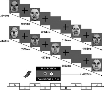
Subjects were presented with 20 facial expression stimuli at 0% (neutral)–50% (mild)–100% (intense) intensities of happy and sad emotion expressions. Separate scans were performed for happy and sad conditions. Subjects were required to determine the sex of the face, ie, an implicit emotion recognition task. Figure 1 depicts the design diagram of the experimental tasks. More detailed information on the task can be found in the online Supplementary Information.
Presentation design for fast implicit facial expression task. Facial expressions were excerpted from the POFA set of photographs, courtesy of Dr. Paul Ekman, professor emeritus University of California at San Francisco (written permission from paul.ekman.com of January 2013).

Gradient echo echoplanar imaging (EPI) data were acquired on a Neurovascular GE Signa 1.5 Tesla system (General Electric, Milwaukee, WI, USA), which was equipped with 40 m/mT high-speed gradients, at Maudsley Hospital, London. A quadrature birdcage headcoil was used for radio frequency (RF) transmission and reception. 180 T2*-weighted images were acquired over 6 minutes for each of the 2 tasks at each of 16 near-axial noncontiguous 7 mm thick planes parallel to the intercommissural (AC-PC) line using the following specifications: TE 40 ms, TR 2000 ms, in-plane resolution 3.44 mm, interslice gap 0.7 mm, flip angle (FA) α 70°, matrix 642, field of view (FOV) 25 cm providing whole brain coverage. During the same session, a high-resolution EPI dataset was acquired with a gradient echo EPI pulse sequence. The structural images were acquired at 43 near-axial 3-mm-thick planes parallel to the AC-PC line: TE 73 ms, TI 180 ms, TR 16,000 ms, in-plane resolution 1.72 mm, interslice gap 0.3 mm, matrix size 1282, FOV 25 cm, FA α 90°. The high resolution EPI dataset was later used to register the fMRI datasets that were acquired from each individual in standard stereotaxic space. The program suite XBAM (http://www.brainmap.it), with mathematical control for signal-to-noise ratio, was used to perform the analysis of fMRI data.
Under the assumption that the subtraction map reflects pure emotion-induced cerebral activation at higher intensity levels, aggregate images of 50–100% of emotional expression were computed after subtracting neutral facial expression activation. The aggregate images were used as a basis for correlation images with clinical trait scales. The aggregate activation maps and correlation images served as a basis for ascertaining regions in which the two groups significantly differ for a clinical trait. Differential linear regression models were set up by inclusion of age and sex as covariates of no interest, at a minimum of 50 random permutations.
Results
Judgment accuracies in the gender decision task for facial expressions were evaluated as the percentage of correct answers in each of the 6 categories (for each happy and sad, neutral, mild, intense). Correct overall answers were 49.54% for DPRD and 51.31% for controls. These rates are in line with other studies that have utilized implicit facial paradigms.Reference Surguladze, Brammer and Keedwell 28 No systematic differences between DPRD and NC emerged for reaction times and response accuracy.
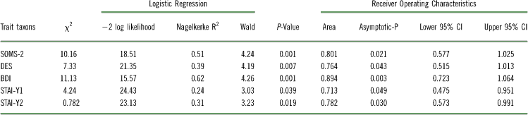
The internal consistencies for the Dissociative Experience Scale, the Screening for Somatoform Disorders, the Beck Depression Inventory, and the State-Trait Anxiety Inventory (DES α = 0.965, SOMS α = 0.783, BDI α = 0.779, STAI Y1 α = 0.919, STAI Y2 α = 0.815) were satisfactory for subsequent analyses. We attempted to predict clinical DSM diagnoses of DPRD by each of these scales. Logistic regression was used to model the linear relationships between BDI, SOMS, DES, and STAI Y1 and Y2, and clinical diagnoses. ROC were utilized to assess the classification sensitivity of the self-report scales with respect to psychiatric diagnosis.
The results are presented in Table 1. Each of the tested personality trait scales was able to predict the clinical DPRD diagnosis, and each also demonstrated sufficient classification specificity for the clinical diagnosis. Both the BDI and SOMS scales have unique variance contributions >50% for the clinical diagnosis, and have areas under the curve >80%. The DES, STAI Y1, and STAI Y2 scales still yield significant regression models; however, in comparison, they are less potent regressors, but are still valid classifiers for clinical diagnosis of DPRD.
Prediction of clinical DPRD diagnosis with scales of personality traits: logistic regression and areas under the ROC curves

Note: N = 21.
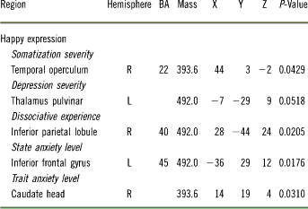
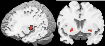
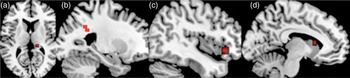
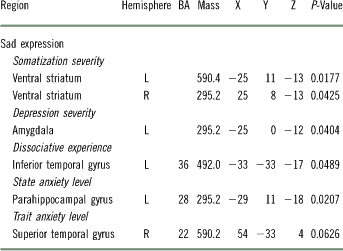
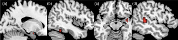
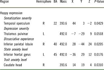
Under stimulation with happy facial expressions (Table 2A, Figure 2 left panel, Figure 3), regions significantly discriminating between the DPD and NC groups were we follows: for somatization severity, the right temporal operculum; for dissociative experience, the right supramarginal gyrus (BA 40); for depression load, the left pulvinar nucleus of the thalamus; for state anxiety level, the left inferior frontal gyrus (BA 45); and for trait anxiety level, the right caput of the caudate nucleus. During processing of sad faces (Table 2B, Figure 2 right panel, Figure 4), DPRD and NC groups differed significantly in the following regions: for somatization, bilateral ventral striatum adjacent to the subgenual cortices (BA 25); for dissociation, the left inferior temporal gyrus (BA 36); for depression, the left amygdala; for state anxiety level, the left parahippocampal gyrus (BA 28); and for trait anxiety level, the right superior temporal gyrus (BA 22).
Comparison of regression slopes: depersonalization disorder > normal control subjects
Note: Mass = volume in mm3, BA = Brodmann Area, XYZ = Talairach coordinates, P-Value tested against 50 random permutations.
Discriminative regions for somatization. Neurological convention. Left panel, right temporal operculum (BA 22), differential region for happy facial expression. Right panel, bilateral ventral striatum, differential region for sad facial expression of emotion.

Differential regions of other traits in happy emotion condition. Radiological convention. (a) Left pulvinar nucleus of the thalamus (BDI, depression); (b) right supramarginal gyrus (DES, dissociation); (c) left inferior frontal gyrus (BA 45) (STAI Y1, state anxiety); (d) right caput caudatis (STAI Y2, trait anxiety).

Comparison of regression slopes: depersonalization disorder > normal control subjects

Note: Mass = volume in mm3, BA = Brodmann Area, XYZ = Talairach coordinates, P-Value tested against 50 random permutations.
Discriminative regions of other traits in sad emotion condition. Radiological convention. (a) left amygdala (BDI, depression); (b) left inferior temporal gyrus (BA 36) (DES, dissociation); (c) left parahippocampal gyrus (BA 28) (STAI Y1, state anxiety); (d) right superior temporal gyrus (BA 22) (STAI Y2, trait anxiety).

Discussion
We aimed to examine the potential neural mechanisms underlying DPRD with respect to the cerebral bases of the traits of depression, anxiety, dissociation, and somatization in relation to normal controls. Using a whole-brain correlational neuroimaging approach, we ascertained the cerebral correlates of these traits as elicited by tasks of facial emotion processing. The main findings in the behavioral domain were the following: the strongest predictors for DPRD diagnosis were depression severity, then somatization severity. Although trait dissociation is also a significant predictor of DPRD, its classification specificity is considerably lower than depression and somatization.
In the neural domain, our results indicate that there are dissociable cerebral bases subserving each of the personality traits in the 2 emotion conditions. For depression, the regions that significantly discern the 2 experimental groups were the thalamus and the amygdala under happy and sad stimulation, respectively. These results are consistent with previous findings that have implicated these limbic structures in depression. For trait somatization, the regions that significantly discriminated between the 2 experimental groups were the temporal operculum in the happy condition and the bilateral ventral striatum in the sad condition. These findings support the notion that trait somatization complaints could follow neural representations of interoception, and they support accumulating evidence that somatization is serotoninergically mediated. The differential association clusters of dissociation were located in the supramarginal and inferior temporal cortices, respectively, which is consistent with our a priori hypotheses. The association of state anxiety with the neural response in the inferior frontal gyrus to happy faces and the parahippocampal gyrus to sad faces replicates published literature for anxiety in affective disorders.Reference Osuch, Ketter and Kimbrell 29 The discriminative regions for trait anxiety are the caudate head and the superior temporal gyrus. This finding also replicates previous findings for anxiety, although these were observed for state anxiety.Reference Chua, Krams, Toni, Passingham and Dolan 20
We will briefly discuss our discriminative regions in the light of recent findings. The operculum is known for its role in unpleasant somatosensation, pain conditioning, and memory retrieval.Reference Ushida, Ikemoto and Tanaka 30 A particular role of the operculum in happiness has been found in smile and laughter processing.Reference Hennenlotter, Schroeder and Erhard 31 The ventral striatum has been demonstrated to constitute the valence appraisal regulatory system,Reference Mitterschiffthaler, Fu, Dalton, Andrew and Williams 32 as it is sensitive to the whole content of the face. Because compassion and sad affects encompass social reward, ventral striatal activity is often found in reversal of the normal pattern.Reference Lee, Seong Whi and Hyung Soo 33 The pulvinar nucleus of the thalamus, which is anatomically connected to the amygdala and orbitofrontal cortex,Reference Blaizot, Mansilla and Insausti 34 is part of the fast-track route that conveys low-frequency facial processing.Reference Vuilleumier, Armony, Driver and Dolan 35 It has been demonstrated to play a key role in the suppression of emotional memory, specifically in reappraisal and suppression of negative emotions.Reference Goldin, McRae, Ramel and Gross 36 Amygdalar correlation is frequently seen in association studies with scales that measure depression.Reference Peluso, Glahn and Matsuo 37 As an indicator for depressive trait vulnerability, amygdalar connectivity with orbitofrontal cortices has been observed to be altered by sad stimulation.Reference Versace, Thompson and Zhou 38 Essential for visuospatial processing, the supramarginal gyrus has been recognized as a key region for happy facial expression processing.Reference Phillips, Bullmore and Howard 39 More recent findings have indicated that the supramarginal gyrus is specifically active in positive word recognition,Reference Hofer, Siedentopf and Ischebeck 40 as well as emotion attribution in normal controls.Reference Sommer, Sodian and Döhnel 41 The lower portion (BA 36) of the inferior temporal gyrus has been implicated in emotion reappraisal.Reference Winecoff, Labar, Madden, Cabeza and Huettel 42 A key role seems to be in valence discrimination and in ambiguity evaluation in nonsalient emotion signals.Reference Leitman, Wolf and Ragland 43 The ventrolateral prefrontal cortex has been found to be central to compassion toward others.Reference Longe, Maratos and Gilbert 44 This also explains its role in negative emotional rumination.Reference Keedwell, Drapier and Surguladze 45 The parahippocampal gyrus has been implicated in impaired emotion regulation. Specifically, it has been found in positive emotion suppression, but has been described as part of the reward circuit,Reference Colibazzi, Posner and Wang 46 along with the caudate and ventral striatum. The caudate head has been implicated in familiarity processing in smile perception.Reference Vrticka, Andersson, Sander and Vuilleumier 47 As part of the motivational control system, it showed inverse correlation with extraversion in happiness states.Reference Suslow, Kugel and Reber 48 The superior temporal gyrus (STG) is a key region in human face processing; because of this, it becomes active as a basis of the familiarity bias in sad emotional memories.Reference Sergerie, Lepage and Armony 49 Modulation of the STG is seen by exposure to sad faces during tryptophan depletion,Reference Daly, Deeley and Hallahan 50 but also during sadness states induced by nonfacial stimuli.Reference Goldin, Hutcherson and Ochsner 51
Limitations
Although this sample comprises a cross-sectional patient sample distributed countrywide, the absolute number of patients should always be regarded with caution. The nonparametric and permutation based inference software should, however, be safe against inflation of coefficients and reflect brain bases with precision. It cannot be excluded that the weighting of the traits (eg, depression) could be biased by comorbidity, although the minor weight of anxiety would speak against assumption of a comorbidity bias. In contrast, the potency of somatization, documented here for the first time, could emphasize the possible consequences of traumaReference van der Kolk, Pelcovitz and Roth 5 in the etiology of DPRD. Due to the lack of trauma data in this sample, however, a final conclusion for this finding is not yet feasible. Finally, it can be regarded as a limitation that not a structured interview for (such as Structured Clinical Interview for DSM-IV Dissociative Disorders (SCID-D)) was employed. However, DSM diagnostic criteria were applied by 2 expert clinicians for this disorder independently.
Conclusion and Recommendations
In sum, the neuroimaging results suggest that DPRD patients may employ abnormal emotion regulation mechanisms, as indicated by the finding that differential regions are structures concerned with cognitive processing of emotion perception, appraisal, and rumination. This implies that DPRD patients may be occupied with cognitive processing and reflection of emotion signals, as a correlate of emotional dampening in this group. Regarding our directions for future research endeavors, we would recommend that a direct comparison of other clinical groups, as mentioned here, would be beneficial in further efforts to clarify the nosological status of DPRD.
Disclosures
The authors do not have anything to disclose.
Supplementary Material
To view supplementary material for this article, please visit http://dx.doi.org/10.1017/S1092852913000588.