Kawasaki disease is an acute self-limited vasculitis of unknown aetiology that occurs in infants and children. It is considered the leading cause of acquired heart disease in children in the developed world.Reference Newburger, Takahashi and Gerber 1 – Reference Newburger, Takahashi and Burns 3 The vasculitis mainly affects coronary arteries, and cardiac sequelae, such as giant coronary aneurysms, are some of the more serious complications.Reference Makino, Nakamura and Yashiro 4 In Japan, giant coronary aneurysms are reported in ~0.3–0.4% of patients with Kawasaki disease. This is a small proportion; however, it is important because the presence of giant coronary aneurysms affects the prognosis of the disease.Reference Sudo, Monobe and Yashiro 5
The aetiology of Kawasaki disease remains unknown and it is not possible to establish strategies to prevent it; therefore, efforts must focus on identifying the risk factors for the development of the most serious outcomes – for example, giant coronary aneurysms–in order to decrease morbidity and mortality in these patients. There are only a few studies identifying risk factors for the development of giant coronary aneurysms.Reference Nakamura, Yashiro and Oki 6 – Reference Nakamura, Yashiro and Uehara 8
In the present study, we aimed to describe patients with Kawasaki disease and giant coronary aneurysms and compare them with patients with Kawasaki disease without giant coronary aneurysms in a Mexican population.
Materials and methods
A retrospective study was conducted at the National Institute of Paediatrics, which is an academic tertiary reference centre in Mexico City, Mexico. The clinical records of patients under 18 years who were diagnosed in the acute phase of Kawasaki disease between August, 1995 and August, 2015 were retrospectively reviewed. The diagnosis of Kawasaki disease was made using the criteria of the American Heart Association. Those who had fever and met more than four of the major criteria were classified as having complete Kawasaki disease. Incomplete cases were defined as those with unexplained fever for 5 days or more, associated with two or three classical features of Kawasaki disease with coronary involvement.Reference Newburger, Takahashi and Gerber 1 – Reference Newburger, Takahashi and Burns 3
Clinical and laboratory findings, in addition to echocardiographic measurements, were recorded. Age, gender, weight, height, days of illness at admission, presence of fever, changes in lips and the oral cavity, changes in palms or soles, and the presence of polymorphous exanthema, bulbar conjunctival injection, cervical lymphadenopathy, Bacille Calmette-Guérin reactivation, incomplete Kawasaki disease, arthralgia, congestive heart failure, as well as gastrointestinal or neurological symptoms were recorded. We defined neurological manifestations in patients if they presented with extreme irritability, somnolence, or with encephalopathy and seizures.
Haemoglobin levels, haematocrit, white blood cell and platelet counts, neutrophil proportion, concentration of C-reactive protein, erythrocyte sedimentation rate, as well as levels of albumin, sodium, bilirubin, aspartate aminotransferase, and alanine aminotransferase were documented. Patients with incomplete records and those who had not undergone laboratory tests or echocardiography at diagnosis were excluded from the study.
We considered a patient to have giant coronary aneurysms if the luminal dimensions of his or her coronary arteries had a body surface area-adjusted z-score⩾10.0.Reference Dallaire and Dahdah 9 , Reference Manlhiot, Millar, Golding and McCrindle 10 As this criterion was published in 2010, patients registered before this date were classified according to the criteria of the American Heart Association. A paediatric cardiologist reviewed all echocardiograms and reclassified them according to body surface area. The presence of myocarditis, pericarditis, or pericardial effusion was also recorded.
Patients with giant coronary aneurysms were compared with all other patients with Kawasaki disease who did not present with giant coronary artery aneurysms.
Continuous variables were expressed as means and standard deviations; a χ2 test was performed to compare categorical variables. Students’ t-test was used for normally distributed continuous variables; p values<0.05 were considered to be significant and the confidence interval (CI) was 95%. Odds ratios and their 95% CIs were calculated to define the risk factors. Statistical analysis was performed using the SPSS program, version 21 (SPSS, Chicago, Illinois, United Sates of America).
The Institutional Research and Ethics Committee approved the study.
Results
Over the 20-year period, a total of 416 patients were diagnosed with Kawasaki disease at our Institution. During the acute stage of the disease, there were 34 patients who had giant coronary aneurysms (z-score⩾10), with an incidence of 8.1%. Of the patients with giant coronary aneurysms, 21 (61.76%) were male and 13 (38.23%) were female. The mean age at diagnosis of Kawasaki disease was 19.47±16.94 months, with an age range of 2–66 months. The average number of days from the onset of symptoms to diagnosis in patients with giant coronary aneurysms was 15.88±9.38 days, compared with 8.65±5.15 days in patients without giant coronary aneurysms.
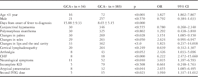
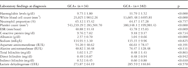
The complete clinical manifestations and laboratory results in both groups are shown in Tables 1 and 2.
Comparison of the clinical manifestations of patients with or without giant coronary aneurysms (GCA) in the acute phase of Kawasaki disease (KD) in Mexican children.

CHF=congestive heart failure; CI=confidence intervals; IVIG=intravenous immunoglobulin; OR=odds ratio
Laboratory results in patients with or without giant coronary aneurysms (GCA) in the acute phase of Kawasaki Disease in Mexican Children.

ESR=erythrocyte sedimentation rate
Of the 34 patients with giant coronary aneurysms, 30 (88.23%) were treated with intravenous immunoglobulin; only five patients (14.70%) required a second dose of immunoglobulin because of persistent fever after 36 hours. In 21 cases (61.76%) immunoglobulin was administered along with systemic corticosteroids.
The echocardiogram at diagnosis showed myocarditis – that is, depressed left ventricular ejection fraction, diastolic dysfunction, altered echogenicity of the papillary muscles, and, in recent years, alterations in two-dimensional strain echocardiograms – in 10 cases (29.41%), pericarditis and pericardial effusion in 10 patients (29.4%), and myocardial hypokinesia in five patients (14.7%). Myocardial hypokinesia was found in five patients who presented with myocardial infarction, and the areas of regional wall motion abnormalities represented the areas of compromised coronary perfusion.
The most affected artery was the left main coronary artery in 21 patients, followed by the proximal right coronary artery in 20 cases and the left anterior descending coronary artery in 11 patients. The circumflex artery was affected only in four patients.
In the multivariate analysis, patients younger than 1 year (p<0.001) and those who had more days of fever at admission (p<0.000) were found to be associated with a significantly higher risk for developing giant coronary aneurysms.
Anaemia (p<0.000), leucocytosis (p<0.000), thrombocytosis (p<0.000), and hypoalbuminaemia (p<0.000) were laboratory abnormalities that were significantly present in patients with giant coronary artery aneurysms at the time of diagnosis.
In all, three patients (8.82%) died during the acute stage of the disease. The cause of death in all of them was Kawasaki disease-related shock and acute myocardial infarction.
Discussion
Kawasaki disease is an acute self-limited vasculitis in children, characterised by vasculitis of small- and medium-sized arteries, with a predilection for coronary arteries.Reference Newburger, Takahashi and Gerber 1 – Reference Newburger, Takahashi and Burns 3
Its aetiology remains unknown, and many aspects of the disease mimic infectious processes; however, to date, no unique agent has been proven to cause Kawasaki disease. A hypothesis proposes that many infectious agents trigger a final common pathway in genetically susceptible hosts.Reference Newburger, Takahashi and Burns 3
Kawasaki disease is highly prevalent in Japan, with an annual incidence of 264.8 cases per 100,000 children <5 years old.Reference Makino, Nakamura and Yashiro 4 In the United States of America, the incidence is race-specific.Reference Uehara and Belay 11 In Mexico, there is no epidemiological record of this disease; thus, the incidence of Kawasaki disease in Mexico is unknown but it might be similar to the incidence reported in Latin America: that is, 3–3.9 cases per 100,000 children <5 years old.Reference Budnik, Hirsch, Fernández, Yánez and Zamorano 12 , Reference Martínez-Ruíz, del Castillo and Borque 13 The largest series of cases reported so far in Mexico is by Garrido-Garcia et al,Reference Garrido-García, Soto-Blanquel and Espinosa-Rosales 14 at the National Institute of Paediatrics, with 338 cases over a period of 18 years.
Cardiac sequelae, such as giant coronary aneurysms, are one of the more serious outcomes. In Japan, this complication is reported in ~0.18% of the patients during the acute phase of Kawasaki disease.Reference Makino, Nakamura and Yashiro 4 In Korea, the incidence of giant coronary aneurysms was 0.25–2%.Reference Kim, Hong and Yun 15 , Reference Lee, Kim, Kwon, Bae and Noh 16 Despite the low incidence, they are a serious cause of concern because their presence affects the prognosis of the disease by increasing the risk for myocardial ischaemia, myocardial infarction, or sudden death.Reference Sudo, Monobe and Yashiro 5
In our study, the incidence of giant coronary aneurysms during the acute phase of Kawasaki disease was 8.1%, higher than that reported in studies from countries with a high prevalence of the disease, but similar to the incidence reported by Schroh et alReference Schroh, Domínguez and Laghezza 17 in Spain, where the incidence of giant coronary aneurysms was found to be 11.1%.
In Japan, as mentioned previously, Kawasaki disease is a common pathology; therefore, physicians have a high index of suspicion, allowing early diagnosis and administration of intravenous immunoglobulin during the initial days of the disease, contrary to what happens in our country. It is well documented that early treatment with immunoglobulin significantly decreases the incidence of coronary aneurysms.Reference Makino, Nakamura and Yashiro 4 , Reference Nakamura, Yashiro and Uehara 7 This may explain the difference in the incidence of giant coronary aneurysms in Asian countries compared with Latin American countries.
In the 22nd Nationwide Survey in Japan, Makino et alReference Makino, Nakamura and Yashiro 4 reported an incidence of 0.18% for giant coronary aneurysms during the acute phase of the disease; however, of the 26,691 patients included in this study, 86.1% received immunoglobulin in the first 6 days after symptom onset. In our study, the mean number of days from the onset of symptoms to diagnosis – and therefore treatment – in patients with giant coronary aneurysms was 15.88±9.38, and in 88.2% of the patients with giant coronary aneurysms the diagnosis was made after 10 days of the onset of fever. In our study, a longer duration of symptoms until diagnosis and treatment seems to be related to the development of giant coronary aneurysms (p<0.000).
Despite the importance of giant coronary artery aneurysms, there are a few studies evaluating risk factors for their development. In 2010, Sudo et al described risk factors for developing giant coronary aneurysms and found that being younger than 1 year or older than 5 years, receiving a total dose of immunoglobulin >2500 mg/kg, and the use of corticosteroids following the administration of intravenous immunoglobulin were risk factors for giant coronary aneurysms.Reference Sudo, Monobe and Yashiro 5 , Reference Morikawa, Ohashi and Harada 18
Nakamura et alReference Nakamura, Yashiro and Uehara 7 reported that being younger than 1 year, having leucocytosis, having a low serum concentration of sodium, as well as having low haemoglobin and elevated alanine aminotransferase levels were risk factors for the development of giant coronary aneurysms.
In our study, we found, similar to that reported by Sudo and Nakamura,Reference Sudo, Monobe and Yashiro 5 – Reference Nakamura, Yashiro and Uehara 8 that patients younger than 1 year had a significantly higher risk for the development of giant coronary aneurysms (p<0.001, OR=3.827, 95% CI 1.86–7.87). Interestingly, patients with giant coronary aneurysms presented atypically,Reference Ha, Jang and Lee 19 , Reference Lin, Hsu and Fu 20 and with neurological symptoms such as severe irritability, somnolence, or seizures, which were seen in three patients with a normal cerebral spinal fluid. Neurological symptoms that presented in the patients with giant coronary artery aneurysms may be a reflection of a more severe systemic inflammation with intracranial vasculitis. Haemorrhagic cerebral infarction has been reported as a complication of Kawasaki disease with giant coronary aneurysms.Reference Samada, Shiraishi, Sato and Momoi 21 Some of the clinical features associated with Kawasaki disease, such as changes in palms and soles, reached statistical difference in the giant coronary aneurysm group, but that could be due to a delayed diagnosis.
As reported by Nakamura, in our study the presence of anaemia (<9.75±1.8 mg/dl), leucocytosis (>21,025±9032 mm3), and additional administration of intravenous immunoglobulin were associated with the diagnosis of giant coronary aneurysms.Reference Nakamura, Yashiro and Oki 6 , Reference Nakamura, Yashiro and Uehara 7
We found that thrombocytosis and hypoalbuminaemia were two factors found in patients with giant coronary aneurysms compared with patients without them. These two findings were also not previously reported.Reference Al-Mashham, Sinclair and Duncan 22 , Reference Patil, Shirodkar, Pinto and Dalvi 23
Some extracoronary echocardiographic findings, such as myocarditis, pericardial effusion, pericarditis, and hypokinesia, were significantly associated with the development of giant coronary aneurysms and were associated with persistent and severe myocardial inflammation. A similar finding had been reported by Lega et al,Reference Lega, Bozio and Cimaz 24 who found that children with coronary artery lesions were more likely to have pericardial effusion or mitral regurgitation during the acute phase of the disease; however, this association was reported for coronary artery lesions and not specifically for giant coronary aneurysms. Lega et al also reported that patients with Kawasaki disease-related shock more often had coronary artery lesions, dilatation, or coronary aneurysms (p=0.04) compared with patients without haemodynamically unstable Kawasaki disease.Reference Lega, Bozio and Cimaz 24 In our series of cases, three patients died, and in all of them the cause of death was Kawasaki disease-related shock; the three patients also had giant coronary aneurysms.
As mentioned previously, the importance of giant coronary aneurysms lies in the morbidity and mortality associated with them, with cardiac sequelae being the cause of mortality in all deaths due to Kawasaki disease. In Japan, mortality due to Kawasaki disease is 0.08%, and in the United States of America it increases to 0.17%. In our study, an overall mortality of 0.72% was found, similar to that reported in the United States of AmericaReference Uehara and Belay 11 ; however, this mortality increases to 8.8% if we consider the mortality in only the giant coronary aneurysm group.
Despite these findings associated with the development of giant coronary artery aneurysms, this study had several limitations. First, it is a small sample compared with the Japanese series of Sudo and Nakamura,Reference Sudo, Monobe and Yashiro 5 – Reference Nakamura, Yashiro and Uehara 8 though case series are smaller in Europe and Latin America. In Chile, Budnik et alReference Budnik, Hirsch, Fernández, Yánez and Zamorano 12 reported 32 cases of giant coronary artery aneurysms after Kawasaki disease over a period of 8 years at one tertiary reference centre. In Spain, Schroh et al evaluated 150 children diagnosed with Kawasaki disease over a period of 16 years,Reference Schroh, Domínguez and Laghezza 17 and in France Lega et alReference Lega, Bozio and Cimaz 24 studied 194 patients with Kawasaki disease between 1988 and 2007, also in a tertiary reference centre. To our knowledge, this is the largest case series conducted in a single hospital in Latin America.
Acknowledgements
None.
Financial Support
This research received no specific grant from any funding agency, commercial or not-for-profit sectors.
Conflicts of Interest
None.
Ethical Standards
This present study was approved by the Institutional Research and Ethics Committee with the number 2015/39.