Balloon aortic valvoplasty, first described in the early 1980s,Reference Labadibi, Jiunn-Ren and Walls1, Reference Labadibi2 is a widely used technique in the treatment of valvar aortic stenosis in children. Nowadays, it constitutes the therapeutic procedure of choice in most centers for the treatment of congenital aortic stenosis.Reference Khalid, Luxenberd and Sable3 Reports on short, mid, and long term results have been published, documenting its security and effectiveness, but nearly all reports describe patients with isolated congenital aortic stenosis, excluding those with associated lesions involving the left side of the heart. Since the typical clinical practice in any particular institution involves aortic valvar stenosis both with and without such associated lesions, we sought to report the longitudinal experience over a 20-year span in a single institution. The purpose of our study was to describe and analyze the characteristics and outcomes of a consecutive cohort of newborns and children, including patients with associated lesions involving the left heart, and to compare our data with previously published experiences.
Patients and methods
Population studied
We included all patients who underwent balloon aortic valvoplasty at our institution from November 20th, 1986, to November 30th, 2006. We did not exclude any patient because of presence of associated cardiac defects or for previous surgical interventions on the aortic valve. Starting in 1996, balloon aortic valvoplasty became the primary therapeutic option in our institution, as opposed to a primary surgical approach during the previous years. The population included 102 boys and 41 girls, which we divided into groups of newborns for those under 1 month, infants for those aged from 1 to 12 months, and children aged above 12 months.
Indications for valvoplasty
During the neonatal period, balloon aortic valvoplasty was performed for critical or severe stenosis. It was also performed in 6 babies with hypoplastic left heart syndrome prior to any surgical palliation. For infants and children, this procedure was indicated in general when the peak transvalvar Doppler gradient was over 70 mmHg, the mean gradient was over 50 mmHg, or when the peak gradient was over 50 mmHg with either symptoms, electrocardiographic alterations involving the ST segment and/or the T wave, or evidence of left ventricular hypertrophy on the electrocardiogram or echocardiography. From a functional perspective, aortic regurgitation of moderate or higher degree was considered a contraindication for balloon aortic valvoplasty.
Methods
The retrospective, longitudinal and observational study was carried out by reviewing the medical records of all patients who underwent a balloon aortic valvoplasty at our institution during the selected period. We described and analyzed biometric data at the time of initial balloon aortic valvoplasty, including gender, age, weight, height, and body surface area. We also recorded, when applicable, the presence of any associated cardiac anomalies or additional therapeutic approaches on the valve.
Most interventions were performed under deep sedation, with the exception of the neonates and the critically ill patients. Balloon aortic valvoplasty technique was performed in retrograde fashion as previously described in the literature, using an antegrade approach via the oval foramen in selected neonatal cases. Following initial left ventriculography, we measured the diameter of the aortic valve, selecting a balloon with a diameter at 80 to 90% of the measured value. When the reduction in gradient obtained was below 50%, and the degree of aortic insufficiency remained less than moderate, the procedure was continued by sequential progressive dilations using larger balloons, up to 100 to 110% of the aortic diameter. We evaluated left ventricle systolic and end-diastolic pressures, aortic systolic pressure, and the gradient from the left ventricle to the aorta before and after the valvoplasty.
The morphology of the aortic valve was based on echocardiography description, and was classified as unicuspid and unicommissural, truly bifoliate, bifoliate by fusion, or trifoliate. The degree of aortic regurgitation was also based on description of the colour Doppler findings using a scale of 5.Reference Perry, Helmcke, Nanda, Byard and Soto4 The aortic regurgitation was recorded before and immediately after the valvoplasty, and throughout the period of follow-up. Peak and mean gradients across the valve were also based on Doppler estimates prior to, after the intervention, and throughout the period of follow-up.
Follow-up
For each patient, follow-up started with the first catheter intervention, and finished with the last available control, either at our hospital or at our ancillary clinics, or the termination of this study. Death of a patient, loss to follow-up, or transfer to adult cardiac care also represented endpoints for follow-up.
Statistical analysis
Data are expressed as mean ± standard deviations for continuous data. The 95% confidence intervals are provided when applicable. Non parametric Mann-Whittney analysis was used in case of abnormal distributions. Student’s t-test was performed to compare continuous data with normal distribution. Signed rank tests or paired t-tests were also used when appropriate. The rank sum test was used for comparison of grades. Fisher’s exact test, and the χ2 test with Yates correction for continuity, was used for data with a categorical distribution. Kaplan-Meier actuarial curves were created to assess survival, with comparison among groups established using the Wilcoxon rank sum test. Multinomial regression statistics were introduced to account for confounding variables.
Results
Baseline characteristics
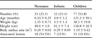
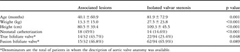
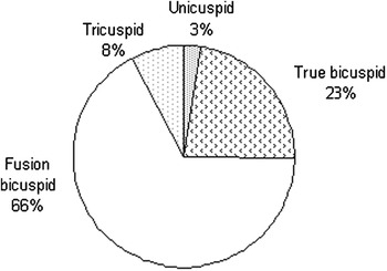
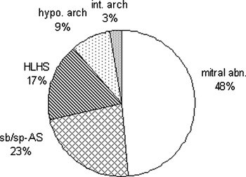
Between November, 1986, and November, 2006, 161 percutaneous procedures were completed in 143 patients, including 102 boys (71.3%) and 41 girls (28.7%) (Table 1). The morphology of the aortic valve was well described by echocardiography in 126 of the patients (88.1% – Fig. 1). Isolated aortic valvar stenosis was present in 102 (71.3%) patients, with 41 (28.6%) having associated lesions involving the left heart (Fig. 2). Among the associated lesions, non-valvar aortic obstruction was present in 31 patients (21.6%), with additional lesions observed in 10 other patients (7%). There was a significant difference between patients with and those without associated lesions (Table 2).
Baseline characteristics per age groups upon initial catheterization.

Baseline characteristics according to the presence or the absence of associated cardiac lesions.

*Denominators are the total of patients in whom the description of aortic valve anatomy was available.
Distribution of anatomy.

Distribution of associated lesions involving the left heart. Abbreviations: mitral abn., mitral abnormalities; sb/sp-AS, sub-valvar or supra-valvar aortic stenosis; HLHS, hypoplastic left heart syndrome; hypo. arch, hypoplastic aortic arch; int. arch, interrupted aortic arch.

Procedural results
In 129 patients (90.8%) the balloon aortic valvoplasty was the first approach, while surgical valvotomy had been performed prior to initial catheter intervention in 13 (9.2%), 10 of these prior to 1996, and 3 afterwards. During the 20-year study period, 16 patients (11.2%) underwent a second percutaneous dilation, 2 patients underwent a third dilation, and 34 (24%) required subsequent aortic valvar surgery.
The ratio of the balloon to the aortic diameter was 98.8% ± 33.6% (95% confidence intervals from 93.3 to 104.3) in the entire population, with no significant difference between age groups (p = 0.13). There were no significant differences between first procedures versus reinterventions (96.6% ± 29.3% versus 110.2% ± 53.6%; p = 0.17), or between prior surgical interventions or native dilations (95.7% ± 7.1% versus 97.4% ± 31.4%; p = 0.8).
In 54 cases (33.8%), the procedure was performed using sequentially progressive dilations during the same procedure. The use of such sequential dilation was similarly for all age groups, and for primary and secondary valvoplasties. Reintervention, in 9 of 54 patients (16.6%) was less often required when sequential progressive dilation was necessary compared to 18 of 89 cases (20.2%) where a single dilation was sufficient, albeit that this difference is not statistically significant (p = 0.759).
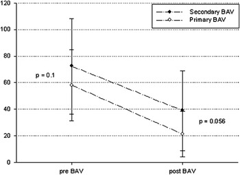
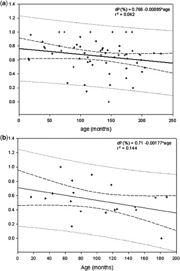
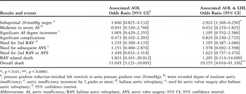
The peak to peak transvalvar gradient decreased significantly, by 60% ± 24% (p < 0.01), with no difference between age groups. Invasive measurements during the procedure are summarized in Figure 3. There was a significant difference between reduction in gradient achieved at primary versus secondary interventions following surgical valvotomy (63.3% ± 24% versus 47% ± 23%; p < 0.03). Nevertheless, comparison of the final gradient achieved between these groups was borderline (Fig. 4). On the other hand, and with respect to valvar anatomy, the reduction in gradient was more significant in those with fused bifoliate valves compared to those having truly bifoliate valves (66% ± 17% versus 53% ± 30%; p = 0.01). The Pearson correlation analysis, performed to determine any effect of age on response to angioplasty, showed no statistical significance (Fig. 5). Despite the apparent cluster of better results for children under 6 years of age (Fig. 5a), this observation yielded no statistical significance according to Mann-Whitney statistical analysis (p = 0.35 and p = 0.72, respectively for patients with isolated stenosis and those with associated lesions). Patients with additional supra- or subvalvar stenosis in general had lower reductions in gradient than those with strictly valvar stenosis (65% ± 22% versus 48% ± 20%, p = 0.004), irrespective of the age at intervention. In Table 3, we summarise the impact of associated lesions on the estimated odds ratios for the variable outcomes.
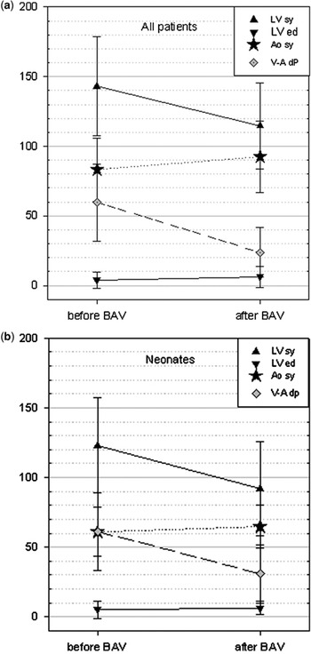
Invasive haemodynamic pressure measurements before and after balloon dilation, in all subjects (Fig. 3a) and neonates (Fig. 3b), showed significant reduction in left ventricular systolic (LV-sy) pressure and in left ventricle to aortic (LV-Ao) pressure gradient (p < 0.01). Aortic systolic (Ao-sy) pressure increased following dilation (p < 0.01). LVsy, left ventricle systolic pressure; LVed, left ventricle end diastolic pressure; Ao sy, aortic systolic pressure; V-A dP, ventricle-aorta pressure gradient; BAV, balloon aortic valvoplasty.

Measured peak-to-peak pressure gradients showed significant drops both for patients with prior aortic valvar surgery (secondary BAV) and no previous surgery (primary BAV) (p < 0.001 each). There was a trend towards a better pressure gradient reduction for patients undergoing primary valvoplasty, with no statistically significant difference (p = 0.056).

Pearson regression analysis for children over 1 year old shows no significant variation of pressure gradient reduction (dP %) in relation with age. Despite the apparent trend for better improvement in the youngest, the slope is near zero, both in patients with isolated stenosis (Fig. 5a) and those with associated lesions (Fig. 5b). Full line, mean regression line; dashed lines, 95% confidence interval of the mean; dotted lines, prediction interval.

Estimated effect of the presence of significant associated aortic obstructive lesions (AOL) or aortic obstructive lesions and left heart lesions (LHL) on variable outcomes.

*, p < 0.01; **, p < 0.0001.
a: pressure gradient reduction (residual left ventricle to aorta pressure gradient over 20 mmHg); b: worst recorded degree of resultant aortic insufficiency; c: aortic insufficiency increment by 2 grades or more; d: balloon aortic valvoplasty; e: need for aortic valvar surgery after balloon aortic valvoplasty; f: 95% confidence interval.
Abbreviations: AI, aortic insufficiency; BAV, balloon aortic valvoplasty; AVS, aortic valve surgery; 95% CI, 95% confidence interval.
The initial drop of pressure gradient increased significantly during follow-up (Fig. 6). For patients who required percutaneous reintervention, the residual gradient was comparable between the initial and the subsequent procedures (34.2 ± 19.8 mmHg versus 35.7 ± 25.0 mmHg; p = 0.73) as well as the reduction in gradient (61.0% ± 33.2% versus 55.8% ± 29.6%; p = 0.33).
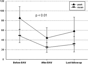
Estimated peak instantaneous and mean left ventricle to aortic pressure gradients showed significant drop immediately after the intervention. A significant rise of these measurements were also recorded at the last follow-up (p < 0.01).

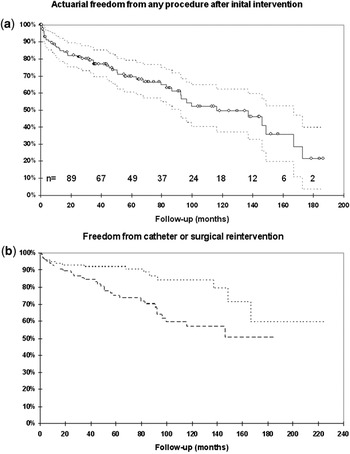
The actuarial analysis for survival yielded a freedom from catheter reintervention of 93.7%, 91.7%, and 83.9% at 1, 5, and 10 years respectively, a freedom from surgical intervention of 91%, 75%, and 56.7% for the same intervals, and a freedom from any reintervention of 86.7%, 69.6%, and 49.5% (Figs 7a and 7b). The freedom from catheter reintervention was significantly advantageous for children and infants compared to neonates (p = 0.005). The freedom from surgical intervention was comparable for all three age groups (p = 0.685), albeit that the freedom from both reinterventions remained lower (p = 0.022) in neonates. With respect to aortic valvar anatomy, there was a clear advantage for valves with 2 leaflets, whether deemed truly bifoliate or bifoliate by fusion, in comparison to trifoliate dysplastic valves or unicuspid and unicommissural valves (p = 0.023, 0.025, and 0.005 for catheter, surgical, or both reinterventions). Male gender was associated with a significantly higher incidence of surgical reintervention compared to female gender (p = 0.05), but conferred no difference with regards to catheter reintervention (p = 0.323). With respect to the presence of associated lesions involving the left heart, the advantage was for isolated aortic valvar stenosis (p = 0.023) in terms of need for percutaneous reintervention. In contrast, there was no significant difference in terms of surgical reintervention on the aortic valve (p = 0.847).
(a) Actuarial freedom from surgical or catheter reintervention following initial balloon valvoplasty. The solid line represents actuarial mean, circles represent censored observations dotted lines represent the 95th confidence intervals. Subsequent to 143 initial subjects, number of observed subjects (n) is provided at each 20-month follow-up intervals. (b) Survival from reintervention by balloon valvoplasty (dotted line) or surgical intervention (dashed line) on the aortic valve following initial balloon intervention.

Complications
Aortic regurgitation
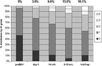
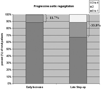
The assessment of aortic valvar insufficiency was based on echocardiographic description for subsequent mid-term and long-term follow-up comparison. We used the scale of zero for none, through trivial, mild, and moderate form to 4 for severe regurgitation to chart any increase in the degree of aortic regurgitation occurring within 24 hours of valvoplasty, and subsequently until the last available follow-up (Fig. 8). We found an increase in aortic regurgitation of 2 grades or more in 16% of patients at the early assessment, with this increasing to 38% at the last follow-up (Fig. 9). There were no significant differences between patients with and without associated lesions, at 16.1% versus 17.1% (p = 0.888), or for gradual worsening, at 3.2% versus 16.9% (p = 0.097). Following intervention, the prevalence of moderate to severe aortic insufficiency increased steadily, up to 16.1% at the last follow-up. There was no correlation noted between the ratio of the balloon to the aortic diameter and the degree of post-procedural aortic regurgitation, neither by linear regression nor by ANOVA between the intensity of regurgitation among the groups (p > 0.50–0.80).
Distribution of aortic regurgitation at different phases of follow-up. A 0 to 4 scale is reported (0 for absent, 1 for trivial, 2 for mild, 3 for moderate, and 4 for severe regurgitation). Percentage values above bars are cumulative for grade 3 and 4 for each bar. (AI, aortic insufficiency; BAV, balloon aortic valvoplasty).

Increase of aortic valve regurgitation as reported by echocardiography, shortly after catheter intervention (early increase) and at late follow-up (late step-up). The degree of change from baseline is categorized as no change or 1 grade worse (0 to 1), 2 grades worse (2), or 3 to 4 grades worse (3–4). Scale is percentage of total available data.

Electrocardiographic changes
Changes were recorded following 13 procedures, with 11 of these (78.6%) performed in neonates, representing one-third of the neonates. Left bundle branch block was observed in 5 patients, ventricular tachycardia or fibrillation in 3 patients, complete atrioventricular block in 3 patients, supraventricular tachycardia in 2 patients, Mobitz-I second degree atrioventricular block in 1 patient, and ST depression in the left precordial leads in a final patient. Treatment was successful, consisting of electrical cardioversion for ventricular arrhythmia, transient electric stimulation for complete atrioventricular block, and administration of adenosine for those with supraventricular tachycardia.
Other complications
Of the total 161 procedures, 20 serious complications (12.4%) were recorded in 15 procedures (9.3%), these proving fatal in 3 neonates (2.1% of the total population, and 9.1% of the neonates). The serious, non lethal complications were iatrogenic aortic regurgitation graded as moderate or more, observed in 6 patients, severe hypotension requiring medical intervention occurring in 4 patients, ventricular arrhythmias seen in 3 patients, excluding self resolving isolated extrasystoles, damage to the femoral artery requiring surgical intervention in 3 patients, complete atrioventricular block in 1 patient, mitral insufficiency in 1 patient, and cardiac tamponade in a final patient. There were no differences between first catheterizations and reinterventions in terms of incidence of serious complications. The 19 serious complications, nonetheless, occurred more often in the neonates, occurring in 9 patients, compared to 4 instances in infants and 6 in children (p = 0.006).
Mid-term and long-term follow-up
The period of follow-up has now extended to 66.36 ± 56.28 months, and ranges from 1 week to  years, with a median of 58.6 months, and 95% confidence intervals from 9.1 to 113.2 months.
Reintervention
Overall, 61 procedures were performed in 51 patients after the first valvoplasty, including 16 balloon dilations and 45 surgical interventions, including replacement of the aortic valve in 14, Ross or Ross-Konno procedures in 11, valvoplasty in 7, valvotomy in 6, subvalvar resections in 5, cardiac transplantation in 1, and placement of a conduit from the right ventricle to the pulmonary arteries in 1. Of the 16 patients (11.2%) who underwent percutaneous reintervention, 5 were neonates, 3 infants, and 8 children. Among the 51 patients, a second intervention, either by catheter or surgery, was necessary for 46 (32.4%) at an interval of 48.5 ± 49.7 months, with 95% confidence intervals from 33.7 to 63.3 months, whereas a third intervention was required after 25 ± 11.87 months, with 95% confidence intervals from 10.3 to 40.0 months in 5 cases. The incidence of reintervention was comparable between the age groups (p = 0.63). The interval between the initial valvoplasty and reintervention was significantly shorter for neonates, at 2.4 ± 0.8 months, when compared to infants, at 107.2 ± 78.9 months, or children at 66.5 ± 47.0 months (p = 0.02). Multinomial regression analysis identified aortic valvar anatomy as the main predictive variable of reintervention, specifically for the truly bifoliate (p = 0.007) and trifoliate valves (p = 0.01).
Percutaneous reintervention was more often required in the neonate, in patients with significant associated left heart lesions, or with trifoliate aortic valves. The estimated freedom from reintervention at 1 year was 75% versus 95.2% and 98.6% for neonates versus infants and children (p = 0.005), 88.6% versus 96.9% for those with and without associated left heart lesions (p < 0.05), and 71.4% versus 93.9% and 95.1% for those with trifoliate valves as opposed to the 2 forms of bifoliate valves (p < 0.05). Freedom from percutaneous intervention dropped further to 86.4% and 72.7% after intervals of 5 and 10 years for those with aortic stenosis and associated left heart lesions compared to 95.2% and 91.7% for those with isolated stenosis (p = 0.023).
A surgical reintervention was more often required in boys, with a freedom from surgical intervention of 88.4%, 68.4%, and 52.8% compared to 97.0%, 89.9%, and 64.5% for girls at intervals of 1, 5, and 10 years (p = 0.05). Similarly, surgical intervention was more likely to be required in those with trifoliate valves, with an estimated freedom from intervention of 75% and −50%, compared to 90.6% and 69.2%, and 96.9% and 83% at intervals of 1 and 5 years for those with truly bifoliate and fused bifoliate valves, respectively (p < 0.05).
Mortality
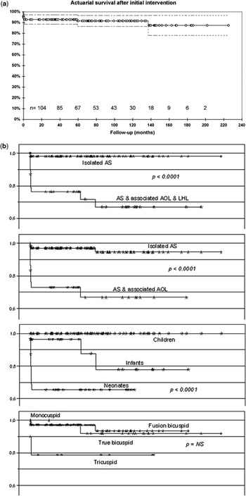
Of the 143 patients, 13 (9.1%) died during the follow-up (Fig. 10a), 11 (84.6%) within one month of the intervention, and 2 at 5 and at  years. Among those dying early, 4 had isolated aortic stenosis, and 7 had severe associated left heart lesions, specifically hypoplastic left heart syndrome in 4, significant mitral insufficiency in 2, and interruption of the aortic arch with perimembranous ventricular septal defect in 1. The death was considered to be directly related to the valvoplasty in 3 cases, representing 2.1% of the complete series, and 9.1% of the neonates. Death was secondary to cardiac tamponade in one patient. In the second case, a patient with moderate hypoplastic left ventricle died 17 days after an attempt at functionally univentricular repair following percutaneous dilation complicated by severe valve regurgitation. The third death, which was not secondary to a cardiac or valvar complication, occurred immediately after the termination of the procedure due to severe spasm and avulsion of the femoral artery during withdrawal of the introducer. In the complete series, overall mortality within 1 month was 30.3% for neonates. Separating our experience chronologically, neonatal deaths occurred in 9 of 15 patients (60%) treated prior to 1998, and in 1 of 18 patients (5.5%) treated subsequently. The risk of overall mortality varied according to the relationship with anatomy, age, and the presence or absence of associated left heart lesions (Fig. 10b). Multinomial regression analysis identified neonatal age and the association with cardiovascular malformation as the main causative variables predicting a high risk of mortality (p = 0.016). The overall risk of death varied between 27.5% and 29.0% for those having aortic obstructive lesions with or without left heart lesions, as opposed to 1.9 and 3.6% for those without such lesions (p < 0.0001), albeit that there was no statistical difference for death related to valvoplasty, at 2.5% and 3.2% for those with associated lesions versus 1.8% and 1.9% for those without such lesions; p = NS).
(a) Actuarial survival (freedom from death) following the initial catheter intervention in the total cohort. The solid line represents actuarial mean, circles represent censored observations, dotted lines represent the 95th confidence intervals. (b) Actuarial survival (freedom from death) following the initial catheter intervention according to the association or non association with other cardiovascular anomalies (AOL, aortic obstructive lesions; LHL, left heart lesions), age group, and anatomic description of the aortic valve.

Discussion
We have collated the results over 20 years for treatment in a single institution of 143 consecutive children with isolated and complex aortic stenosis treated by percutaneous intervention. Although some have reported results of balloon aortic valvoplasty for those with aortic valvar stenosis associated with aortic coarctation,Reference Mc Elhinney, Lock, Keane, Moran and Colan5 most previous accounts of this procedure performed in children have excluded patients with significant associated lesions involving the left heart. Although we did not offer balloon dilation to all patients presenting in the early years of our experience, with only 7 patients treated in this fashion during the first 3 years, we did not exclude this initial phase of our experience. Hence, we describe a consecutive series of patients assembled on the basis of intention to treat.
In terms of immediate reduction in gradient, our overall results are consistent with those previously reported.Reference Perry, Helmcke, Nanda, Byard and Soto4, Reference Gatzoulis, Rigby, Shinebourne and Redington6–Reference Rao, Thapar, Wilson, Levy and Chopra9 We noted, however, several differences in terms of predicted outcomes. For instance, while previous data suggested that children over 14 years old with high pre-procedural left ventricle end diastolic pressure were less likely to respond to valvoplasty in terms of relief of the gradient,Reference Kusa, Bialkowski and Szkutnik7 we failed to find any age after which there is a reduced reduction. In contrast, we found effectiveness to be unrelated to age or to haemodynamic parameters prior to dilation, in part because of a potential exclusion bias among adolescent patients with very thickened or calcified valves. More interestingly, we found that a better reduction was more likely achieved in those with trifoliate valves functioning in bifoliate fashion because of fusion of leaflets when compared to those we deemed to possess true bifoliate valves. To our knowledge, such a relationship has not previously been reported.
Our results oppose preceding publications suggesting comparable effectiveness between patients who had a previous surgical valvotomy and primary balloon aortic valvoplasty.Reference Meliones, Beekman, Rocchini and Lacina10–Reference Sreeram, Kitchiner, Williams and Jackson12 Repeated valvoplasty also seems to be efficient, with no extra morbidity or mortality compared to the first intervention in the same patient.Reference Kuhn, Latson, Cheatham, Fletcher and Foreman13–Reference Shim, Lloyd and Beekman15 In our experience, nonetheless, better results were recorded for primary balloon aortic valvoplasty, as well as in the first intervention compared to repeated interventions.
The early experience with balloon valvoplasty, achieved from 1984 through 1995, demonstrated the correlation between the ratio of the diameter of the balloon to the aorta and the relief achieved in gradient.Reference McCrindle8, Reference Sholler, Keane and Perry11, Reference Phillips, Gerlis, Wilson and Walker14 It also set a correlation between higher ratios and new onset or increased aortic regurgitation, which lead to improved practices and guidelines. Our findings now corroborate the most recent studies,Reference Kusa, Bialkowski and Szkutnik7, Reference Balmer, Beghetti, Fasnacht, Friedli and Arbenz16–Reference Reich, Tax and Marek18 most likely because of the narrow range of the ratio of balloon to aorta used in our practice. In contrast, we failed to endorse the described association between bifoliate valves due to fused leaflets and a higher risk of aortic regurgitation.Reference Reich, Tax and Marek18 Independently, a new onset or a worsening in aortic regurgitation was well observed in our series, again endorsing previous accounts.Reference Perry, Helmcke, Nanda, Byard and Soto4, Reference Kusa, Bialkowski and Szkutnik7, Reference Balmer, Beghetti, Fasnacht, Friedli and Arbenz16, Reference Pedra, Pedra and Braga17, Reference Echigo19, Reference Vogel, Benson, Burrows, Smallhorn and Freedom20 An increase of 2 grades or more tended to progress over time, involving one-third of the patients at the latest follow-up. The proportion of patients with resulting severe or moderate regurgitation increased stepwise, from 3.6% to a maximum of 16.1%. In preceding series,Reference McCrindle8, Reference Balmer, Beghetti, Fasnacht, Friedli and Arbenz16–Reference Reich, Tax and Marek18, Reference Fratz, Gildein and Balling21 which included both neonates and older children, the incidence of post-procedural aortic regurgitation of at least moderate grade varied between 7.3% and 22.6%, while at long-term follow up the reported incidence varies between 22.3% and 35% after 5 to 5.3 yearsReference Mc Elhinney, Lock, Keane, Moran and Colan5, Reference Reich, Tax and Marek18 and in 50% at 7.5 years follow-up interval.Reference Pedra, Pedra and Braga17
Such progressive worsening of aortic regurgitation seems inevitable, with the increase in residual transvalvar gradient varying between 0% and 32.1% according to different definitions of residual gradients used by different investigators.Reference Rao, Thapar, Wilson, Levy and Chopra9, Reference Balmer, Beghetti, Fasnacht, Friedli and Arbenz16, Reference Reich, Tax and Marek18 In a very recent series, including both neonates and older children, one-tenth of the patients needed reintervention at long term follow-up due to restenosis.21 The survival rate after valvoplasty at 6 and 14.4 years varies between 93% and 100% in older children,Reference Reich, Tax and Marek18, Reference Fratz, Gildein and Balling21–Reference Moore, Egito, Mowrey, Perry, Lock and Keane23 whereas it varies between 71–74.6%Reference Mc Elhinney, Lock, Keane, Moran and Colan5, Reference Fratz, Gildein and Balling21 and 71%Reference Reich, Tax and Marek18 at 10 and 14.4 years respectively when performed in the neonatal period. The freedom from reintervention at mid and long-term follow up varies between 46 and 76%Reference Reich, Tax and Marek18, Reference Fratz, Gildein and Balling21–Reference Rao24 after 8–14.4 years in older children, whereas it varies between 47–57.6%Reference Mc Elhinney, Lock, Keane, Moran and Colan5, Reference Fratz, Gildein and Balling21, Reference Villalba Nogales, Herráiz Sarachaga, Bermúdez-Cañete Fernández, Maitre Azcárate, Mora de Oñate and González Rocafort25 and 26%Reference Reich, Tax and Marek18 at 5–10 years and 14.4 years respectively when performed in the neonatal period. These data have been cited in favour of using percutaneous intervention as the initial approach compared to surgery for those with aortic stenosis despite the potential complications. Transient electrocardiographic alterations, for instance, are well documented,Reference Sholler, Keane and Perry11, Reference Kuhn, Latson, Cheatham, Fletcher and Foreman13 with the highest incidence noted in neonates (34.6%).Reference Villalba Nogales, Herráiz Sarachaga, Bermúdez-Cañete Fernández, Maitre Azcárate, Mora de Oñate and González Rocafort25 This corroborates with our own reported incidence of 33%. As with our experience, others have reported left bundle branch block, ventricular tachycardia or fibrillation, complete atrioventricular block, supraventricular tachycardia and sinus bradycardia.Reference Sholler, Keane and Perry11, Reference Kuhn, Latson, Cheatham, Fletcher and Foreman13, Reference Villalba Nogales, Herráiz Sarachaga, Bermúdez-Cañete Fernández, Maitre Azcárate, Mora de Oñate and González Rocafort25 Iatrogenic insufficiency, severe hypotension, arrhythmias and vessel damage are considered as the main serious complications.Reference Kusa, Bialkowski and Szkutnik7, Reference Sholler, Keane and Perry11, Reference Kuhn, Latson, Cheatham, Fletcher and Foreman13, Reference Balmer, Beghetti, Fasnacht, Friedli and Arbenz16, Reference Borghi, Agnoletti, Valsecchi and Carminatti26
Even including our early experience, which started in 1986, our overall mortality rate of 2.1% is low, and comparable to other reports.Reference Perry, Helmcke, Nanda, Byard and Soto4, Reference Kusa, Bialkowski and Szkutnik7, Reference McCrindle8, Reference Balmer, Beghetti, Fasnacht, Friedli and Arbenz16, Reference Borghi, Agnoletti, Valsecchi and Carminatti26 As expected, death is most frequent in neonates. Our own rates of death are lower than some previous experiences, which were as high as 34%.Reference Mc Elhinney, Lock, Keane, Moran and Colan5, Reference Gatzoulis, Rigby, Shinebourne and Redington6, Reference Sholler, Keane and Perry11, Reference Echigo19, Reference Fratz, Gildein and Balling21, Reference Villalba Nogales, Herráiz Sarachaga, Bermúdez-Cañete Fernández, Maitre Azcárate, Mora de Oñate and González Rocafort25 The range in rates of mortality, nonetheless, is considerable. These rates, therefore, must be interpreted cautiously, either due to relatively low number of cases,Reference Gatzoulis, Rigby, Shinebourne and Redington6, Reference Sholler, Keane and Perry11 or because recorded deaths were reported between 1 week and 1 month following the procedure.Reference Fratz, Gildein and Balling21, Reference Villalba Nogales, Herráiz Sarachaga, Bermúdez-Cañete Fernández, Maitre Azcárate, Mora de Oñate and González Rocafort25 Since 1998, our rate of death within 1 month in neonates is in line with recent studies,Reference Mc Elhinney, Lock, Keane, Moran and Colan5, Reference Fratz, Gildein and Balling21 despite our inclusion of patients with significant associated lesions.
Conclusions
When based on current recommendations for the size of the balloon relative to the aorta, we conclude that balloon valvoplasty is a safe and effective method for the treatment of congenital aortic valve stenosis. The association with additional obstructive lesions does not significantly affect the main outcomes. The achieved relief in gradient is satisfactory, with an expected progressive increase of regurgitation or transvalvar gradient requiring reintervention without additional mortality or significant morbidity. Prior aortic surgery, reintervention and the anatomy of the valve predict a lower reduction in gradient. The risk of major complications is related to the young age at intervention rather than to the potential associated cardiac lesions. Mortality, which is higher in patients with associated lesions, is not directly linked to the procedure itself. This leads us to suggest that balloon valvoplasty, as an initial approach, provides excellent long-term survival, delaying the definitive surgical treatment on the valve which may be required, bearing in mind that the association of cardiovascular lesions is not a prohibitive condition for balloon aortic valvoplasty.