Introduction
Vocal polyps are benign, round, and sessile or pedicled lesions located at the free borders of the vocal folds. They are commonly unilateral and vary in size. Vocal abuse or overuse, infectious disease of the upper airways, allergy, smoking and reflux are associated with vocal polyps.Reference Martins, Defaveri, Domingues and de Albuquerque e Silva1 Vocal polyps are more frequent in men and surgery is the preferred treatment option.Reference Altman2 Repetitive vocal trauma is a leading causes of vocal polyp formation. Vocal overuse, abuse and misuse lead to mechanical stress and trauma to the vocal fold, resulting in wound formation. Subsequent wound healing and remodelling of the lamina propria and vocal fold epithelium cause vocal fold nodule, polyp and cyst formation. Thus, vocal overuse, abuse and misuse are central factors in vocal polyp development; however, other aetiological factors are also believed to have synergistic effects on vocal polyp pathogenesis.Reference Martins, Defaveri, Domingues and de Albuquerque e Silva1–Reference Johns3
Diagnosis of benign vocal fold lesions depends mainly on their clinical appearance. There is no consensus on histological diagnostic criteria for these lesions. Epithelial hyperplasia, basement membrane thickening, oedema, vascular proliferation or ectasia, and extracellular amyloid-like fibrin deposition are common histopathological findings for benign vocal fold masses. Vascular proliferation and extracellular fibrin deposition are reported to be more common in polyps than in nodules.Reference Cipriani, Martin, Corey, Portugal, Caballero and Lester4 Furthermore, in a histological study of vocal polyps, Martins et al. reported vessel proliferation in 92.86 per cent.Reference Martins, Defaveri, Domingues and de Albuquerque e Silva1 In addition, an immunohistochemical study of vocal polyps demonstrated anti-laminin, anti-fibronectin and anti-collagen IV immunostaining in many cells surrounding the lamina propria. All of these findings suggest that extracellular matrix remodelling and angiogenesis are important for vocal polyp formation.
A disintegrin and metalloproteinase proteins (‘ADAMs’) comprise a family of zinc-dependent transmembrane and secreted proteins that function in cell adhesion, migration, proteolysis and signalling. Dysregulation of ADAM family members is associated with cancer, cardiovascular disease, asthma and Alzheimer's disease.Reference Edwards, Handsley and Pennington5 ADAM-33 is involved in the pathogenesis of respiratory system diseases, such as asthma and nasal polyposis.Reference Erbek, Erinanc, Erbek, Topal and Kiyici6–Reference Puxeddu, Pang, Harvey, Haitchi, Nicholas and Yoshisue8 Its role in asthma was recently revealed to occur via angiogenesis and airway remodelling processesReference Erbek, Erinanc, Erbek, Topal and Kiyici6–Reference Puxeddu, Pang, Harvey, Haitchi, Nicholas and Yoshisue8; these processes may also be involved in vocal polyp pathogenesis. To our knowledge, ADAM-33 expression has not yet been studied in vocal fold polyps. This study aimed to investigate ADAM-33 expression in vocal polyps and correlate it with patients’ clinical characteristics.
Materials and methods
This study included 32 patients who were diagnosed with vocal polyps and operated on at the Otolaryngology Department, Baskent University, Turkey. The study protocol was approved by the Ethics Committee of Baskent University. Data on age, sex, symptom duration, polyp side, smoking and associated symptoms were obtained from patients’ medical charts. Patients with incomplete charts were excluded. Vocal polyps were clinically diagnosed by videolaryngoscopy and during surgery, and confirmed histologically by assessment under light microscopy. Controls were normal vocal fold tissue samples obtained from 36 patients during laryngeal squamous cell carcinoma surgery: histopathologically non-tumoural normal vocal fold tissue was removed from sites as far from the tumour as possible.
Immunohistochemical staining
Immunohistochemical staining was performed using the streptavidin–biotin method. Briefly, 3 mm tissue sections were deparaffinised in xylene and dehydrated using a graded ethanol series. Sections were then heated in citrate buffer (10 μm, pH 6.0) at 120°C for 20–25 minutes for antigen retrieval and rinsed 3 times in distilled water. Endogenous peroxidase activity was blocked by incubation for 30 minutes in 0.3 per cent hydrogen peroxidase. Sections were then incubated in a 1:100 dilution of primary anti-ADAM-33 antibody (C-20 rabbit polyclonal; Santa Cruz Biotechnology: Santa Cruz, California, USA) overnight at room temperature, washed in phosphate-buffered saline for 5 minutes, and incubated in biotinylated goat anti-polyvalent secondary antibody (Thermo Scientific LabVision, Fremont, California, USA) for 10 minutes at room temperature. Sections were washed again in phosphate-buffered saline and then incubated in streptavidin peroxidase (Thermo Scientific LabVision) for 15 minutes at room temperature. Immunoreactivity was visualised using the chromogen, 3-amino-9-ethylcarbazole (Dako, Glostrup, Denmark). Sections were counterstained with Mayer's haematoxylin solution and mounted in Entellan (Merck, Darmstadt, Germany). ADAM-33 expression was scored as: negative, 0; weak, 1; moderate, 2; or strong, 3. The intensity of immunostaining in epithelial cells, stroma and vessels was compared in vocal polyps and control tissues.
Statistical method
Statistical analyses were performed using SPSS software program version 15.0 (SSPS Inc, Chicago, Illinois, USA). ADAM-33 staining scores for the epithelium, stroma and mesenchymal cells of vessels in vocal polyp and control tissues were compared using the chi-square test. The chi-square test was also used to evaluate the relationship of ADAM-33 immunostaining scores with clinical parameters (sex, smoking and symptom duration) associated symptoms (vocal overuse, reflux, sinonasal symptoms and allergy). A p value of less than 0.05 was considered statistically significant.
Results
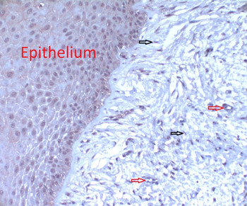
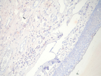
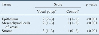
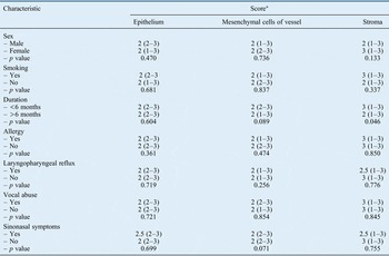
There were 27 men (84 per cent) and 5 women (16 per cent) in the vocal polyp group (median age 49 years, range 17–73 years), and 34 men (94 per cent) and 2 women (6 per cent) in the control group (median age 63 years, range 38–79 years). In all, 18 (56 per cent) of the polyps were located on the right vocal fold, 12 (38 per cent) on the left and 2 (6 per cent) bilaterally. The mean symptom duration was 13.48 months (range 1–48 months). ADAM-33 staining scores in vocal polyp and control tissue is summarised in Table I. All epithelial, stromal and vascular staining scores were significantly higher in polyp tissues than in control tissue (p < 0.001). Figures 1 and 2 show ADAM-33 immunohistochemical staining of vocal fold polyp and control tissue sections, respectively. Patients were separated into 2 groups: those with symptom duration of more than 6 months (16 patients, 50 per cent) and those with symptom duration of less than 6 months (16 patients, 50 per cent). When ADAM-33 staining scores were compared between these groups, stromal ADAM-33 staining was found to be significantly higher in the second group (p < 0.05). Active smokers and individuals who had quit smoking less than two months before presentation were classified as smokers. There were 24 smokers (75 per cent) in the vocal polyp group. There was no significant difference in the ADAM-33 staining scores of smokers and non-smokers (Table II). A total of 22 out of 32 patients (69 per cent) had a history of vocal overuse (these individuals had to use their voices for long periods of time at work). In all, 56 per cent (18/32) of patients had reflux, 31 per cent (10/32) had sinonasal symptoms and 19 per cent (6/32) had allergies. There was more than one predisposing factor in 17 out of 32 patients (53 per cent). However, there were no significant differences in ADAM-33 staining scores when stratified according to the presence of predisposing factors such as vocal overuse, reflux symptoms, sinonasal symptoms and allergy (Table II). Predisposing factors for vocal polyp formation were not analysed in the control group.
Photomicrograph showing ADAM-33 staining of fibroblasts (black arrows), vessels (red arrows) and epithelial cells within the stroma of vocal fold polyp tissue (magnification ×400).

Photomicrograph showing the weak, general ADAM-33 immunostaining in normal laryngeal tissue (magnification ×400).

Adam-33 immunostaining scores in vocal polyp and control tissue

*Values represent the median and range.
Correlation Of Patients’ Clinical Characteristics With Adam-33 Immunostaining Scores

*Values represent the median (range)
Discussion
ADAM proteins contain disintegrin and metalloproteinase domains.Reference Wolfsberg, Primakoff, Myles and White9 Nearly 40 ADAM family members have been described, and 25 of these are expressed in humans.Reference Paulissen, Rocks, Gueders, Crahay, Quesada-Calvo and Bekaert10 ADAMs are involved in many physiological and pathological processes.Reference Edwards, Handsley and Pennington5, Reference Erbek, Erinanc, Erbek, Topal and Kiyici6, Reference Topal, Erinanc, Ozer, Canpolat, Celik and Erbek11, Reference van Goor, Melenhorst, Turner and Holgate12 They have roles in sperm–egg binding and fusion, trophoblast invasion and matrix degradation during pregnancy, and in angiogenesis, and neovascularisation. They are also involved in pathological processes such as cancer, inflammation, neurodegeneration and fibrosis.Reference van Goor, Melenhorst, Turner and Holgate12 ADAMs are generally expressed in inflammatory cells and so may play an important role in initiating repair mechanisms in the airways.Reference Dijkstra, Postma, Noordhoek, Lodewijk, Kauffman, ten Hacken and Timens13 ADAM-33 is mainly expressed in airway smooth muscle cells and fibroblasts. This protein is also expressed in endothelial cells, where it may have a role in cell adhesion via mediating critical protein–protein interactions, thereby possibly activating inflammatory cells and promoting angiogenesis.Reference Edwards, Handsley and Pennington5, Reference Paulissen, Rocks, Gueders, Crahay, Quesada-Calvo and Bekaert10, Reference Dijkstra, Postma, Noordhoek, Lodewijk, Kauffman, ten Hacken and Timens13, Reference Garlisi, Zou, Devito, Tian, Zhu and Liu14 Puxeddu et al. showed that increased expression of soluble forms of ADAM-33 induced angiogenesis and endothelial cell differentiation in asthmatic patients.Reference Puxeddu, Pang, Harvey, Haitchi, Nicholas and Yoshisue8 Erbek et al. showed increased ADAM-33 expression in nasal polyp tissues and proposed it to be related to tissue remodelling.Reference Erbek, Erinanc, Erbek, Topal and Kiyici6
ADAM-33 over-expression in a range of respiratory diseases suggests that it promotes angiogenesis, which in turn facilitates inflammation and promotes airway remodelling.Reference Erbek, Erinanc, Erbek, Topal and Kiyici6, Reference Puxeddu, Pang, Harvey, Haitchi, Nicholas and Yoshisue8, Reference Shaffiq, Haitchi, Pang, Alangari, Jones and Marshall15 As in many other respiratory diseases, basic morphological changes present in vocal polyps include oedema, increased blood vessel numbers and inflammation of the lamina propria.Reference Martins, Defaveri, Domingues and de Albuquerque e Silva1 Although the exact mechanism of vocal polyp formation is unclear, it is speculated that vibratory trauma to the membranous vocal folds may disturb microcirculation and damage superficial vessels, leading to extravasation of blood, which eventually results in inflammatory reactions and malformed neovascularised masses with hyalinised stroma.Reference Altman2, Reference Johns3
Only a few clinical studies into the role of extracellular matrix and soluble mediators in the vocal polyp pathogenesis have included controls. A possible reason for this may be the difficulty of obtaining control tissue. Pathological specimens of vocal fold polyps are usually very small and do not include normal vocal fold tissue. Obtaining normal vocal fold tissue from healthy individuals to use as controls is impossible because of ethical problems. Thus, we used normal vocal fold tissues obtained from laryngectomy specimens, as described previously by Karahan et al.Reference Karahan, Baspinar, Yariktas and Kapucuoglu16 These authors recently investigated the role of matrix metalloproteinases (MMP-2 and MMP-9) and cyclooxygenase-2 (COX-2) in vocal polyp pathogenesis and suggested that these proteins might play a role in vocal polyp development. These proteins are also involved in inflammation, tissue remodelling and angiogenesis. In this study, immunohistochemical analysis revealed ADAM-33 over-expression in vocal polyps; we speculate that this might play a role in vocal polyp pathogenesis via promoting tissue remodelling and angiogenesis.
Martins et al. investigated clinical, morphological and immunohistochemical characteristics of vocal polyps.Reference Martins, Defaveri, Domingues and de Albuquerque e Silva1 These authors analysed the symptoms associated with vocal polyps in 76 patients. In that case series, 51 per cent of patients were smokers and vocal overuse was the most frequently detected aetiological factor for vocal polyp formation (61 per cent). Gastroesophageal symptoms (47 per cent) and sinonasal symptoms (32 per cent) were also reported as other associated symptoms. Similarly, vocal overuse was identified as the most common aetiological factor (69 per cent) in vocal polyp formation in our study. Reflux symptoms were present in 56 per cent of patients and sinonasal symptoms in 31 per cent. These findings further suggest that predisposing factors might lead to laryngeal mucosa inflammation and thus contribute to vocal polyp formation.
In this study, ADAM-33 staining scores of vocal polyp patients were unaffected by the presence of predisposing factors. A former study comparing pathological and normal tissues obtained from the same patients showed that ADAM-33 staining scores were significantly higher in pathological tissues than in normal tissues.Reference Topal, Erinanc, Ozer, Canpolat, Celik and Erbek11 This finding suggests that the ADAM-33 expression is increased in pathological tissues. Furthermore, ADAM-33 scores were not associated with the presence or absence of predisposing factors in the vocal polyp tissues. Therefore, we did not analyse the effect of predisposing factors in the control tissues.
• Wound healing and tissue remodelling are important in vocal fold nodule formation
• ADAM-33 participates in respiratory system pathogenesis via promoting angiogenesis and airway remodelling
• ADAM-33 over-expression in vocal polyps may indicate an early tissue response to vocal trauma
• ADAM-33-related angiogenesis and tissue remodelling may be important factors in vocal polyp development
• ADAM-33 might be a useful therapeutic target for vocal polyps
Duflo et al. analysed the gene expression profiles of vocal polyp and Reinke's oedema tissues.Reference Duflo, Thibeault, Li, Smith, Schade and Hess17 They demonstrated over-expression of genes associated with extracellular matrix remodelling, cell growth and repair processes. In contrast, they did not find any differences in the expression of genes involved in reflux and smoking. In our study, the pattern of ADAM-33 over-expression was consistent with these findings. Predisposing factors such as voice overuse, reflux symptoms, sinonasal symptoms, smoking and allergy were not associated with ADAM-33 immunostaining. Only patients with symptom duration of less than six months had significantly higher ADAM-33 staining scores in the stroma. This finding suggests that stromal fibroblasts have an important role in the early stages of vocal polyp formation via participating in tissue remodelling, and that ADAM-33 over-expression may indicate an early tissue response to vocal trauma. However, further research involving a larger series of benign vocal fold lesions is required to support this correlation.
Conclusion
This is the first study to show increased ADAM-33 expression in vocal polyp tissue. The findings suggest that ADAM-33-related angiogenesis and tissue remodelling may be an important factor in the development of chronic inflammation of the vocal folds. Thus, ADAM-33 might be a useful therapeutic target for vocal polyps.
Acknowledgements
The authors received financial support for this study from Baskent University Research Foundation (KA10/78). The authors have no financial interest related to the work or any financial support provided by companies toward completion of the work.