Introduction
The sea urchin embryo has been designated by the National Institutes of Health as a model system to study processes such as cellular interactions in the early stages of the developing embryo. Over the past several decades numerous adhesion assays have been performed on dissociated sea urchin embryo cells using microplate assays (McClay & Fink, Reference McClay and Fink1982; Fink & McClay, Reference Fink and McClay1985; Adelson & Humphreys, Reference Adelson and Humphreys1988; Wessel et al., Reference Wessel, Berg, Adelson, Cannon and McClay1998). Such quantitative adhesion microassays were used to determine attachment of cells to sea urchin extracellular matrix components such as hyalin compared to other substrates (Wessel et al., Reference Wessel, Berg, Adelson, Cannon and McClay1998). Here, a microplate assay has been developed for quantitatively examining the effects of exogenous hyalin on whole embryos of the sea urchin Strongylocentrotus purpuratus.
Hyalin glycoprotein (330 kDa) is a major constituent of the hyaline layer, an extracellular matrix surrounding the sea urchin embryo from fertilization to metamorphosis (Rimsay et al., Reference Rimsay and Robinson2003). The hyaline layer is formed during the cortical reaction immediately following fertilization and lies between the vitelline-derived fertilization envelope and the plasma membrane. This extracellular matrix and its major component serve as an adhesive substrate that maintains organization and orientation of cells during cleavage and gastrulation (Spiegel & Speigel, Reference Spiegel and Spiegel1979; Adelson et al., Reference Adelson, Alliegro and McClay1992).
Early studies that suggested the adhesive properties of the hyaline layer involved treatment of embryos with calcium-free seawater. In the absence of calcium, the hyaline layer does not form, blastomeres lose their adhesiveness to one another and the embryo dissociates into single cells. When dissociated cells are returned to artificial seawater, they regenerate the hyaline layer and the dissociated cells reaggregate to form almost normal embryos. Upon return to artificial seawater, the proteins in the hyaline layer are re-established on the surface of cells following reaggregation (Gustafson, Reference Gustafson1963; Spiegel & Spiegel, Reference Spiegel and Spiegel1978a, Reference Spiegel and Spiegelb; McCarthy & Spiegel, Reference McCarthy and Spiegel1983).
A number of cell adhesion changes accompany gastrulation as well. Micromere descendants undergo three simultaneous changes in adhesive affinity: (1) they lose affinity for proteins of the hyaline layer, including the protein hyalin; (2) they lose affinity for the monolayers of embryonic epithelial cells; and (3) they gain an affinity for the basal lamina (Hardin, Reference Hardin1996). Prior to gastrulation, cells of the three presumptive germ layers—ectoderm, endoderm and mesoderm—have an affinity for hyalin. Early in gastrulation primary mesenchyme cells lose contact with the hyaline layer and ingress into the blastocoel. Later the endoderm changes its relative affinity for hyalin as the vegetal plate invaginates to form the archenteron. However, ectoderm cells continue to retain an affinity for hyalin throughout development (Fink & McClay, Reference Fink and McClay1985; Wessel, 1998).
Treatment of embryos with the monoclonal antibody (McA Tg-HYL), specific for hyalin protein, inhibits archenteron invagination and arm rudiment formation. Observations of treated embryos revealed areas of separation of the hyaline layer from the embryonic cells, suggesting that McA Tg-HYL interferes with binding of cells to the hyaline layer (Adelson & Humphreys, Reference Adelson and Humphreys1988). Such evidence implies that matrix proteins such as hyalin play more than just structural roles and suggests their importance in events such as gastrulation. The present microassay study was conducted to examine and quantify the effect(s) of exogenous hyalin on gastrulation in the embryos of the sea urchin S. purpuratus.
Materials and Methods
Solutions
Artificial seawater (ASW; 423 mM NaCl, 9 mM KCl, 9.3 mM CaCl2, 22.9 mM MgCl2, 25.5 mM MgSO4, 2.1 mM NaHCO3, pH 8.0) was prepared by using the Marine Biological Laboratory (Woods Hole, MA, USA) formula. Low calcium seawater (LCSW) was prepared by reducing the calcium concentration to 1.5 mM (Bidwell & Spotte, Reference Bidwell and Spotte1985).
Preparation of hyalin
Adult S. purpuratus sea urchins were obtained from Marinus Scientific (Long Beach, CA, USA). Gametes were obtained by intracoelomic injection of 0.55 M KCl. Eggs were collected by inverting the female over a beaker of artificial seawater at 11 °C. Sperm were collected ‘dry’ in 100 × 15 mm plastic Petri plates and held on ice. Eggs were rinsed through 202 μm Nitex mesh and washed three times with large volumes of artificial seawater prior to acid dejellying. The dejellying procedure involved bringing a suspension of 0.5% eggs rapidly to pH 5.5–5.7 with 1 N HCl, letting the suspension incubate for 2 min without stirring and then returning the suspension to pH 8.0 with 2 M Tris base. The dejellied eggs were washed three times with large volumes of artificial seawater and their vitelline envelopes were disrupted with 0.01 M dithiothreitol (DTT), 0.1 M Tris base, pH 9.2 for 3 min. Eggs were then washed extensively with 0.01 M Tris–seawater, pH 8.0. Four volumes of eggs were inseminated with 1 volume of dilute sperm (1 ml sperm/25 ml 0.01 M Tris–seawater, pH 8.0). At 45–90 s postinsemination, the suspension was diluted into eight volumes of artificial seawater and the hyaline layers were allowed to develop for 45 min while the eggs settled. Hyalin protein was isolated and purified by the method described by Gray et al. (Reference Gray, Justice, Nagel and Carroll1986) with the following variations. The supernatant seawater containing embryos with fully formed hyaline layers was aspirated leaving a mat of loosely adherent cells. The hyaline layers were dissolved from the egg surfaces by the addition of 50 ml of 0.475 M NaCl, 0.025 M KCl. Embryos were stirred in this medium for 15 min until the hyaline layers appeared to be substantially reduced. Embryos were allowed to settle down and the supernatant solution containing crude hyalin proteins was collected. Crude hyalin proteins were centrifuged in a Sorvall SA600 rotor at 15 000 rpm for 15 min at 4 °C, to remove residual sperm and contaminants. The supernatant solution containing purified hyalin protein was collected and stored on ice.
Electrophoresis
Electrophoresis was performed under non-denaturing conditions using Native Next Gel Agarose Electrophoresis Kit (Amresco) on horizontal submarine slabs of 2% agarose. The gels were fixed with 40% methanol, 10% acetic acid and 50% water and stained in 50 ml of Coomassie Brilliant Blue G250 (Sigma).
Concentration measurements
Protein concentration measurements were made using a Shimadzu Biospec spectrophotometer at 280 nm and 260 nm.
Embryo preparation and culture
Gametes of S. purpuratus sea urchins were extracted as described above. Eggs were washed three times with 500 ml of artificial seawater, pH 8.0. Freshly diluted sperm (1.2 ml concentrated sperm/5 ml artificial seawater, pH 8.0) were added to 6 ml of eggs suspended in 500 ml of artificial seawater. The embryos were washed twice with 500 ml artificial seawater, pH 8.0 to remove excess sperm. The embryos were then transferred to a Pyrex tray and incubated at 15 °C for 24 h.
Microplate assay
Using wide-mouthed pipette tips, 25 μl of 24-hour-old hatched S. purpuratus embryos were transferred to five wells of a 96-well polystyrene flat-bottom microplate. On average, there were about 11–15 embryos per well. Since the embryos had hatched and were swimming, a consistent sample size (number of embryos/well) could not be obtained. The embryos in each well were incubated at 15 °C with 25 μl of different hyalin concentrations (0.00 mg/ml, 0.036 mg/ml, 0.075 mg/ml, 0.150 mg/ml and 0.225 mg/ml) diluted in low calcium seawater. The microplating was repeated nine more times for each treatment. After an additional 24 h, the embryos were fixed by addition of 5.5 μl 10% formaldehyde. Embryos in each well were then observed using a Zeiss Axiolab photomicroscope and the archenteron morphologies (complete, unattached, no invagination and exogastrula) were tallied. For each treatment, the total sample size was obtained by combining the number of embryos in each well for the 10 replicates. The specific archenteron morphologies observed for the 10 replicates were also combined and recorded as mean percentages ± standard error of the mean (SEM).
Results
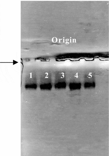
Hyalin was isolated and purified as described by Gray et al. (Reference Gray, Justice, Nagel and Carroll1986). Electrophoretic examination of hyalin preparations (0.3 mg/ml) on 2% agarose gels yielded the results shown in Fig. 1. Hyalin preparations showed one major band suggesting the purity of the protein. This is a standard method of determining hyalin purity (Gray et al., Reference Gray, Justice, Nagel and Carroll1986). In examining the effects of exogenous hyalin on gastrulation, 24-h S. purpuratus embryo samples were incubated for 24 h with increasing hyalin concentrations. Upon transfer of the embryos to the microplate wells, a precisely consistent sample size was not obtained due the fact that the embryos were hatched and swimming. The total sample size of embryos obtained for 10 replicates treated with the different hyalin concentrations of 0.00 mg/ml, 0.036 mg/ml, 0.075 mg/ml, 0.150 mg/ml and 0.225 mg/ml was as follows respectively: 114, 141, 154, 137 and 109.
2% agarose gel electrophoresis of hyalin preparations (lanes 1–5). The origin is indicated by an arrow. The direction of migration, towards the positive pole, is from top to bottom.

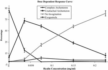
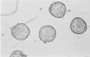
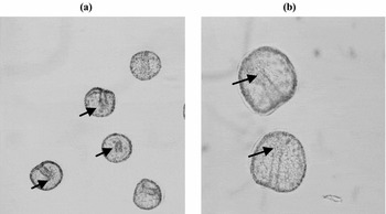
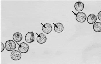
Figure 2 depicts the dose-dependent response curve for the microassay experiment. Mean percentage ±SEM of each morphology (complete archenteron, unattached archenteron, no invagination and exogastrula) was obtained and graphed as a function of hyalin concentration. In absence of hyalin, 91.07 ± 3.82% of embryos developed complete attached archenterons. At lower hyalin concentrations—0.036 mg/ml and 0.075 mg/ml—72.93 ± 4.34% and 63.25 ± 2.89% of the embryos developed unattached archenterons respectively. With increasing hyalin concentrations—0.15 mg/ml and 0.225 mg/ml—62.82 ± 2.36% and 85.91 ± 4.76% of the embryos did not invaginate respectively. Percentage of exogastrulas did not change significantly. No invagination, unattached archenteron and complete archenteron morphologies are shown in Figs 3, 4 and 5 respectively. Embryos were alive in all experiments as indicated by cilia movement.
Dose-dependent response curve. The graph shows the percentage of 48-h S. purpuratus embryos that developed either complete archenterons, unattached archenterons, did not invaginate or exogastrulated when incubated with increasing hyalin concentrations diluted in low calcium seawater. Data are plotted as mean percentage ± SEM of 10 experiments. The total sample size obtained for the five treatments is as follows: 114, 141, 154, 137 and 109.

Typical 48-h S. purpuratus embryos treated with high hyalin concentrations (0.15–0.255 mg/ml) diluted in low calcium seawater at 24 h. Note absence of archenterons. Magnification ×200.

Typical 48-h S. purpuratus embryos treated with low hyalin concentrations (0.036–0.075 mg/ml) diluted in low calcium seawater at 24 h showing mostly unattached archenterons as indicated by the arrows. Magnification a, ×100; b, ×200.

Typical 48-h S. purpuratus embryos incubated in low calcium seawater, pH 8.0, at 24 h in absence of hyalin. Arrows indicate complete archenterons attached to the blastocoel roof. Magnification ×100.

Discussion
Many studies have shown that the hyaline layer and its components serve as an anchoring substrate for epithelial sheets (Citkowitz, Reference Citkowitz1971; McClay & Fink, Reference McClay and Fink1982; Adelson & Humphreys, Reference Adelson and Humphreys1988; Hardin, Reference Hardin1996; Kimberly & Hardin, Reference Kimberly and Hardin1998). In this study, the effects of exogenous hyalin on archenteron morphology of whole, living gastrulating embryos were quantified. As depicted in Fig. 2, there is a significant decrease of the ‘complete archenteron’ morphology and a significant increase of the ‘no invagination’ morphology with increasing hyalin concentrations. Most notably, there is a rise and fall of the ‘unattached’ archenteron morphology with increasing hyalin concentrations. For the first time, we show that exogenous hyalin not only prevents invagination but also at important lower concentrations (0.036–0.075 mg/ml) inhibits archenteron elongation and/or attachment to the blastocoel roof.
We hypothesize that exogenous hyalin prevents invagination by changing the stiffness of the hyaline layer. Invagination is accomplished through mechanical processes of apical constriction. Apical constriction involves contraction of vegetal plate cells at the outer edge making them wedge-shaped (narrower at the apical face). This change in cell shape is initially sufficient to pull the outer surface of the cell sheet inwards to maintain primary invagination, as shown by a computer simulation in which apical constriction results in invagination (Wolpert et al., Reference Wolpert, Beddington, Brockes, Jessell, Lawrence, Meyerowittz and Lawrence1998).
Attachment of cells to the hyaline layer is required for apical constriction to take place. Studies have indicated that the hyaline layer develops blisters and wrinkles over the vegetal plate coincident with invagination of the endoderm (Kimberly & Hardin, Reference Kimberly and Hardin1998). Furthermore, the stiffness of the hyaline layer has a strong influence on the behavior of apical constriction (Davidson et al., Reference Davidson, Koehl, Keller and Oster1995). The computer-simulated mechanical analysis of Davidson et al. (Reference Davidson, Koehl, Keller and Oster1995) suggested that stiffness of the extracellular matrix is crucial for apical constriction to produce an invagination. Realistic invaginations occur only if the hyaline layer is of equal or lesser stiffness than the cell layer.
We propose that high concentrations of exogenous hyalin upon addition to gastrulating embryos may form an aggregate with the hyaline layer. This in turn could serve as an added source of stiffness for the hyaline layer disrupting the balance of stiffness between the extracellular matrix and the cell layer. We can speculate that the hyaline layer will no longer deform as easily as the cell layers and as a result apical constriction and primary invagination do not take place. Further work is needed to confirm our proposed model.
Perhaps more important is our finding that at low concentrations (0.036–0.075 mg/ml), exogenous hyalin inhibits archenteron elongation and/or attachment to the blastocoel roof (Fig. 4). It is possible that hyalin protein can enter the blastocoel and interfere with the interaction between the tip of the archenteron and the blastocoel roof. It has been previously shown that, under low calcium conditions, large proteins can enter the blastocoel of living embryos (Latham et al., Reference Latham, Martinez, Cazares, Hamburger, Tully and Oppenheimer1998; Itza & Mozingo, Reference Itza and Mozingo2005). Under low calcium conditions, hyalin (330 kDa) may be able to enter the blastocoel and block the interaction between the advancing tip of the archenteron and the blastocoel roof. This speculation can be further tested by examining the presence of hyalin in the embryo interior by using confocal microscopy and observing where labelled exogenous hyalin localizes.
The assay used in this study allows precise quantification of the effects of substances such as hyalin on the morphogenesis of intact living embryos. The microassay can be used to study the effects of many other molecular probes on the development of the sea urchin embryo.
Acknowledgements
We would like to thank Dr Cathy Coyle-Thompson for her valuable input on the microassay experiments and Dr Stan T. Metzenberg for his suggestions on the agarose gel electrophoresis. This work was supported by grants from NIH NIGMS, SCORE, RISE, MARC, ITQ Program and the Joseph Drown Foundation.