Implications
Early hatched chicks remain longer times inside the incubator after hatching compared with chick that hatched later hours of incubation. The present study showed that egg storage duration affected intestine development and gene expression of nutrient transporters of early hatched chicks. It could be expected that nutrient absorption process at the end of the hatch window would be more efficient in chicks from eggs stored for shorter periods compared with chicks from eggs stored for longer periods. The results reveal the importance of the feed access for early hatched chicks from eggs stored longer durations for the productivity of broilers.
Introduction
In commercial hatcheries, it is common to store eggs for 3 to 7 days. However, hatcheries may need longer storage duration depending on the supply of hatching egg and market demand for chicks. It is a well-known fact that longer egg storage reduces hatchability, impairs embryo development (Uddin and Hamidu, Reference Uni, Tako, Gal-Garber and Sklan2014), leads to higher embryonic mortalities by activating apoptotic cell death mechanisms and leads to reduced chick quality (Meijerhof et al., Reference Meijerhof, Noordhuzen and Leenstra1994; Christensen et al., Reference Christensen, Wineland, Fasenko and Donaldson2001; Tona et al., Reference Tona, Bamelis, De Ketelaer, Bruggeman, Moraes, Buyse, Onagbesan and Decuypere2003; Yalcin and Siegel, Reference Yalcin, Izzetoglu and Aktaş2003; Reijrink et al., Reference Reijrink, Meijerhof, Kemp, Graat and van den Brand2009; Hamidu et al., Reference Hamidu, Uddin, Fasenko, Guan and Barreda2011). Longer egg storage duration results in a longer incubation time (Christensen et al., Reference Christensen, Wineland, Fasenko and Donaldson2002). Thus, mixing eggs from different storage period affects the hatch spread, which is referred to 12 to 48 h of hatch window (Decuypere et al., Reference Decuypere and Bruggeman2001; Careghi et al., Reference Careghi, Tona, Onagbesan, Buyse, Decuypere and Bruggeman2005). It was reported that 80% of the chicks from eggs stored for 3 days hatched before 490 h of incubation, whereas this number was obtained at 500 h of incubation for chicks from eggs stored for 18 days (Tona et al., Reference Tona, Bamelis, De Ketelaer, Bruggeman, Moraes, Buyse, Onagbesan and Decuypere2003). Even under standardized egg storage conditions 30 h of hatch spread is still common (van de Ven et al., Reference van de Ven, van Wagenberg, Decuypere, Kemp and van den Brand2011). This means that the time spent in the incubator from hatching to pulling is longer for early hatched than late hatched chicks. This leads to a delayed access to first feed for early hatched chicks (Decuypere et al., Reference Decuypere and Bruggeman2001; Decuypere and Bruggeman, Reference Decuypere, Tona, Bruggeman and Bamelis2007). In other words, early hatched chicks remain without nutrients and water for a longer time, which results in a reduction in chick weight, yolk weight (Tona et al., Reference Tona, Bamelis, De Ketelaer, Bruggeman, Moraes, Buyse, Onagbesan and Decuypere2003; Yalcin et al., Reference Yalcin and Siegel2013) and depresses intestine mucosal development for several days post-hatch (Uni et al., Reference Uddin and Hamidu1998).
As the intestine is the primary nutrient supply organ, early development of digestive functions enables it to better utilize nutrients. Maturation of the small intestine is characterized by increased intestine weight, villus number and size, intestinal enzyme activity and increased nutrient transporter activity as well as RNA or DNA content (Geyra et al., Reference Geyra, Uni and Sklan2001; Uni et al., Reference Uni, Ganot and Sklan2003; Yalcin et al., Reference Yalcin and Siegel2013; Miska et al., Reference Miska, Fetterer and Wong2014). Ingested proteins and carbohydrates are hydrolyzed in the lumen of the small intestine and products are retrieved by enterocytes involving nutrient transporters that are responsible for absorption of peptides, amino acids and monosaccharides. Proteins are broken down to oligopeptides and free amino acids and then passed through the epithelial lining of the small intestine reaching the blood stream via oligopeptide and amino acid transporters such as PepT1 (H+-dependent peptide transporter) (Chen et al., Reference Chen, Pan, Wong and Webb2002). Carbohydrates are broken down into monosaccharides and absorbed by the action of Na+-dependent monosaccharide transporters such as SGLT1 (sodium–glucose co-transporter) and GLUTS (glucose transporter type 5) (Sklan et al., Reference Sklan, Geyra, Tako, Gal-Gerber and Uni2003). Expressions of PepT1 and SGLT1 influence the development of intestinal digestive and absorptive functions. As intestinal development during embryogenesis has a long-term influence on digestive and absorptive capacity in chickens, previous studies in chicks have concentrated on the presence of PepT1 and SGLT1 during embryonic growth (Uni et al., Reference Uni, Ganot and Sklan2003; Li et al., Reference Li, Gilbert, Zhang, Crasta, Emmerson, Webb and Wong2008; Speier et al., Reference Speier, Yadgary, Uniz and Wong2012; Miska et al., Reference Miska, Fetterer and Wong2014). Their upregulation between 18 days of incubation and 14 days post-hatch indicates the importance of those transporters for post-hatch growth and optimum development (Gilbert et al., Reference Gilbert, Li, Emmerson, Webb and Wong2007; Li et al., Reference Li, Gilbert, Zhang, Crasta, Emmerson, Webb and Wong2008; Mott et al., Reference Mott, Siegel, Webb and Wong2008).
Recent studies demonstrated that physiological differences exist between early and late hatching chicks, that is, early hatched chicks found less developed than later hatched chicks at the end of hatch window (van de Ven et al., Reference van de Ven, van Wagenberg, Decuypere, Kemp and van den Brand2011 and Reference van de Ven, van Wagenberg, Debonne, Decuypere, Kemp and van den Brand2013). The studies on hatch window so far have not taken into consideration the changes in gene expression of nutrient transporters in chicks during the hatch window. Therefore, the present study aimed to evaluate the combined effects of egg storage duration and 30 h of the time spent in the incubator on gene expression of nutrient transporters and intestine morphological parameters of early hatched broiler chicks.
Material and methods
Experimental procedures were approved by the Ege University Animal Care and Ethics Committee with the Turkish Code of Practice for the Care and Use of Animals for Scientific Purposes (2012-026).
A total of 1218 eggs obtained from 38-week-old Ross 308 broiler breeder flocks were used. To standardize pre-incubation factors, eggs were collected from a single broiler breeder flock. In order to incubate all eggs at the same time, eggs were collected in 11-day interval; therefore, half of eggs were stored for 3 days (ES3), whereas the other half was stored for 14 days (ES14). Average egg weight was 62.12±0.21 g. The storage conditions were 18°C and 14°C for 3 and 14 days stored eggs, respectively, with 75% relative humidity. Different storage temperatures were chosen, as these temperatures emulate current industry conditions to optimize hatchability (Meijerhof, Reference Meijerhof1992; Schulte-Drüggelte, Reference Schulte-Drüggelte2011). All eggs were numbered and placed into a Combi Incubator C82 (Pas Reform, Zeddam, The Netherlands). The incubation temperature was 37.7°C during the first 18 days and 36.7°C thereafter, with a relative humidity of 58%. There were seven replicate egg trays with 87 eggs for each treatment.
Sample collection and morphological measurements
At hatch
Eggs were checked between 475 and 480 h of incubation and hatched chicks from both egg storage durations were recorded as early hatched chicks.
The 40 hatched chicks at 480 h from each egg storage duration were color coded and weighed; chick length and rectal temperature were measured. The 20 chicks/egg storage duration were randomly chosen, were placed back in the incubator and allowed to remain in the incubator during the hatch window.
The remaining 20 chicks/egg storage duration were killed by cervical dislocation, and residual yolk sac and small intestine were dissected. The small intestine was separated into duodenum, jejunum, ileum and length of intestine parts and weights of residual yolk sac and intestine parts were measured. About 2 cm sampled from the midpoint of the jejunum from six randomly selected chicks were immediately rinsed in phosphate-buffered saline, frozen in liquid nitrogen and stored at −80°C until RNA extraction and analysis. A 2 cm of the jejunum was also sampled from eight chicks for histological measurements.
At the end of hatch window
At 510 h of incubation, the same measurements were conducted with the chicks (early hatched 20 chicks/egg storage duration) kept in the incubator. Therefore, the hatch window period was 30 h for chicks, being similar to previous studies (van de Ven et al., Reference van de Ven, van Wagenberg, Debonne, Decuypere, Kemp and van den Brand2013).
The chicks sampled for intestinal measurements at hatch and end of hatch window were dried at 110°C for 24 h and their dry matter content was calculated as the differences between wet and dry weights divided by wet weight.
Histological measurements
Tissue samples of chicks were gently flushed with 0.9% NaCl to remove intestinal contents and fixed in fresh 70% alcohol. All samples were dehydrated, cleared and embedded in paraffin. Serial sections (5 μm) were counted and mounted on a slide, deparaffinized in xylene, dehydrated in a graded alcohol series, and stained with hematoxylin and eosin. Sections were examined for villus length (from the top of the villi to the villus crypt junction) and villus width (at half height of villi) by light microscopy using computer software (SigmaScan, Point Richmond, CA, USA). Values were means of 12 villi/chick.
Goblet cell counts of chicks were performed by staining sections with alcian blue (pH 2.5, 1052340010; Merck, Darmstadt, Germany), periodic acid (0.5%, P7875; Sigma, Taufkirchen, Germany) and Schiff (3952016; Sigma). The slides were deparaffinized, rehydrated and stained with alcian blue solution for 30 min. This was followed by incubation in periodic acid for 20 min and in Schiff’s reagent for 20 min. Slides were then washed in distilled water between each incubation period, dehydrated, cleared and mounted in entellan. The number of goblet cells along the villi was counted by light microscopy. Values are means of goblet cells from 12 villi/chick.
Real-time PCR analysis
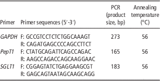
Total RNA was extracted from 20 to 30 mg jejunum tissues using TRIzol Reagent (Invitrogen, Carlsbad, California, USA). RNA samples were resuspended in DNase/RNase-free H2O and the optical densities were measured at 260 nm with the NanoDrop ND-1000 spectrophotometer (NanoDrop Technologies, Thermo Fisher Scientific, Waltham, Massachusettes, USA). cDNA synthesis kit (NEB, Ipswich, Massachusettes, USA) ProtoScript First Strand cDNA was used to transcribe total RNA samples according to manufacturer’s recommended protocol. PCR reaction was prepared with Quick-load Taq 2X Master Mix (NEB). PCR conditions were 95°C for 10 min for initial denaturation, and 34 cycles of 95°C for 10 s, 56°C for 30 s, 72°C for 30 s for denaturation annealing and extension and final extension of 10 min at 72°C. Primers were in-house designed from Primer 3 software (Table 1). Gene expressions of PepT1 and SGLT1 of chicks were calculated using the ΔΔC t method to that of glyceraldehyde-3-phosphate dehydrogenase expression as the endogenous control.
Chicken primer sequences and their expected product size

bp=base pair; GAPDH=glyceraldehyde-3-phosphate dehydrogenase; PepT1: H+-dependent peptide transporter; SGLT1: sodium–glucose co-transporter.
Statistical analyses
All data were analyzed by using JMP software from SAS, version 5.0 (SAS Institute, 2003). Data for chick weights, lengths and rectal temperatures were analyzed by using a mixed model repeated-measures ANOVA. Data for yolk sac, dry matter content and intestine measurements were analyzed with a model that included storage duration and hatch window and their interactions. Least square means were compared using Tukey’s test. Differences were considered significant at P<0.05, unless otherwise stated.
Results
Hatching time and morphologic and histologic measurements
At 480 h of incubation, 53.3% of ES3 chicks hatched while it was only 21.2% for ES14 chicks (P=0.034) (data not shown).
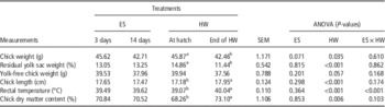
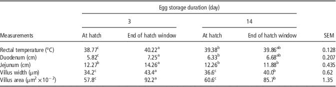
There was no effect of egg storage duration on chicks weight, relative residual yolk sac weight, yolk-free chick weight, length, rectal temperature and dry matter content (Table 2). Chick weight and residual yolk sac weight significantly reduced (7.4% and 23.0%, respectively) during hatch window, whereas chick length and chick dry matter content increased (4.5% and 7.1%, respectively) (Table 2). A significant storage duration by hatch window interaction showed that chicks from ES3 had higher rectal temperatures at the end of the hatch window compared with at hatch; however, there was no change in rectal temperature of chicks from ES14 during hatch window (Table 3).
Effect of egg storage duration (ES) and 30 h of hatch window (HW) on weight, residual yolk sac weight, length, rectal temperature and dry matter content of early hatched chicks

a,bMeans in the same row within a measurement and treatment with no common superscript differ significantly (P<0.05).
Egg storage duration and 30 h of hatch window interaction for rectal temperature, lengths of duodenum and jejunum, and villus width and area of early hatched chicks

a,b,cMeans in the same column within a measurement with no common superscript differ significantly (P<0.05).
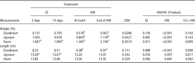
Storage durations had no effect on weights of duodenum and jejunum of chicks. Ileum weights of ES14 chicks were heavier than those from ES3 (Table 4). The weights of intestine segments increased during the hatch window. There was a significant egg storage duration by hatch window interaction for the lengths of duodenum and jejunum (Table 3). This interaction showed that during the hatch window, the lengths of duodenum and jejunum increased in ES3 chicks but not in the ES14 chicks. At the end of the hatch window, jejunum length of ES3 chicks was longer than ES14 chicks (Table 3). Neither egg storage duration nor hatch window affected ileum length (Table 4).
Effect of egg storage duration (ES) and 30 h hatch window (HW) on weights and lengths of small intestine segments of early hatched chicks

a,bMeans in the same row within a measurement and treatment with no common superscript differ significantly (P<0.05).
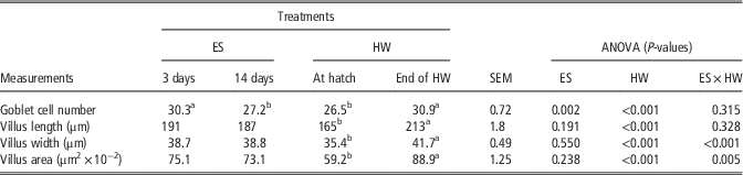
Chicks from ES3 had higher numbers of goblet cells than ES14 chicks (Table 5). During hatch window, villus length, width and area increased by 29.1%, 17.8% and 50.2%, respectively; however, the interaction between storage duration and hatch window revealed that the increase in villus width and surface area was greater in chicks from ES3 than ES14 (Table 3).
Effect of egg storage duration (ES) and 30 h hatch window (HW) on goblet cell number, villus length, width and area of early hatched chicks

a,bMeans in the same row within a measurement and treatment with no common superscript differ significantly (P<0.05).
Gene expression of nutrient transporters
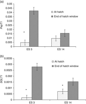
Expression of PepT1 was influenced by egg storage duration. There was much higher transcript expression of PepT1 in ES3 chicks compared with ES14 chicks (average PepT1 abundance was 0.0232 and 0.0125, for ES3 and ES14, respectively, P=0.037, data not shown in the tables). Although there was a significant message transcript upregulation in PepT1 (P<0.001) at the end of hatch window, a significant interaction (P=0.004) between egg storage duration and hatch window implicated that the increase in PepT1 expression was only significant for ES3 chicks, whereas PepT1 expression in the jejunum of ES14 chicks did not show any change during the hatch window (Figure 1a). Thus, higher PepT1 expression was observed for ES3 than ES14 chicks at the end of hatch window. Fold increase of PepT1 at the end of hatch window was 10.2-fold for ES3 chicks and 2.3-fold for ES14 chicks (data not shown).
PepT1 (a) and SGLT1 (b) gene expressions in the jejunum of early hatched chicks at hatch and end of hatch window. Gene expressions were calculated using the ΔΔC t method to that of glyceraldehyde-3-phosphate dehydrogenase expression as the endogenous control. Interaction between egg storage duration (ES) and hatch window was significant for PepT1 (P=0.004). PepT1=H+-dependent peptide transporter; SGLT1=sodium–glucose co-transporter.

Egg storage duration had no effect on SGLT1 expression (average SGLT1 abundance was 0.00148 and 0.00112, for ES3 and ES14, respectively, P=0.463, data not shown in the tables). Higher expressions of SGLT1 level were observed in both ES3 and ES14 chicks at the end of the hatch window compared with at hatch (mean increase was from 0.00045 to 0.0022, P=0.002) (Figure 1b). The differences between ES3 and ES14 chicks for the expression of SGLT1 approached significant (P=0.083) at the end of the hatch window. Fold increase of SGLT1 was 17.6 and 3.3-fold for ES3 and ES14 chicks, respectively, at the end of hatch window (data not shown).
Discussion
Chick development could be influenced by a variety of factors during incubation including egg and hatch window. Recent studies showed that early hatched chicks differ from late hatched chicks from a metabolic point of view. However, gene expression of nutrient transporters during hatch window is still not explored. Therefore, this study aimed to determine the changes from hatch to the end of the hatch window in intestinal development and expression of PepT1 and SGLT1 genes in early hatched broiler chicks obtained from eggs stored for 3 or 14 days before incubation.
Hatching time and morphologic and histologic measurements
The delay in hatching from eggs stored for longer periods supports the findings of Tona et al. (Reference Tona, Bamelis, De Ketelaer, Bruggeman, Moraes, Buyse, Onagbesan and Decuypere2003). As observed in previous studies (Decuypere et al., Reference Decuypere and Bruggeman2001; van de Ven et al., Reference van de Ven, van Wagenberg, Debonne, Decuypere, Kemp and van den Brand2013; Yalcin et al., Reference Yalcin and Siegel2013), there was a decrease in chick weight at the end of the hatch window that coincided with the increase in dry matter content of chicks showing longer hatch windows resulting in significant BW loss. The decrease in relative yolk sac weight at the end of hatch window is explained by the nutrient transfer from yolk sac into intestine (Noy and Sklan, Reference Noy and Sklan2001; Yadgari et al., Reference Yadgari, Yair and Uni2011). This transfer of yolk sac helps early growth of small intestine after hatching, regardless of access to food (Noy and Sklan, Reference Noy and Sklan1999; Lamot et al., Reference Lamot, van de Linde, Molenaar, van der Pol, Wijtten, Kemp and van den Brand2014). The relative weight increases of the small intestine segments were 64.7%, 46.5% and 47.8% for duodenum, jejunum and ileum, respectively, during the hatch window and was independent of egg storage duration. These results also indicated that digestive system of chicks either from eggs stored for shorter or for longer storage duration continue to develop after hatch, irrespective of feed access (Lamot et al., Reference Lamot, van de Linde, Molenaar, van der Pol, Wijtten, Kemp and van den Brand2014).
However, egg storage duration affected the length of jejunum at the end of the hatch window, suggesting that shorter egg storage durations led to much longer jejunum. With the larger villus width and area in the jejunum of chicks from ES3 compared with ES14 at the end of the hatch window, the results may explain better growth rate of chicks obtained from eggs stored for shorter durations (Tona et al., Reference Tona, Bamelis, De Ketelaer, Bruggeman, Moraes, Buyse, Onagbesan and Decuypere2003).
Gene expression of nutrient transporters
The increased nutrient transport maintains embryo growth within the normal range until hatch. At 2 days before hatching, Na-independent anaerobic metabolism provides most of the energy, but sodium is vital for glucose transfer 2 days after hatching (Moran, Reference Moran1985). The SGLT1 mRNA transcript remain high by 19 days of incubation and decreased at day of hatch and then upregulated after the ingestion of carbohydrates up to day 7 (Sklan et al., Reference Sklan, Geyra, Tako, Gal-Gerber and Uni2003; Uni et al., Reference Uni, Ganot and Sklan2003). Chen et al. (Reference Chen, Pan, Wong and Webb2002) reported that the peptide transporters were mainly expressed in the small intestine of broilers. The expression of PepT1 was regulated by developmental stage during embryonic growth and its mRNA level increased from day 16 to hatch with an abrupt rise just before hatch (Chen et al., Reference Chen, Pan, Wong, Bloomquist and Webb2005; Gilbert et al., Reference Gilbert, Li, Emmerson, Webb and Wong2007; Speier et al., Reference Speier, Yadgary, Uniz and Wong2012). In the present study, greater PepT1 expression than SGLT1 probably related to the importance of proteins during development and may be necessary to maximize amino acid assimilation when the feed become available (Mott et al., Reference Mott, Siegel, Webb and Wong2008). It was previously described that genes that are important for functional developments should have the highest expression levels at early life (Schokker et al., Reference Schokker, Hoekman, Smits and Rebel2009). On the other hand, it was also reported an increase in PepT1 expression in response to starvation in rats (Ihara et al., Reference Ihara, Tsujikawa, Fujiyama and Bamba2000) and chickens (Mott et al., Reference Mott, Siegel, Webb and Wong2008). In the present study, PepT1 expression was greater in ES3 chicks than those from ES14 chicks from 480 h of incubation to the end of hatch window at 514 h. In addition, compared with ES14 chicks, ES3 chicks exhibited greater SGLT1 expression at the end of the hatch window. Enhanced villus surface area along with upregulated expression of nutrient transporters of ES3 chicks at the end of hatch window appears to positively contribute to the nutrient absorption and digestion as reported previously (Li et al., Reference Li, Gilbert, Zhang, Crasta, Emmerson, Webb and Wong2008). Our findings suggested that ES3 chicks would have a greater aptitude for absorption of proteins and carbohydrates when food intake begins compared with ES14 chicks. The fold increase in the expression of SGLT1 was higher compared with PepT1 at the end of hatch window.
In conclusion, these results established that development of small intestine and nutrient transporters of early hatched chicks were influenced by egg storage duration and hatch window. The PepT1 and SGLT1 expressed at significantly higher levels in the jejunum of ES3 compared with ES14 chicks at the end of the hatch window. When taken together data regarding to villus development, duodenum and jejunum lengths indicated a higher intestinal absorptive capacity of early hatched ES3 than ES14 when access to feed at the end of the hatch window. Therefore, due to downregulated nutrient transporters for chicks from eggs that were stored for longer periods coupled with less-developed small intestine could lead to depressed growth. Our findings also reveal the importance of early feeding of those chicks from eggs stored longer durations. In this study, only early hatched chicks were studied; therefore, it remains unknown if these differences exist between late hatched ES3 and ES14 chicks.
Acknowledgment
This research was supported by TUBITAK (Project No. 112 0 220).