INTRODUCTION
The Oncaeidae is a speciose family of copepods, that contributes to zooplankton communities in a wide range of marine environments, from low to high latitudes (Star & Mullin, Reference Star and Mullin1981; Cowles et al., Reference Cowles, Roman, Gauzens and Copley1987; Paffenhöffer, Reference Paffenhöfer1993; Richter, Reference Richter1994; Metz, Reference Metz1995) and from near surface waters to the deep sea (Boxshall, Reference Boxshall1977a; Wishner, Reference Wishner1979; Roman et al., Reference Roman, Gauzens and Cowles1985; Yamaguchi et al., Reference Yamaguchi, Watonabe, Ishida, Harimoto, Furusawa, Suzuki, Ishizaka, Ikeda and Takahashi2002). The taxonomic diversity of the Oncaeidae, as re-defined by Huys & Böttger-Schnack (Reference Huys and Böttger-Schnack1997), has been increasingly recognized during recent years (Shmeleva, Reference Shmeleva1969; Boxshall, Reference Boxshall1977b; Heron, Reference Heron1977; Heron et al., Reference Heron, English and Damkaer1984; Heron & Bradford-Grieve, Reference Heron and Bradford-Grieve1995; Böttger-Schnack, Reference Böttger-Schnack1999, Reference Böttger-Schnack2001; Heron & Frost, Reference Heron and Frost2000). The generic level classification of this family is under review (Böttger-Schnack & Huys, Reference Böttger-Schnack and Huys1998), but Boxshall & Halsey (Reference Boxshall and Halsey2004) listed seven valid genera, Archioncaea Böttger-Schnack & Huys, Monothula Böttger-Schnack & Huys, Spinoncaea Böttger-Schnack, Oncaea Philippi, Triconia Böttger-Schnack, Epicalymma Heron and Conaea Giesbrecht. These genera have been recorded from distant localities in the world ocean, including the Mediterranean (Shmeleva, Reference Shmeleva1966, Reference Shmeleva1968, Reference Shmeleva1969; Malt et al., Reference Malt, Lakkis and Ziedanz1989; Böttger-Schnack, Reference Böttger-Schnack1997; Kršinic, Reference Kršinic1998), the Red Sea and Arabian Sea (Böttger-Schnack, Reference Böttger-Schnack1990a, Reference Böttger-Schnackb, Reference Böttger-Schnack1995, Reference Böttger-Schnack1996), the north-west and north-east Pacific (Heron & Frost, Reference Heron and Frost2000; Nishibe & Ikeda, Reference Nishibe and Ikeda2004) and the Antarctic (Metz, Reference Metz1995).
The genus Triconia was established by Böttger-Schnack (Reference Böttger-Schnack1999) to accommodate eleven species of Oncaeidae characterized by the presence of a conical process on the distal margin of the endopod of swimming leg 4. It corresponds to the Oncaea conifera Giesbrecht and Oncaea similis Sars groups recognized in the phylogenetic analysis of Böttger-Schnack & Huys (Reference Böttger-Schnack and Huys1998). Triconia umerus (Böttger-Schnack & Boxshall) and T. hawii (Böttger-Schnack & Boxshall), originally placed in the genus Oncaea, were based on females collected in the Red Sea (Böttger-Schnack & Boxshall, Reference Böttger-Schnack and Boxshall1990). Böttger-Schnack (Reference Böttger-Schnack1999) subsequently redescribed both species, transferred them to Triconia, and described the males for the first time.
The oncaeids of the Mediterranean Sea are relatively well known: forty-seven species have been reported and many small-size Oncaea species (less than 0.6 mm) were first described from the Adriatic (Shmeleva, Reference Shmeleva1966, Reference Shmeleva1968, Reference Shmeleva1969, Reference Shmeleva1979). Triconia is represented by only eight species in the Mediterranean whereas 12 species have been reported from the Red Sea. Identification of oncaeid species requires detailed investigation of numerous characters, including mouthparts and surface ornamentation, which are difficult to examine in these small copepods. Recent detailed taxonomic studies (Heron & Bradford-Grieve, Reference Heron and Bradford-Grieve1995; Heron & Frost, Reference Heron and Frost2000, Böttger-Schnack, Reference Böttger-Schnack2001) have demonstrated that many apparently well known species, such as Triconia conifera (Giesbrecht) and Oncaea media Giesbrecht, represent complexes of closely related yet distinct species. A recent phylogeographical study of Oncaea venusta Philippi revealed genetically distinct clades at five different locations in the Indo-West Pacific (Elvers et al., Reference Elvers, Böttger-Schnack, Blohm and Hagen2006).
Temporal changes in the community of oncaeid copepods at a fixed coastal station in the Gulf of Naples (Tyrrhenian Sea) have been investigated over seasonal and inter-annual scales (Mazzocchi & Aguzzi, Reference Mazzocchi and Aguzzi1998). This study has shown for the first time the presence of Triconia umerus, T. hawii and T. rufa (Boxshall & Böttger-Schnack) in the western Mediterranean. The record of T. hawii is the first for the Mediterranean Sea. These new records highlight the importance of improved taxonomic resolution of oncaeids in time-series studies in providing further evidence of faunistic change.
MATERIALS AND METHODS
Mesozooplankton samples were collected between 08:00 h and 12:00 h to minimize the variability due to vertical migration. Vertical hauls were taken from 50 m depth to the surface using a Nansen net (113 cm mouth diameter, 200 µm mesh aperture). The samples were fixed immediately after collection and preserved in a 4% buffered formaldehyde–seawater solution. The samples examined were part of an on-going time-series study of the pelagic system at a fixed station Marechiara (Station MC) in the Gulf of Naples which commenced in January 1984 (Ribera d'Alcalà et al., Reference Ribera d'Alcalà, Conversano, Corato, Licandro, Mangoni, Marino, Mazzocchi, Modigh, Montressor, Nardella, Saggiomo, Sarno and Zingone2004).
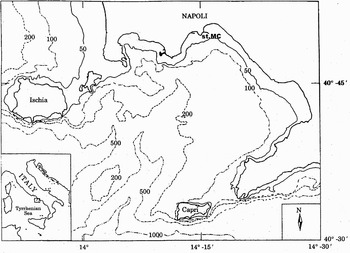
The sampling site (40°48.5′N 14°15′E) is located two nautical miles from the coast, close to the 80 m isobath (Figure 1). The Gulf of Naples is a wide embayment (870 km2) in the Tyrrhenian Sea (western Mediterranean). The littoral area is influenced by land runoff from a densely populated region. However, due to the general physiography and bottom topography, the inner shelf area is strongly coupled with offshore Tyrrhenian waters. These features result in the coexistence of two subsystems within the Gulf: a wide oligotrophic area with Tyrrhenian characteristics and a narrower eutrophic coastal zone. The Marechiara station is located at the boundary between the two subsystems (Ribera d'Alcalà et al., Reference Ribera d'Alcalà, Conversano, Corato, Licandro, Mangoni, Marino, Mazzocchi, Modigh, Montressor, Nardella, Saggiomo, Sarno and Zingone2004).
Location of the sampling site (Station MC) of the long-term study conducted in the Gulf of Naples (Tyrrhenian Sea, western Mediterranean).

Oncaeids from samples taken at the Marechiara station on 31 January and 8 February 2005 were sorted and examined. About 20 females of each species were identified under a Wild M3C stereomicroscope. Specimens were dissected in lactic acid and examined as temporary mounts in lactophenol. Measurements were taken under the microscope using an ocular micrometer. Total body length was measured dorsally from the tip of prosome to the distal end of the caudal ramus.
Diagnostic morphological details were verified using scanning electron microscopy (SEM). Specimens, pre-fixed in formaldehyde, were rinsed with distilled water and left overnight to enhance body turgidity. They were post-fixed for 5 h in 2.5% gluteraldehyde at room temperature before dehydration through graded methanol series. Specimens were critical-point dried and coated with gold-palladium before being mounted on stubs. We compared the general body shape of Triconia umerus and T. hawii from the Gulf of Naples with the same species from the Red Sea (type material from collections of the Natural History Museum, London).
RESULTS
In addition to nine common species of Oncaeidae (Oncaea curta Sars, O. scottodicarloi Heron & Bradford-Grieve, O. media, O. mediterranea (Claus), O. venusta, Monothula subtilis (Giesbrecht), Triconia conifera, T. dentipes (Giesbrecht) and T. minuta (Giesbrecht)), three other Triconia species were present. Two of these, Triconia umerus and T. rufa, have already been reported from the eastern Mediterranean (Böttger-Schnack, Reference Böttger-Schnack1997) but the third species, Triconia hawii, has not been reported before from the Mediterranean Sea. These species were first discovered in the Gulf of Naples in April 2004. Their abundances were lower than those of other oncaeid species at the same station. Abundance ranged from 0.2 to 3.2 ind m−3 for all three species, but the mesh size (200 µm) of the net used to collect the meso-zooplankton was too large to efficiently collect these microcopepods. They can be reliably collected using 55 µm mesh nets (Böttger-Schnack, Reference Böttger-Schnack1997, Reference Böttger-Schnack1999).
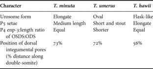
Triconia conifera is a large and distinctive species, but the other five species of Triconia present in the Gulf of Naples are all of similar size, with adult female body length between about 450 and 600 µm. The females of these species can most readily be distinguished by combinations of fine-scale characters, including body length, shape of urosome, relative lengths of setal elements on leg 5, and the positions of the gonopores and integumental pores on the dorsal surface of the genital double-somite. These characters are summarized for the three most similar species in Table 1 and are briefly outlined in a series of differential diagnoses based specifically on Gulf of Naples material taken in the 0–50 m depth horizon.
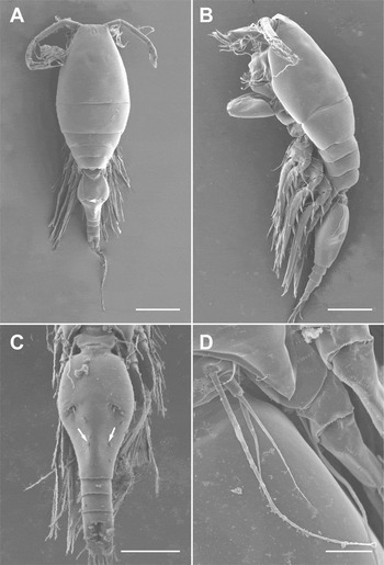
Triconia dentipes (Giesbrecht, Reference Giesbrecht1891). (A) Entire, dorsal view showing position of gonopores (arrowed); (B) entire, lateral view showing long surface seta of leg 4 reaching to level of gonopore (arrowed); (C) urosome, dorsal view showing dorsal integumental pores (arrowed); (D) leg 5. Scale bars: A, B, D, 100 µm; C, 50 µm.

Morphological characters separating females of Triconia minuta, T. umerus and T. hawii in the Gulf of Naples.

OSDS, outer subdistal spine; ODS, outer distal spine.
Body length 478 µm. Prosome slender; second pedigerous somite lacking any dorsal process. Genital double-somite (in dorsal view) about 1.5 times longer than wide, with evenly convex lateral margins; gonopores located at about 47% of distance along double-somite from anterior margin. Pair of integumental pores located at about 78% of distance along double-somite from anterior margin. Surface of double-somite lacking ornamentation of spinules. Leg 5 with surface seta (derived from incorporated basis) just extending to level of gonopores; exopodal setal elements of similar length, short, not reaching gonopores. Live specimens with distinctive coloration, with orange pigment located anteriorly in narrow band across cephalosome.
REMARKS
Triconia dentipes was originally described from Gulf of Naples material (Giesbrecht, Reference Giesbrecht1891). Giesbrecht's figures showed the same distinctive morphological characters, especially the flask-like form of the genital double-somite (cf. plate 47, figure 7; our Figure 2C). This species was reported from other Mediterranean areas and the Red Sea by Böttger-Schnack (Reference Böttger-Schnack1999) who discussed the differences between Giesbrecht's drawing (1892, plate 47, figure 7) and her redescription (1999, figure 13C) with reference to the length of the terminal accessory seta on the caudal ramus. In accord with Böttger-Schnack (Reference Böttger-Schnack1999), our specimens of T. dentipes from the Gulf of Naples have a long terminal accessory seta on the caudal ramus (Figure 2D).
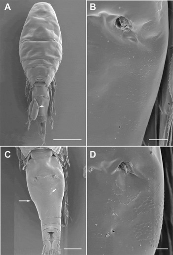
Triconia minuta (Giesbrecht, Reference Giesbrecht1892). (A) Entire, dorsal view showing position of dorsal integumental pore (arrowed); (B) genital double-somite spinular ornamentation. Triconia umerus (Böttger-Schnack & Boxshall, Reference Böttger-Schnack and Boxshall1990); (C) genital double-somite, dorsal view showing position of integumental pore (small arrow) and marginal spinule row at angle of shoulder of double-somite (large arrow); (D) same, showing spinular ornamentation and location of integumental pore. Scale bars: A, 100 µm; B, C, D, 20 µm.

Body length 474 µm. Prosome slender; second pedigerous somite lacking any dorsal process. Genital double-somite (in dorsal view) about 1.6 times longer than wide, with evenly convex lateral margins; gonopores located at about 33% of distance along double-somite from anterior margin. Pair of integumental pores located at about 73% of distance along double-somite from anterior margin. Surface of double-somite ornamented with lateral patches of spinules extending posteriorly from level of gonopores. Leg 5 with surface seta (derived from incorporated basis) short, not reaching level of gonopores; exopodal setal elements of similar length, short, not reaching gonopores.
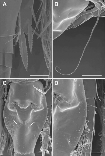
Triconia umerus (Böttger-Schnack & Boxshall, Reference Böttger-Schnack and Boxshall1990). (A) Leg 4, tip endopod showing relative lengths of distal spines Triconia rufa (Boxshall & Böttger, Reference Boxshall and Böttger1987); (B) leg 5, showing extreme length of outer exopodal seta Triconia hawii (Böttger-Schnack & Boxshall, Reference Böttger-Schnack and Boxshall1990); (C) genital double-somite, dorsal view showing position of integumental pores (arrowed) (the highlighted transverse ridges adjacent to the gonopores are artefacts of the drying process); (D) genital double-somite, dorsal view of right side showing spinular ornamentation. Scale bars: A, B, C, D, 20 µm.

Body length 580 µm. Prosome slender; second pedigerous somite lacking any dorsal process. Genital double-somite (in dorsal view) about 1.5 times longer than wide, bottle-shaped with distinct ‘bottleneck’ narrowing posteriorly; gonopores located at about 35% of distance along double-somite from anterior margin. Pair of integumental pores located at about 72% of distance along double-somite from anterior margin. Lateral surface of double-somite ornamented with patches of surface spinules just posterior to gonopores, including defined row of spinules at ‘shoulders’ of bottleneck. Leg 5 with plumose surface seta (derived from incorporated basis) strong and short, not reaching level of gonopores; exopodal setal elements of similar length, short, not reaching level of gonopores.
REMARKS
Triconia umerus is very similar to T. minuta but is larger and can be distinguished easily by the orange coloration on the urosome and anterior part of the prosome. In T. umerus the row of small denticles on the margin of genital double-somite is visible under the stereomicroscope.
Body length 497 µm. Prosome very slender; second pedigerous somite with rounded dorsal process visible in lateral view. Genital double-somite (in dorsal view) about 1.6 times longer than wide, bottle-shaped with distinct ‘bottleneck’ narrowing posteriorly; gonopores located at about 30% of distance along double-somite from anterior margin. Pair of integumental pores located at about 60% of distance along double-somite from anterior margin. Surface of double-somite ornamented with lateral patches of spinules extending from level of gonopores to ‘shoulders’ of bottleneck. Leg 5 with very long, curved surface seta (derived from incorporated basis) reaching beyond gonopores as far as ‘shoulders’ of bottleneck; exopodal setal elements distinctly different in length, one elongate and one short, not reaching gonopores: exopodal segment with small spinule.
REMARKS
In this species the ornamentation of surface spinules on the genital double-somite is not visible under the stereomicroscope. The extreme length of the surface seta derived from the incorporated basis of the fifth leg is characteristic for T. rufa.
Body length 497 µm. Prosome slender; second pedigerous somite lacking any dorsal process. Genital double-somite (in dorsal view) about 1.4 times longer than wide, bottle-shaped with distinct ‘bottleneck’ narrowing posteriorly; gonopores located at about 30% of distance along double-somite from anterior margin. Pair of integumental pores located at about 56% of distance along double-somite from anterior margin. Surface of double-somite ornamented with extensive lateral patches of spinules. Leg 5 with surface seta (derived from incorporated basis) short, not reaching level of gonopores; exopodal setal elements of similar length, short, not reaching gonopores.
COMPARISONS
Some new morphological details are revealed here. Most notably the presence of an ornamentation of surface spinules on the lateral margins of the genital double-somite in T. minuta and T. hawii, that has not previously been reported. Similar ornamentation has been reported for both T. umerus and T. rufa (Böttger-Schnack, Reference Böttger-Schnack1999) but it is difficult to observe by light microscopy and we infer that it has previously been overlooked in T. minuta and T. hawii.
The specimens collected in the Gulf of Naples were compared with data on populations from the Red Sea. Small differences were observed between females from the two localities in size and in body proportions (cf. Table 2). However, size differences are unreliable because of differences in mesh sizes used between the Red Sea survey and the Gulf of Naples study. The differences in body proportions are not regarded here as significant and, indeed, Böttger-Schnack (Reference Böttger-Schnack1999) has already noted slight differences in urosome shape between material of T. dentipes from the Red Sea and the Adriatic. We also note slight differences in the positions of the gonopores and the dorsal integumental pores along the genital double-somite. We infer that these differences represent geographical variation in all species with the possible exception of T. hawii. Triconia hawii is unusual in exhibiting variation in the position of the gonopores on the genital double-somite: with Böttger-Schnack (Reference Böttger-Schnack1999) showing gonopores located between 33% and 50% of the distance along the double-somite. In other species this character is relatively stable. We consider this degree of variability to indicate a possible taxonomic problem. Also in T. hawii, the dorsal integumental pore is located at about 71% of distance along double-somite from anterior margin in Red Sea material (Böttger-Schnack, Reference Böttger-Schnack1999) compared with only 56% in Gulf of Naples material. In addition, our Gulf of Naples material has spinular ornamentation, not reported by Böttger-Schnack (Reference Böttger-Schnack1999). These results cause some uncertainty over the identity of the T. hawii from the Gulf of Naples. Further taxonomic research is needed to investigate the significance of this level of variability between populations of this species, although the current diagnosis of the species accommodates much of this variability.
A comparison of prosome lengths and body proportions of Triconia umerus and T. hawii from the Gulf of Naples and the type locality (Red Sea).

L:W, length to width ratio.
There are also marked differences in the depth distributions of the species between the Red Sea and the Gulf of Naples. In the Gulf, all five species co-occur in the top 50 m of the water column whereas in the central Red Sea T. minuta and T. umerus both are found primarily in the 50 to 100 m depth horizon, T. dentipes at depths of 150 to 200 m, and both T. hawii and T. rufa much deeper, at 650 to 700 m (Böttger-Schnack, Reference Böttger-Schnack1990a, Reference Böttger-Schnackb, Reference Böttger-Schnack1992).
KEY TO SPECIES OF ONCAEIDAE IN GULF OF NAPLES (ADULT FEMALES ONLY)
1. Large, heavily-chitinized forms with body length in excess of 1.0 mm.……………2
Small forms with body length less than 1.0 mm.……………3
2. Caudal rami about 3.5 times longer than wide; anal somite about 0.63 times long as wide.……………Oncaea venusta
Caudal rami about 3.0 times longer than wide; anal somite about 0.77 times long as wide.……………Oncaea mediterranea
3. Endopod of leg 4 with distal conical process between apical and outer distal margin spines (Figure 4A)……………4
Endopod of leg 4 without distal conical process between apical and outer distal margin spines.……………8
4. In lateral view, prosome with conspicuous dorso-posterior projection on second pedigerous somite.……………Triconia conifera
In lateral view, prosome without conspicuous dorso-posterior projection (bearing small rounded dorsal process in T. rufa only)……………5
5. Prosome 1.9 times as long as urosome (excluding caudal setae); anal somite about as wide as long; body length 0.47 mm ± 0.05 mm.……………Triconia dentipes
Prosome at least 2.3 times as long as urosome (excluding caudal setae); anal somite at least 1.2 times wider than long; body length 0.49 to 0.60 mm.……………6
6. Prosome about 2.7 times as long as urosome (excluding caudal setae); anal somite at least 1.6 times wider than long; body length 0.49 ± 0.07 mm.……………Triconia hawii
Prosome about 2.3 to 2.4 times as long as urosome (excluding caudal setae); anal somite 1.2 to 1.3 times wider than long; body length 0.50–0.57 mm.……………7
7. Genital double-somite about 1.6 times longer than wide and with evenly convex lateral margins; body length 0.50 mm ± 0.06 mm.……………Triconia minuta
Genital double-somite about 1.5 times longer than wide, bottle-shaped, with distinct bottleneck narrowing posteriorly; body length 0.58 mm±0.05 mm.……………Triconia umerus
8. Prosome about 2.0 times as long as urosome (excluding caudal setae); body length 0.44 mm ± 0.08 mm.……………Monothula subtilis
Prosome about 2.5 to 2.8 times as long as urosome (excluding caudal setae); body length in the range 0.62 to 0.82 mm.……………9
9. Prosome 2.8 times as long as urosome (excluding caudal setae); genital double-somite 1.5 times as long; body length 0.69 ± 0.01 mm.……………Oncaea scottodicarloi
Prosome 2.6 times as long as urosome (excluding caudal setae); genital double-somite 1.9 times as long; body length 0.82 ± 0.02 mm.……………Oncaea media
Prosome 2.5 times as long as urosome (excluding caudal setae); genital double-somite 1.6 times as long; body length 0.62 ± 0.08 mm.……………Oncaea curta
DISCUSSION
We report three species belonging to the genus Triconia (Oncaeidae) from the western Mediterranean for the first time, all of which were originally described from the Red Sea. There are three possible explanations of this discovery: firstly, they may represent recent lessepsian migrants through the Suez Canal from the Red Sea, secondly, they may represent invasive species transported into the Naples region anthropogenically, possibly in ballast water, and thirdly, they may have been present but overlooked in previous studies. Using morphology, we have no way of distinguishing between the first two possibilities, which constitute the recent arrival hypothesis. The copepod fauna of the Gulf of Naples has been extensively studied by one of the most accurate and reliable taxonomists in the history of copepodology, Wilhelm Giesbrecht. He described T. minuta from the Gulf of Naples in 1892 and also recorded T. dentipes from the Gulf (Giesbrecht, Reference Giesbrecht1892). These species are similar in size, or smaller than T. hawii, T. umerus and T. rufa, and all co-occur today in the top 50 m of the water column in the Gulf. We regard it as highly unlikely that Giesbrecht would have failed to notice these distinctive species in his comprehensive studies. In addition, these species were missing from time-series samples taken at the same station prior to April 2004 (M.G. Mazzocchi, personal communication). We therefore regard the most likely explanation of these new records from the Mediterranean as due to the recent arrival of these species. The breakdown of the depth segregation that is apparent in the distributions of these species in the central Red Sea may be interpreted as additional evidence of recent arrival.
In total, the number of oncaeid species currently known from the Gulf of Naples is low compared to the 47 species reported for the Mediterranean Sea and the 34 for the Red Sea, but the presence of six Triconia species in the Gulf, including the three new records, is comparable with the eight reported for the whole Mediterranean Sea. The revised genus Triconia (Böttger-Schnack, Reference Böttger-Schnack1999) includes twelve species worldwide, all of which occur in the Red Sea, double the number from the Gulf of Naples.
ACKNOWLEDGEMENTS
The project is a contribution to the work of the MarBEF Network of Excellence ‘Marine Biodiversity and Ecosystem Functioning’ which is funded by the European Union's Sixth Framework Programme (contract No. GOCE-CT-2003-505446). The study was supported by a grant from the MarBEF Taxonomy Clearing System. This publication is contribution No. MPS-07001 of MarBEF. The first author would like to thank Franco and Gennaro Iamunno for technical assistance at the scanning electron microscope, Dr M.G. Mazzocchi Dr V. Saggiomo and the staff of the Area of Environmental Management and Ecology of Temperate and Polar Coastal Areas (MECA), Stazione Zoologica A. Dohrn for their support.