Introduction
Amebiasis caused by infection with the enteric protozoan parasite Entamoeba histolytica is a major parasitic disease affecting people in both developing and developed countries. Entamoeba histolytica causes an estimated 50 million cases of dysentery, colitis and extraintestinal abscesses that result in 40 000–110 000 deaths annually (Stanley, Reference Stanley2003). Laboratory diagnosis of amebiasis is important to start treatment promptly. Intestinal amebiasis is usually diagnosed by detection of cysts or trophozoites. However, there is a need to discriminate between E. histolytica and morphologically identical nonpathogenic species Entamoeba dispar, Entamoeba moshkovskii and Entamoeba bangladeshi by polymerase chain reaction (PCR) analysis or detection of E. histolytica-specific antigens, especially in cases of asymptomatic cyst passers (Haque et al., Reference Haque2000; Heredia et al., Reference Heredia, Fonseca and Lopez2012; Royer et al., Reference Royer2012; Verkerke et al., Reference Verkerke2015). In addition, in extra-intestinal amebiasis, it is not easy to detect trophozoites in a liver abscess. Therefore, a serological test is practical as a sensitive and noninvasive means of diagnosis of the amebic liver abscess. Detection of serum antibody is also effective, even in asymptomatic cyst passers infected with E. histolytica (Tachibana et al., Reference Tachibana2000).
Serological tests based on recombinant proteins of E. histolytica have recently been reported (Stanley et al., Reference Stanley1991; Lotter et al., Reference Lotter1992; Zhang et al., Reference Zhang1992; Shenai et al., Reference Shenai1996; Lee et al., Reference Lee, Park and Yong2000; Ning et al., Reference Ning2013; Saidin et al., Reference Saidin2014). The heavy subunit of galactose- and N-acetyl-D-galactosamine-inhibitable lectin (Hgl), which is the key molecule in amebic adherence to host cells and subsequent pathogenesis, is a useful antigen. Hgl forms a heterodimer with a light subunit (Lgl) via disulfide bonds. The intermediate subunit of the lectin (Igl) is non-covalently associated with the Hgl/Lgl dimer at different lipid raft-like domain compartments and also contributes to adherence (Cheng et al., Reference Cheng2001; Laughlin et al., Reference Laughlin2004; Welter et al., Reference Welter, Goldston and Temesvari2011). Recombinant Igl, and especially the C-terminal region (C-Igl), is a useful antigen for serodiagnosis (Tachibana et al., Reference Tachibana2004). An ELISA for C-Igl can be used to examine a large number of serum samples (Chen et al., Reference Chen2007; Yang et al., Reference Yang2012), and C-Igl is also applicable for simultaneous diagnosis of multi-infectious diseases (Fujii et al., Reference Fujii2014).
A rapid and simple method for serodiagnosis is required in hospital laboratories, and an immunochromatographic kit is one of the most useful methods, even for parasitic diseases (Wang et al., Reference Wang2013; Saidin et al., Reference Saidin2014; Barbosa Junior et al., Reference Barbosa Junior2015; Janwan et al., Reference Janwan2016). However, there is a limitation in the sensitivity of immunochromatography because colloidal gold particles or colored latex beads are used for detection by visibility. Recently, silica nanoparticles containing a fluorescent dye have been developed for antigen detection (Toriyama et al., Reference Toriyama2015). In this study, we used fluorescein silica nanoparticles to detect a specific antibody in serum samples from patients with amebiasis and developed a new immunochromatographic kit for rapid serodiagnosis.
Materials and methods
Serum samples
A total of 205 serum samples were examined. Most were obtained from hospitalized patients and outpatients at Tokai University Hospital, Tokyo Metropolitan Komagome Hospital and St. Luke's International Hospital in Tokyo, and were stored at −80 °C before use (Tachibana et al., Reference Tachibana2004). Sera from 28 patients with a liver abscess and sera from 35 patients with amebic colitis, but without a liver abscess, were used as symptomatic cases. The diagnoses of these patients were based on their clinical symptoms, positive serology in an indirect immunofluorescent antibody (IFA) or indirect hemagglutination (IHA) test, ultrasound or computed tomography examination (liver abscess), endoscopic or microscopic examination (colitis) and prompt response to treatment with metronidazole. Initial sera collected from the patients suspected of having amebiasis were used for the assay. Sera from 17 asymptomatic cyst passers infected with E. histolytica, but not with E. dispar, were also used. Identification of the species was performed by PCR (Tachibana et al., Reference Tachibana1991). In addition, sera from 45 patients with other protozoan diseases, including Blastocystis hominis-infection (7 cases), giardiasis (6 cases), malaria (9 cases), toxoplasmosis (7 cases), leishmaniasis (9 cases) and Chagas disease (7 cases), were examined. Sera from 10 patients with Clostridium difficile infection were also used for evaluation. Sera from 70 healthy Japanese subjects with no evidence of present infections were used as controls. All samples used in this study were anonymized.
Human immunoglobulin
Human monoclonal antibody XEhI-H2, which is specific for E. histolytica Igls, was prepared as previously described (Tachibana et al., Reference Tachibana2009) and used for evaluation. Human immunoglobulin purified from plasma of healthy subjects was used as a control (Tachibana et al., Reference Tachibana2009).
Preparation of the immunochromatographic kit
Recombinant C-Igl was prepared in Escherichia coli as previously described (Tachibana et al., Reference Tachibana2004). Fluorescent silica nanoparticles (Quartz Dot, Furukawa Electric Co. Ltd.) were synthesized by repeated polymerization of fluorescent dye and silica. Particles with a diameter of approximately 280 nm were used. Sulfhydryl groups on the surface of the particles were converted to amino groups, and C-Igl was bound to the amino groups via N-hydroxysuccinimide. Aliquots of the particles were dispensed into plastic tubes and then freeze-died. C-Igl was also sprayed onto a nitrocellulose membrane to form a test line using an XYZ3060 Dispense Platform (BioDot, Irvine, CA). The membrane was treated with a blocking reagent and then dried at 37 °C for 30 min and then at 23 °C overnight. The membrane, sample pad and absorbent pad were assembled on a laminated membrane card. The assembled sheet was cut into 5-mm strips and then placed into a plastic cassette with windows.
Immunochromatographic assay
A total of 80 µL of serum sample and 50 mm borate buffer (pH 8.0) were added to a tube containing freeze-dried silica nanoparticles. After rehydration of the particles, the solution was added to the well of an immunochromatographic cassette. After incubation at 23 °C, the cassette was set in a hand-held immunochromatographic reader (DiaScan α, Otsuka Electronics, Osaka, Japan). The membrane in the cassette was scanned at 891 spots at 20-μm intervals to measure fluorescent intensity. The optimized value was estimated automatically by the reader, and the value displayed in the reader window was recorded. The measurement was repeated twice and an average value was used. Fluorescence intensity data were sent via Bluetooth wireless connection to a personal computer and analysed by DiaScan Utility software, v.1.0 (Otsuka Electronics). Detection of peak fluorescent intensity and comparisons were performed on Microsoft Excel 2010.
Statistics
GraphPad Prism v.6.0 (San Diego, CA) was used for statistical analysis and graphing.
Results
Preparation of the immunochromatographic assay kit
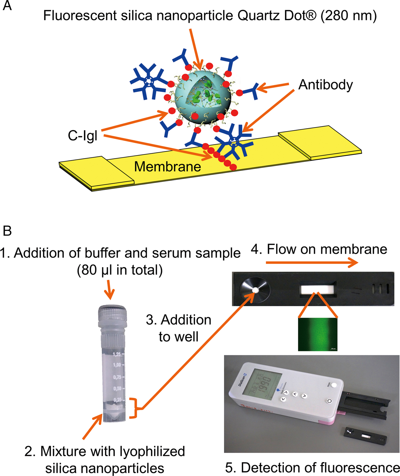
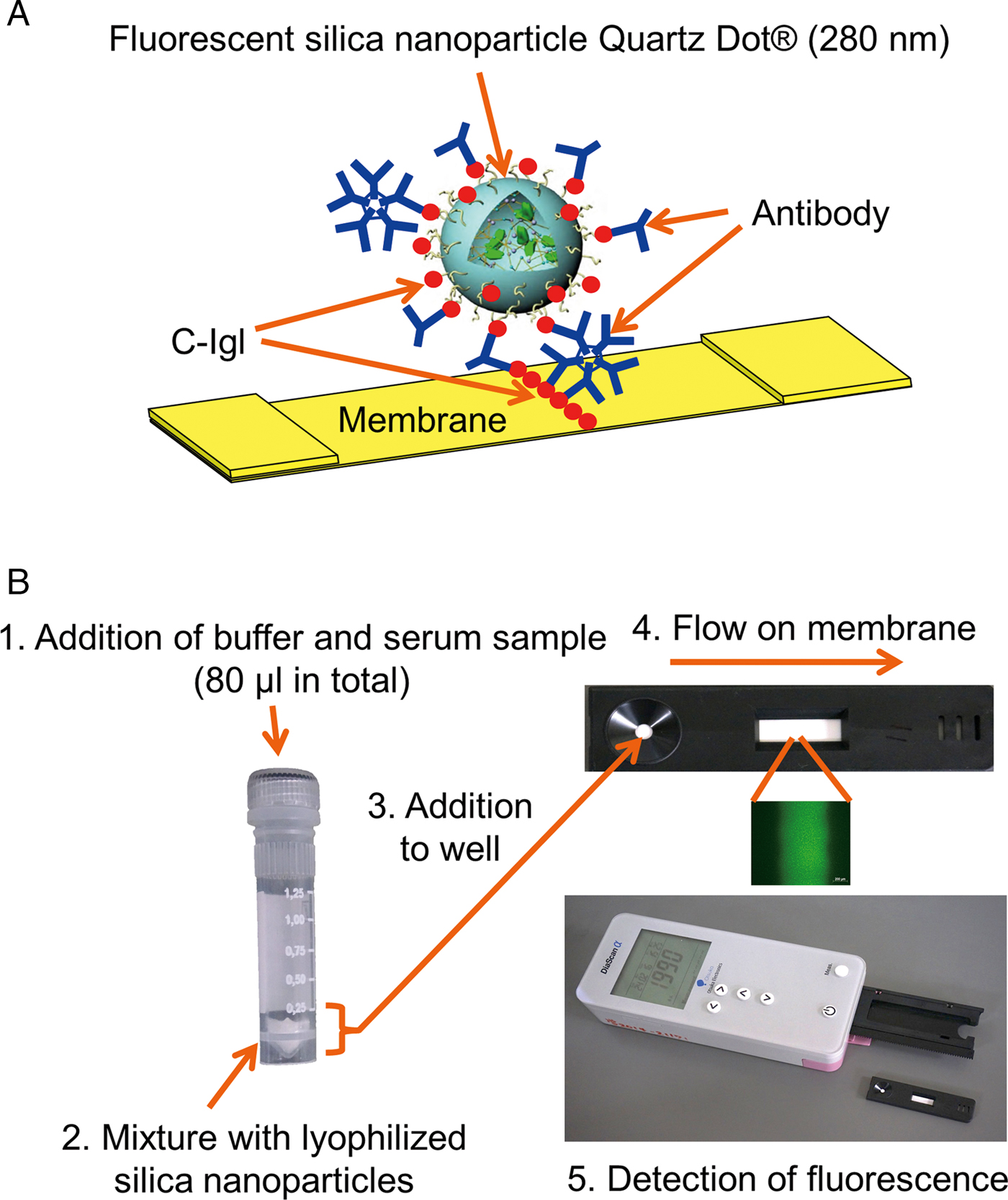
The principles of the detection system for antibodies to C-Igl in the immunochromatographic assay are shown in Fig. 1A. Recombinant C-Igl prepared in Escherichia coli was bound on the surface of silica nanoparticles and also pre-immobilized on a nitrocellulose membrane to form a test line. By evaluation using a human monoclonal antibody, XEhI-H2, specific to Igl, the concentration of C-Igl was optimized to yield distinct fluorescent intensity in comparison with a control with no antibody in the system. The established procedure for the assay kit developed in this study is shown in Fig. 1B. Fluorescence intensity was measured using a hand-held reader.
Schematic chart of immunochromatographic assay kit using fluorescent silica nanoparticles coated with E. histolytica C-Igl for serodiagnosis of amebiasis. (A) Principle of detection of antibodies to C-Igl. If specific antibodies to C-Igl are present, fluorescein silica nanoparticles are captured on the membrane via the antibodies. (B) Procedure of the assay. Numbers 1–5 show each step in the procedure. In step 5, a cassette in the assay kit is set in a tray of hand-held reader DiaScan α (Otsuka Electronics) and then scanned. Intensity is displayed digitally on a window of the reader and scanning data is sent to a personal computer via a Bluetooth wireless connection. The positive fluorescent image was taken using a fluorescein microscope.
Detection limit
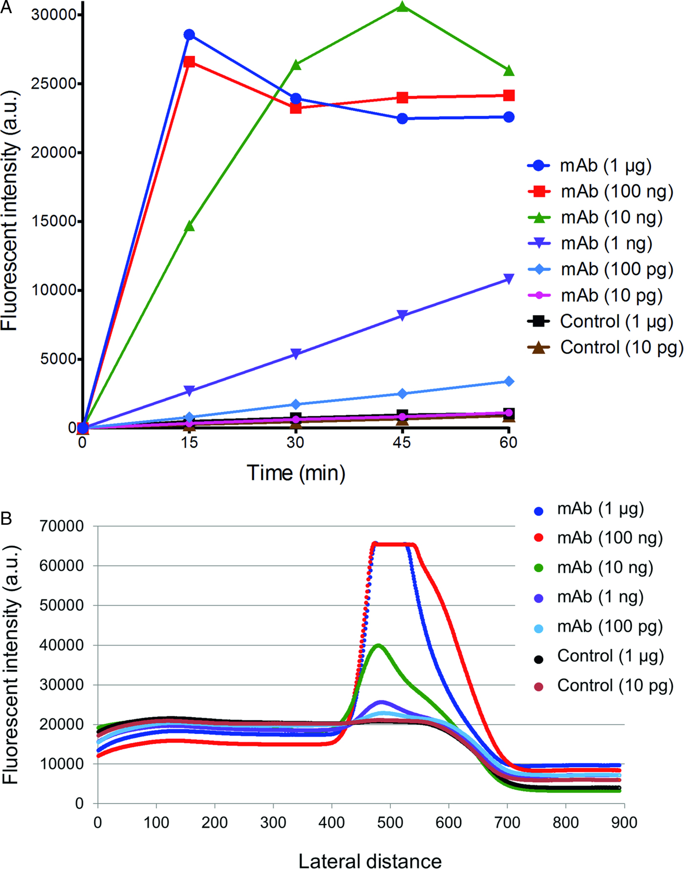
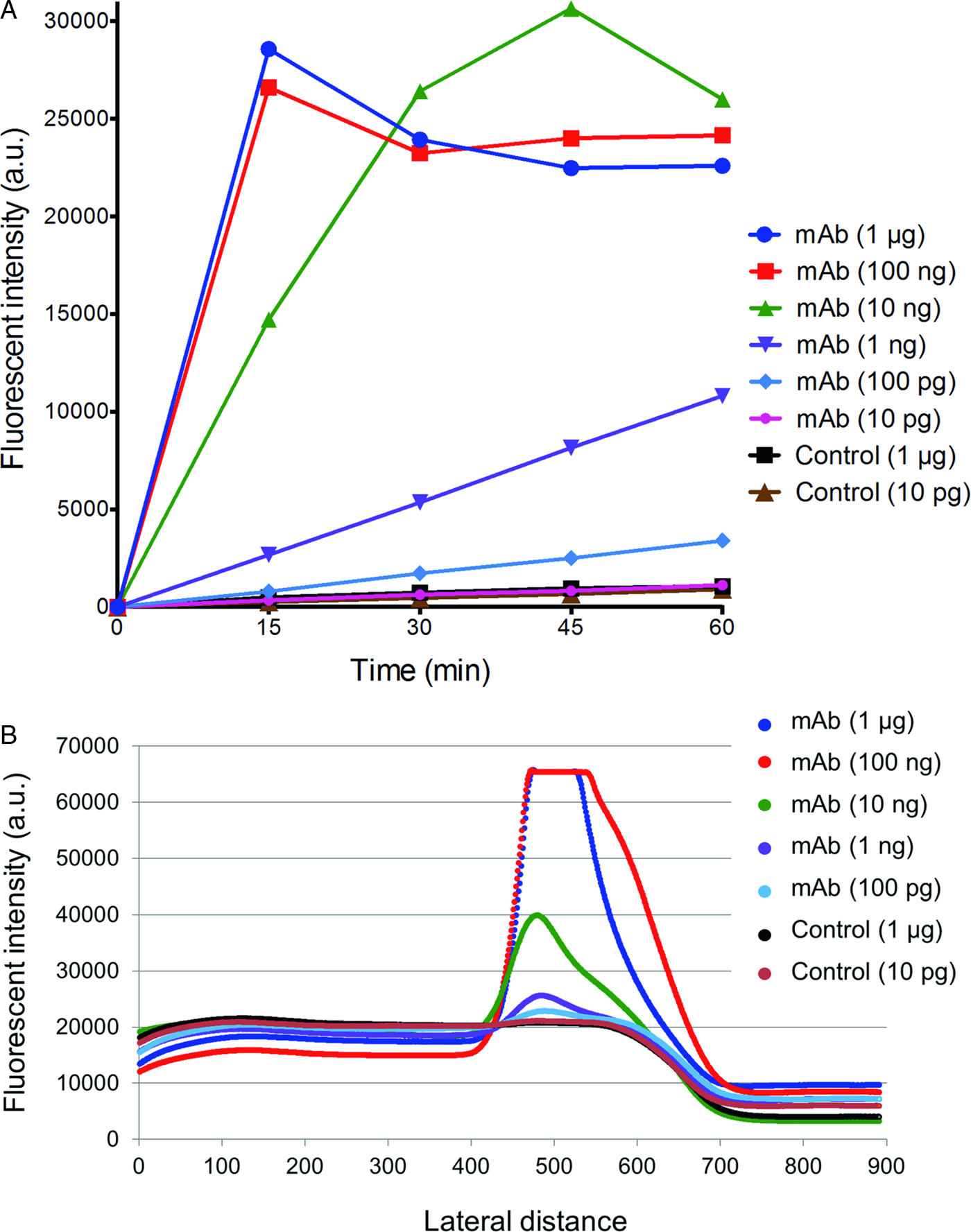
To determine the minimum amount of antibody that can be detected using the kit, various amounts of XEhI-H2 were applied using the kit. As shown in Fig. 2A, the fluorescent intensity increased in a dose- and time-dependent manners. With 100 ng of XEhI-H2, the intensity saturated at 15 min after incubation. Positive intensity was still observed for 100 pg of XEhI-H2. In controls, there was no difference in fluorescent intensity between 1 µg and 10 ng of normal human antibodies. Scanning waveforms of fluorescence from various antibody concentrations at 30 min after incubation are shown in Fig. 2B. The waveform peak was detected for 100 pg of XEhI-H2, while the waveform for the control antibody was flat at 1 µg. Therefore, the minimum amount of antibody detectable by the kit was judged to be 100 pg.
Immunochromatography using a human monoclonal antibody specific to C-Igl. (A) Reactivity of various amounts of human monoclonal antibody XEhI-H2 in the kit. Fluorescein intensity was measured at 15, 30, 45 and 60 min after addition to the well. (B) Scanning waveforms of fluorescence in immunochromatography at 30 min.
Influence of the serum volume and incubation time
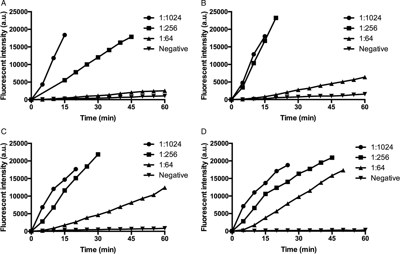
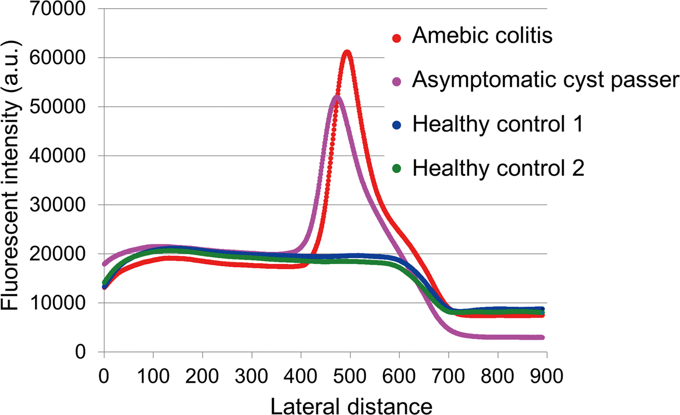
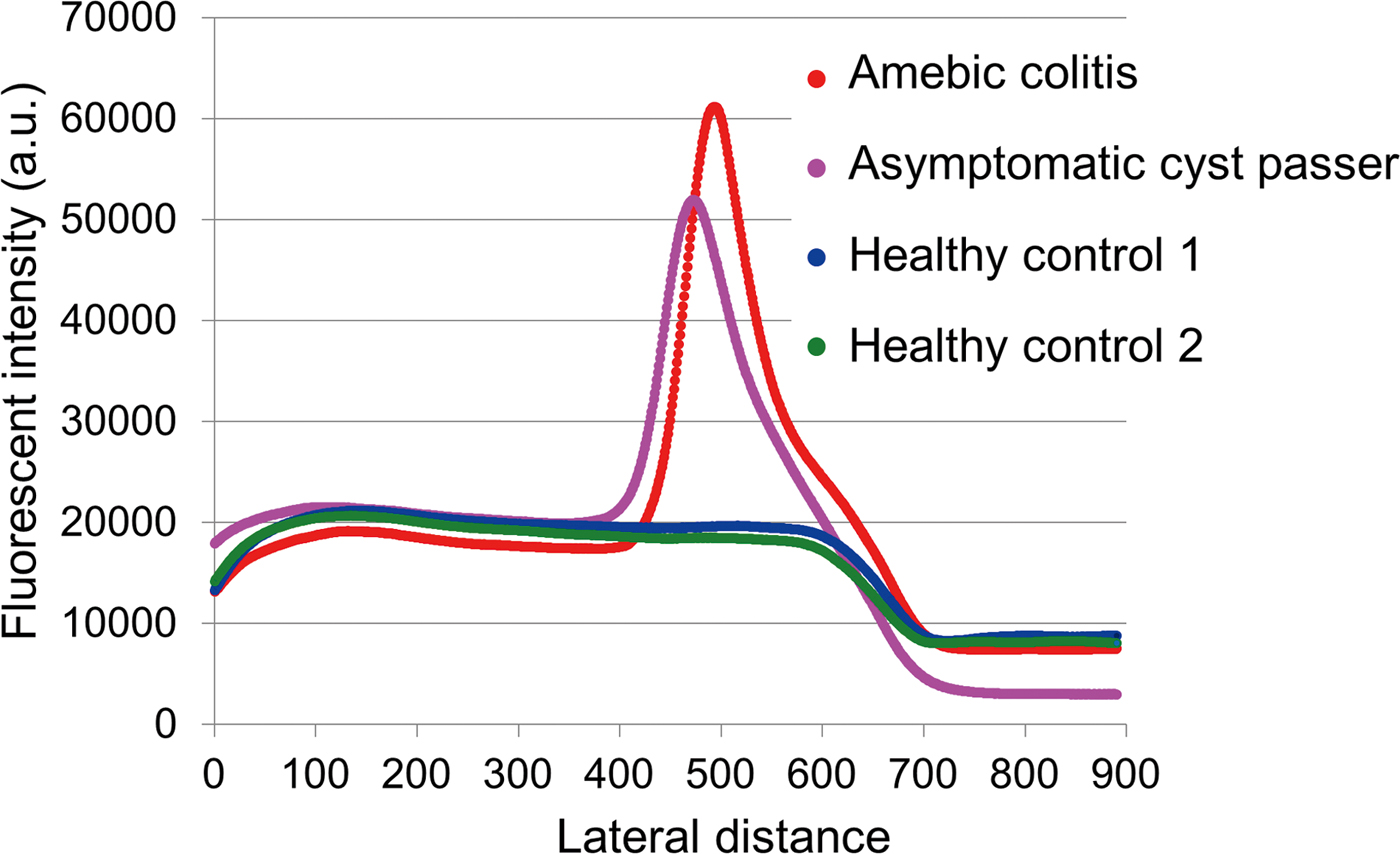
Three serum samples from patients with amebiasis with different antibody titers in an IFA were used to determine the volume of serum required for the kit (Fig. 3). A negative serum in the IFA test was used as a control. For serum with a high IFA titer of 1:1024, strong fluorescence was observed even at 5 µL of the sample (Fig. 3A). To yield an apparent intensity level for serum with a low IFA titer of 1:64, incubation for 30 min with 20 µL of serum or 15 min with 40 µL of serum was adequate (Fig. 3C and D). The condition of 20 µL of serum and incubation for 30 min was finally selected. Scanning waveforms of fluorescence in immunochromatography were confirmed using serum samples from two patients and two healthy controls. Apparent peaks corresponding to the test line were observed for patients with amebic colitis and asymptomatic cyst passers, whereas waveforms in healthy controls were flat (Fig. 4).
Fluorescent intensities from serum samples in the immunochromatographic assay. The serum volumes were 5 µL (A), 10 µL (B), 20 µL (C) and 40 µL (D). Three positive sera with different IFA titers and an IFA-negative serum were used. Fluorescein intensity was measured at 5 min intervals after addition to the well.

Evaluation of scanning waveforms of fluorescence in immunochromatography using human sera. Sera (20 µL) from a patient with amebic colitis, an asymptomatic cyst passer, and two healthy Japanese controls were used. Fluorescent intensity was measured at 15 min after addition to the well.
Evaluation using sera from patients with amebiasis and other infectious diseases
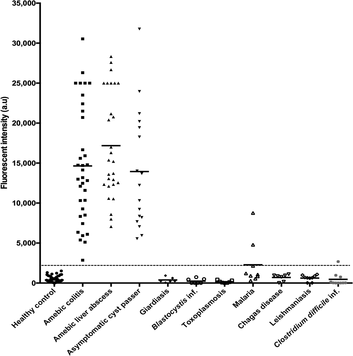
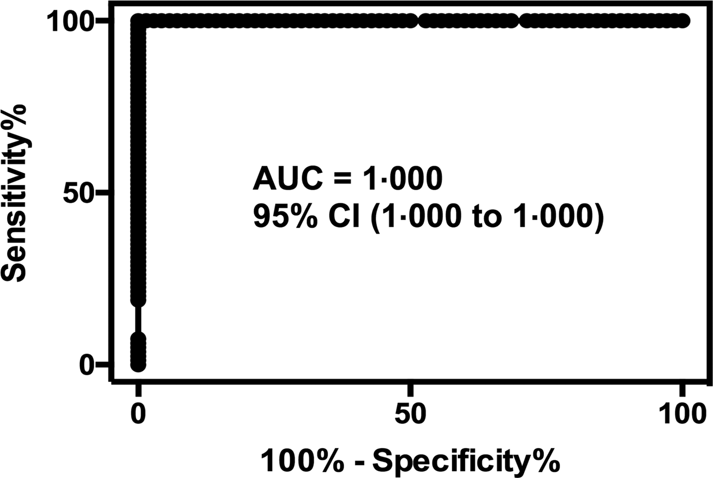
To evaluate the fluorescent immunochromatographic kit, sera from 35 patients with amebic colitis, 28 patients with amebic liver abscess and 17 asymptomatic cyst passers were measured (Fig. 5). Sera from 70 healthy Japanese subjects with no evidence of infectious disease were used as negative controls. Receiver operating characteristic (ROC) curve analysis showed AUC, 1.000; 95% CI, 1.000 to 1.000 (Fig. 6). The cut-off value was determined to be 2,181. To examine cross-reactivity with other pathogens, 55 sera from patients with giardiasis, Blastocystis hominis infection, toxoplasmosis, malaria, Chagas disease, leishmaniasis and Clostridium difficile infection were also evaluated. Two samples from patients with malaria and one from a patient with C. difficile infection were scored as positives (Fig. 5). Based on the analysis of sera from amebiasis and that from healthy controls and other infectious diseases, the sensitivity and specificity of the kit were calculated as 100% and 97.6%, respectively. Since the turnaround time of the immunochromatographic kit was about 30 min, the kit was shown to be useful for rapid serodiagnosis of amebiasis.
Fluorescent intensity of sera from healthy controls and patients with amebiasis and other infectious diseases in the immunochromatographic assay. Horizontal bars show the mean intensity. The broken line shows the cut-off value. For samples with fluorescent intensity in DiaScan α above the measurement range, the value was recorded as 25 000.

Fitted ROC curves from the E. histolytica-infected and healthy control groups.

Discussion
In this study, a new immunochromatographic kit for detection of serum antibody to E. histolytica antigen was developed using fluorescent silica nanoparticles. Quartz Dot is a silica nanoparticle that contains a high concentration of organic dye with high brightness, which makes it possible to lower the detection limit of the kit. Indeed, 100 pg of specific antibody to C-Igl could be detected in the kit with use of monoclonal antibody XEhI-H2. However, since antibodies to C-Igl in real serum samples are polyclonal and recognize several different epitopes of the protein, it is difficult to evaluate the exact detection limit. Recently, it has been reported that an immunochromatographic assay kit using fluorescent silica nanoparticles bound with a monoclonal antibody is useful for detection of Acanthamoeba antigen, even in clinical samples that are negative in direct smear or culture tests (Hiwatashi et al., Reference Hiwatashi1997; Toriyama et al., Reference Toriyama2015). Five trophozoites or 40 cysts in samples have been detected using the assay kit. Another advantage of a fluorescence kit is its more quantitative nature in comparison with an immunochromatographic kit with colloidal gold or latex particles. In the present study, a new hand-held reader was used instead of a fluorescent scope (Toriyama et al., Reference Toriyama2015) and was useful to show the fluorescent intensity of a test line. In addition, diminishing of fluorescent intensity on the membrane over time was negligible, and was <20% after 7 months at room temperature (unpublished data).
We have previously shown that C-Igl is a useful antigen for serodiagnosis of amebiasis in an ELISA (Tachibana et al., Reference Tachibana2004; Chen et al., Reference Chen2007; Yang et al., Reference Yang2012). C-Igl has higher sensitivity and specificity than other fragments of Igl and has been applied in serodiagnostic systems using a microfluidic device and a microsphere-based multiplex assay (Zhao et al., Reference Zhao2013; Fujii et al., Reference Fujii2014). In the present study, all serum samples from patients with amebiasis, including asymptomatic cyst passers, were judged to be positive in our kit, showing that its sensitivity was 100%. However, two sera from patients with malaria and one from a patient with C. difficile infection were also scored as positive. To exclude the possibility that these three patients were coinfected with E. histolytica, the reactivity of these samples to C-Igl was analysed using our previously reported ELISA system (Tachibana et al., Reference Tachibana2004). None of the samples showed positive results and therefore, the reason for these false-positives is unclear. If other infectious disease groups are included in controls in ROC curve analysis (AUC, 0.9981; 95% CI, 0.9944–1.002), the cut-off value becomes 4944 with 98.75% sensitivity and 99.2% specificity. Further evaluation is required using a large number of serum samples, including those from patients with malaria, C. difficile infection and E. dispar infection in addition to healthy controls from endemic areas, to establish an appropriate cut-off.
C-Igl is a candidate vaccine for prevention of amebic liver abscess formation (Min et al., Reference Min2016). Immunization with C-Igl induces humoral immunity with increased expression of IL-4 and IL-10, and it has also been shown that C-Igl possesses both hemolytic and cytotoxic activities (Kato et al., Reference Kato2015, Reference Kato2017). A human monoclonal antibody recognizing C-Igl can inhibit hemolytic activity in vitro and amebic liver abscess formation of hamsters by passive immunization (Tachibana et al., Reference Tachibana2009; Kato et al., Reference Kato2015). The observation that all sera from asymptomatic cyst passers were scored as positive in this study may support the finding that antibodies to C-Igl function protect against invasive amebiasis.
There are several commercial ELISA and IHA kits for serodiagnosis of amebiasis. ELISA is widely thought to be adequate for clinical purposes (Tanyuksel and Petri, Reference Tanyuksel and Petri2003) and an ELISA system is useful for examination of a large number of serum samples simultaneously but is less convenient for a single sample. IHA is easy to perform, but its lower sensitivity may lead to false-negative results in comparison with ELISA (del Carmen Sanchez-Guillen et al., Reference del Carmen Sanchez-Guillen2000). IHA is also a quantitative assay, but the serial dilution of samples is required. The method reported here does not require the dilution steps of serum samples required in both ELISA and IHA. The sensitivity and specificity of commercial ELISA or IHA kits have been reported as 72.2–100% and 90.9–99.1%, respectively (Tanyuksel and Petri, Reference Tanyuksel and Petri2003; Flores et al., Reference Flores2016), and the sensitivity and specificity of this study are comparable with these values. In addition, the short turnaround time of the immunochromatographic kit is an advantage in comparison with ELISA and IHA.
In conclusion, an immunochromatographic kit using fluorescent silica nanoparticles with C-Igl as antigen was shown to be useful for rapid serodiagnosis of amebiasis. To our knowledge, this is the first report of the development of an antibody detection system using fluorescent silica nanoparticles.
Acknowledgements
We thank M. Fukushima, K. Miyoshi and M. Ohkubo for their valuable suggestions.
Financial support
This study was supported by JSPS KAKENHI (grant numbers 26460516, 16H05819, 17K08811 to H.T.).
Declaration of interest
T. Kamada and H. Watanabe are employees of Furukawa Electric Advanced Engineering Co., Ltd. The other authors declare no conflicts of interest.
Ethical standards
The study was approved by the Review Board of Tokai University (17R017).