Introduction
Strongyloidiasis is a cosmopolitan neglected disease caused by the soil-transmitted Strongyloides stercoralis affecting 30 to 100 million people worldwide, particularly in areas with poor sanitation conditions (Olsen et al., Reference Olsen, Lieshout and Marti2009; Mendes et al., Reference Mendes, Minori, Ueta, Miguel and Allegretti2017). The case reports of strongyloidiasis are frequently underestimated as a low sensitivity of conventional diagnostic tests and many infections remain asymptomatic (Requena-Méndez et al., Reference Requena-Méndez, Chiodini and Bisoffi2013; Beknazarova et al., Reference Beknazarova, Whiley and Ross2016). Due to the autoinfective ability, Strongyloides infections have the potential to develop from asymptomatic to severe systemic manifestations, with potentially life-threatening dissemination of larvae to all internal organs, especially in immunocompromised patients (Siddiqui & Berk, Reference Siddiqui and Berk2001; Vadlamudi et al., Reference Vadlamudi, Chi and Krishnaswamy2006; Mejia & Nutman, Reference Mejia and Nutman2012). Therefore, effective and timely treatments are extremely essential for complete elimination of this parasitic nematode (Basile et al., Reference Basile, Simzar and Bentow2010; Bisoffi et al., Reference Bisoffi, Buonfrate and Angheben2011).
Nowadays, management of strongyloidiasis using anthelmintic chemotherapy represents a major global health challenge. In addition to ivermectin, the drug of choice against S. stercoralis, the alternatives currently in use to treat strongyloidiasis are albendazole and its analogues such as thiabendazole. However, several side effects and inconsistent cure rates of these conventional drugs, particularly in immunocompetent patients, lead to discouraged use and reduced compliance of the patients (Bisoffi et al., Reference Bisoffi, Buonfrate and Montresor2013; Luvira et al., Reference Luvira, Watthanakulpanich and Pittisuttithum2014; Mendes et al., Reference Mendes, Minori, Ueta, Miguel and Allegretti2017). Furthermore, like the infective larvae of all nematodes, the Strongyloides larvae are resistant to most synthetic chemical compounds (Ismail et al., Reference Ismail, Ibrahim, Ahmed and Hetta2016). Therefore, the investigation and identification of alternative anti-Strongyloides agents has become a priority and imperative need. During the last few decades, medicinal plants have been accepted as a good source of bioactive compounds, of which potential use against parasitic helminths has gained attention among researchers worldwide (Akhtar et al., Reference Akhtar, Iqbal, Khan and Lateef2000; Rochfort et al., Reference Rochfort, Parker and Dunshea2008; Chan-Pérez et al., Reference Chan-Pérez, Torres-Acosta and Sandoval-Castro2016; El-Refai et al., Reference El-Refai, Atia and Mahmoud2018; Zamilpa et al., Reference Zamilpa, García-Alanís and López-Arellano2018). Effective herbal helminthicides could be of value in not only reducing the harmful effects occurring with the overuse of synthetic drugs, but also preventing and treating helminthic multi-resistance (Nogueira et al., Reference Nogueira, Martins and Duarte2010; El-Sherbini & Osman, Reference El-Sherbini and Osman2013; Zamilpa et al., Reference Zamilpa, García-Alanís and López-Arellano2018).
Piper retrofractum Vahl, commonly known as ‘long pepper’, is a flowering vine in the family Piperaceae, which comprises approximately 1000 species of worldwide distribution. The genus Piper has high commercial, economical and medicinal importance (Kubo et al., Reference Kubo, Ishii and Ishino2013; Bao et al., Reference Bao, Ochir, Sun, Borjihan and Yamagishi2014; Muharini et al., Reference Muharini, Liu, Lin and Proksch2015). Piper retrofractum is native to South-East Asia, widely distributed in tropical and subtropical regions, and mostly cultivated in Indonesia and Thailand (Lee, Reference Lee2005; Lim, Reference Lim2012). Apart from its utilization as a food spice and seasoning, the fruit of P. retrofractum has been used in traditional medicine for anti-flatulent, expectorant, anti-tussive, anti-fungal, uterus-contractile, sedative-hypnotic, appetizing, counter-irritant properties, as well as for treating hyperlipidaemia and coronary heart diseases (Tewtrakul et al., Reference Tewtrakul, Hase, Kadota, Namba and Komatsu2000; - Bao et al., 2012; Hieu et al., Reference Hieu, Thang, Hoi and Ogunwande2014). This plant is also listed as a source of herbal drugs in the Thai Herbal Pharmacopoeia (DMSC, 2019). The phytochemical study of P. retrofractum products revealed various chemical compounds, with a variety of pharmaceutical and biological properties such as anti-oxidant, anti-diabetic, anti-obesity, antifungal, antibacterial, antimalarial, insecticidal and cytotoxic activities (Chansang et al., Reference Chansang, Zahiri and Bansiddhi2005; Lim, Reference Lim2012; Nidhi et al., Reference Nidhi, Mittal and Menghani2012; Luyen et al., Reference Luyen, Tai and Thao2014). However, in a review of the available literature, there are few studies on the anthelmintic potential of P. retrofractum, particularly against S. stercoralis. In our research program directed toward the discovery of herbal helminthicides as alternatives to conventional synthetic drugs, P. retrofractum fruit hexane extract (PHE) has been shown to possess promising helminthicidal activity against the third-stage larvae (L3) of S. stercoralis (Sangkhantree et al., Reference Sangkhantree, Riyong and Jitpakdi2018). To better understand the nematocidal effect of PHE, its chemical constituents and impact on symptom and structural alterations of S. stercoralis were investigated in this study. The information obtained has enhanced our basic knowledge and contributes to the design of an alternative that may be useful for the future management of S. stercoralis.
Materials and methods
Preparation of PHE
Dried fruit of P. retrofractum was acquired commercially from medicinal plant suppliers in Chiang Mai province, northern Thailand. This plant material was authenticated by Miss Wannaree Charoensup, an expert in botanical taxonomy from the Department of Pharmaceutical Science, Faculty of Pharmacy, Chiang Mai University (CMU). A reference voucher, PARA-PI-006-Fr/1, was deposited at the Department of Parasitology, Faculty of Medicine, CMU. Preparation of PHE was performed according to Sangkhantree et al. (Reference Sangkhantree, Riyong and Jitpakdi2018). Briefly, half a kilogram of P. retrofractum fruit powder was extracted by at least two days of maceration with 2 l of hexane at room temperature. The hexane-extract mixture was filtered and concentrated in a rotary evaporator at 40°C before finally freeze-drying at −55°C. The resulting PHE was kept at −20°C until required for further investigations.
Chemical analysis of PHE
Qualitative analyses of the chemical components in PHE were carried out by gas chromatography–mass spectrometry (GC–MS) in an Hewlett-Packard 7890A gas chromatographer (Agilent Technologies, Waldbronn, Germany) equipped with a DB-5MS capillary column (30 m × 0.25 mm ID × 0.25 µm film thickness). Helium was used as the carrier gas (1.0 ml/minute), with a 1.0 µl injection of 1% solution of PHE in dichloromethane (CH2Cl2) heated in an injector to 250°C by operating in split mode (split ratio 5:1). The oven temperature varied from 50 to 300°C at a rate of 5°C/minute. A mass detector was operated by electron ionization (70 Ev) with a mass analyser maintained at 150°C and ionization source at 230°C. The retention times were determined in relation to a homologous series of n-alkanes (C8–C40) under the same operating conditions. Identification of the PHE components was performed by comparing both mass spectra and retention indices with the NIST 2008 and Wiley 8NO8 edition spectral database and data from the literature.
Drugs and chemicals
Ivermectin obtained commercially from Sigma-Aldrich (St Louis, Missouri, USA) was used as a reference drug. All other chemicals and reagents used were of the analytical grade purchased from local agencies in Chiang Mai province, Thailand.
Maintenance of infective third-stage S. stercoralis larvae
Infective L3 of S. stercoralis were recovered from faecal culture of patients admitted to Maharaj Nakorn Chiang Mai Hospital and submitted for routine parasitological examination at the Parasitology Laboratory, Department of Parasitology, Faculty of Medicine, CMU. Culture of S. stercoralis was carried out according to the modified Harada–Mori technique (Harada & Mori, Reference Harada and Mori1955), which is a filter-paper culture method utilizing the water tropism of Strongyloides larvae to concentrate them. Briefly, filter paper (15 × 18 cm) deposited with fresh faeces was inserted into a plastic bag (17 × 23 cm) containing 2–3 ml of distilled water. The sealed plastic bag was incubated in a dark place at 28–30°C for 7–10 days. The water sediment is screened daily under a light microscope to look for living larvae. The L3 of S. stercoralis were collected by washing three times with 10 ml of 0.1 M phosphate-buffered saline solution (PBS; pH 7.4) and centrifuged at 5000 × g rpm for 5 min. These larvae were then resuspended and used as test organisms in anthelmintic bioassays.
In vitro nematocidal bioassays of PHE
Nematocidal activity was evaluated in vitro by exposing the L3 of S. stercoralis to a graded series of four concentrations of PHE (0.015, 0.031, 0.062 and 0.125 mg/ml) in a well of Petri-dish culture (Boonmars et al., Reference Boonmars, Khunkitti, Sithithaworn and Fujimaki2005). The anthelmintic potential of ivermectin, a reference drug, was also obtained by exposing S. stercoralis L3 to a range of drug dilutions (0.004, 0.008, 0.016, 0.032 and 0.064 µg/ml) under identical conditions. The desired dosage of each test material was prepared by diluting with Tween 80 in 0.1 M PBS, pH 7.4 (T80-PBS). While approximately 100 larvae represented the test group by being exposed in 1 ml of test solution, the control group was maintained in T80-PBS only. All procedures were performed under aseptic conditions using a laminar flow hood. Procedures in each group were performed in duplicate, with the larvae incubated at 37°C in a humidified incubator under a 5% CO2 atmosphere. After 24 h incubation, mortality of the worms was observed under an inverted microscope. The larvae without motility were considered dead after trying to stimulate them with light for 5 min. Each test was enumerated by two independent readers. Each assay was performed in two independent experiments in four replicates. The dead larvae in each group were averaged and expressed as percentage mortality.
Determination of symptom and structural alterations in PHE-treated S. stercoralis L3
Symptoms of S. stercoralis L3 treated with LC99 of PHE was observed and compared with those of ivermectin (LC99)-treated and control specimens during exposure under light microscopy (LM). These specimens were also evaluated for structural alterations using LM, scanning electron microscopy (SEM) and transmission electron microscopy (TEM). Morphological changes of the whole organism were observed and photographed. The treated and control specimens of S. stercoralis L3 were processed for the SEM study by following the method of Choochote et al. (Reference Choochote, Maleewong, Sucharit, Pipitgool and Khamboonruang1987). In brief, larval specimens were fixed overnight in 2.5% glutaraldehyde in 0.1 M PBS, pH 7.4 at 4°C, washed with PBS (10 min, two changes) and then post-fixed (1 h) in 1% osmium tetroxide at room temperature. Dehydration was performed via a passage through a gradual ethanol series – that is, 30%, 50%, 70%, 80% (10 min), 95% (15 min, two changes) and absolute ethanol (10 min, two changes). The specimens were finally dried by a critical point dryer, mounted on aluminium stubs, sputter coated with gold and examined at 15 kV in a JEOL JSM-6610LV SEM (JEOL Ltd., Tokyo, Japan).
The treated and control S. stercoralis L3 were processed for TEM analysis by following the method of Sukontason et al. (Reference Sukontason, Chaiwong, Chaisri, Kurahashi, Sanford and Sukontason2011). Larval specimens were fixed in a fixative agent consisting of 2.5% glutaraldehyde at 4°C for 24 h. The fixed specimens were subjected to post-fixation in 1% osmium tetroxide and dehydrated in a graded alcohol series. The specimens remained in each concentration for 12 h, followed by absolute alcohol for two separate 12-h periods. After that, the specimens were placed in acetone for 2 h before being transferred into ratios of resin to acetone of 1:3 for 24 h, 1:1 for 24 h and 3:1 for 24 h, sequentially. This was followed by treating twice for 3 h each time with pure resin. Each specimen was then embedded in Spurr's resin by placing them into a plastic block and incubating at 70°C for 24 h. A semi-thin section (0.5 µm) of each sample was made with a glass knife on an Ultramicrotome (Boeckeler®, Tucson, Arizona, USA). Ultrathin sections (90 nm) were made from the same embedding blocks. Serial sections were collected from copper slot grids before staining with uranyl acetate and lead citrate for viewing under a JEOL JEM-2200FS TEM (JEOL Ltd., Tokyo, Japan), operated at 200 kV.
Statistical analysis
Bioassay tests were discarded if they had >20% control mortality and then repeated in the nematocidal investigation. If 5–20% control mortality was observed, the percentage mortality (% M) was corrected by Abbott's formula (Abbott, Reference Abbott1925):

The corrected data were analysed by regression analysis using the computeried statistical program SPSS (version 19.0; IBM Crop., Armonk, New York, USA). The lethal values of 50%, 95% and 99% (LC50, LC95 and LC99, respectively), with corresponding 95% confidence intervals were calculated using the chi-square method in each bioassay for assessing the significance and measuring the difference between the test samples.
Results
Chemical compositions of PHE
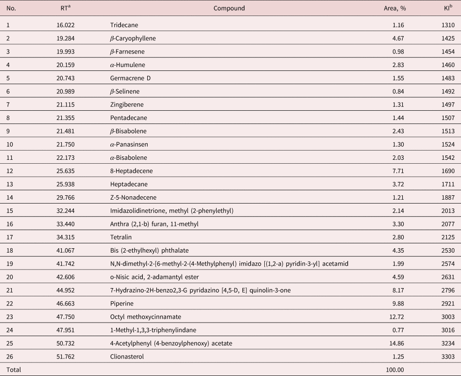
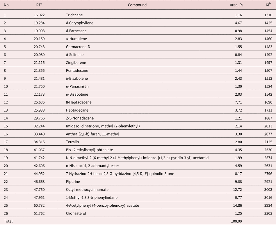
Reddish-brown viscous extract of P. retrofractum fruit, with a pungent odour and yield of 6.32% (w/w) was obtained from hexane extraction. GC–MS analysis of the chemical constituents of PHE revealed the presence of 26 compounds, accounting for 100% of all the compositions (table 1). The main components of PHE were 4-acetylphenyl (4-benzoylphenoxy) acetate (14.86%) and octyl methoxycinnamate (12.72%), with minor constituents of piperine (9.88%), 7-hydrazino-2H-benzo2,3-G pyridazino [4,5-D, E] quinolin-3-one (8.17%) and 8-heptadecene (7.71%).
Chemical constituents of PHE as determined by GC–MS analysis.
a Retention time (minute).
b Kovats index relative to n-alkane (C8–C40) on a DB-5MS column.
In vitro nematocidal efficacy of PHE and ivermectin against S. stercoralis L3
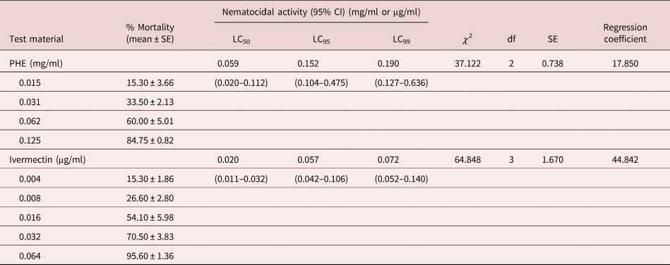
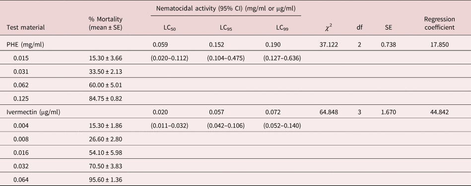
The 50%, 95% and 99% in vitro lethal concentrations (LC50, LC90 and LC99, respectively) against L3 of S. stercoralis attributed to the effect of PHE and ivermectin at 24 h post incubation are shown in table 2. It was evident that exposure to serial concentrations of PHE and ivermectin produced dose-dependent mortality of S. stercoralis L3. Based on the values of larvicide-LC50, PHE (LC50 value = 0.059 mg/ml) had significantly lower nematocidal efficacy than ivermectin, which exerted an LC50 value of 0.020 µg/ml against S. stercoralis L3 (P < 0.05).
Nematocidal activity of Piper retrofractum fruit hexane extract (PHE) in comparison with ivermectin against Strongyloides stercoralis L3.
df, degree of freedom; SE, standard error; CI, confidence interval.
Symptom and structural changes of PHE- and ivermectin-treated S. stercoralis L3
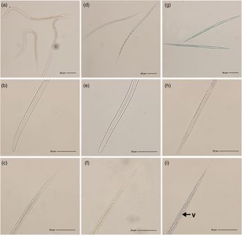
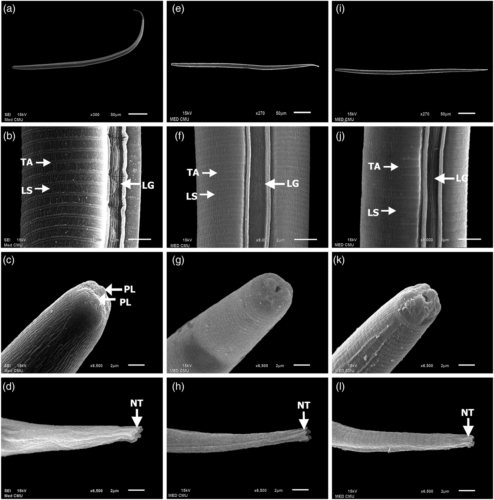
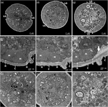
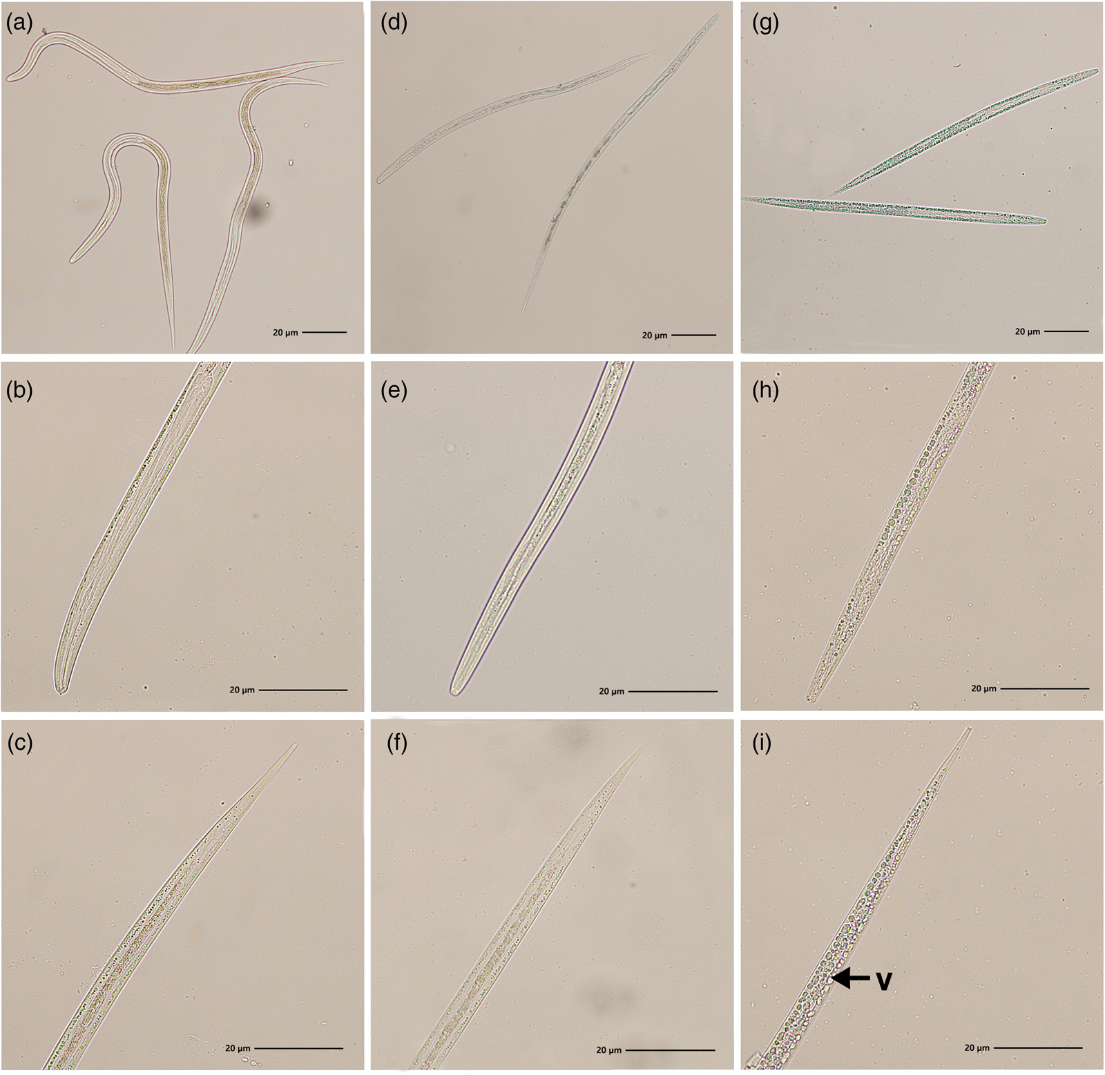
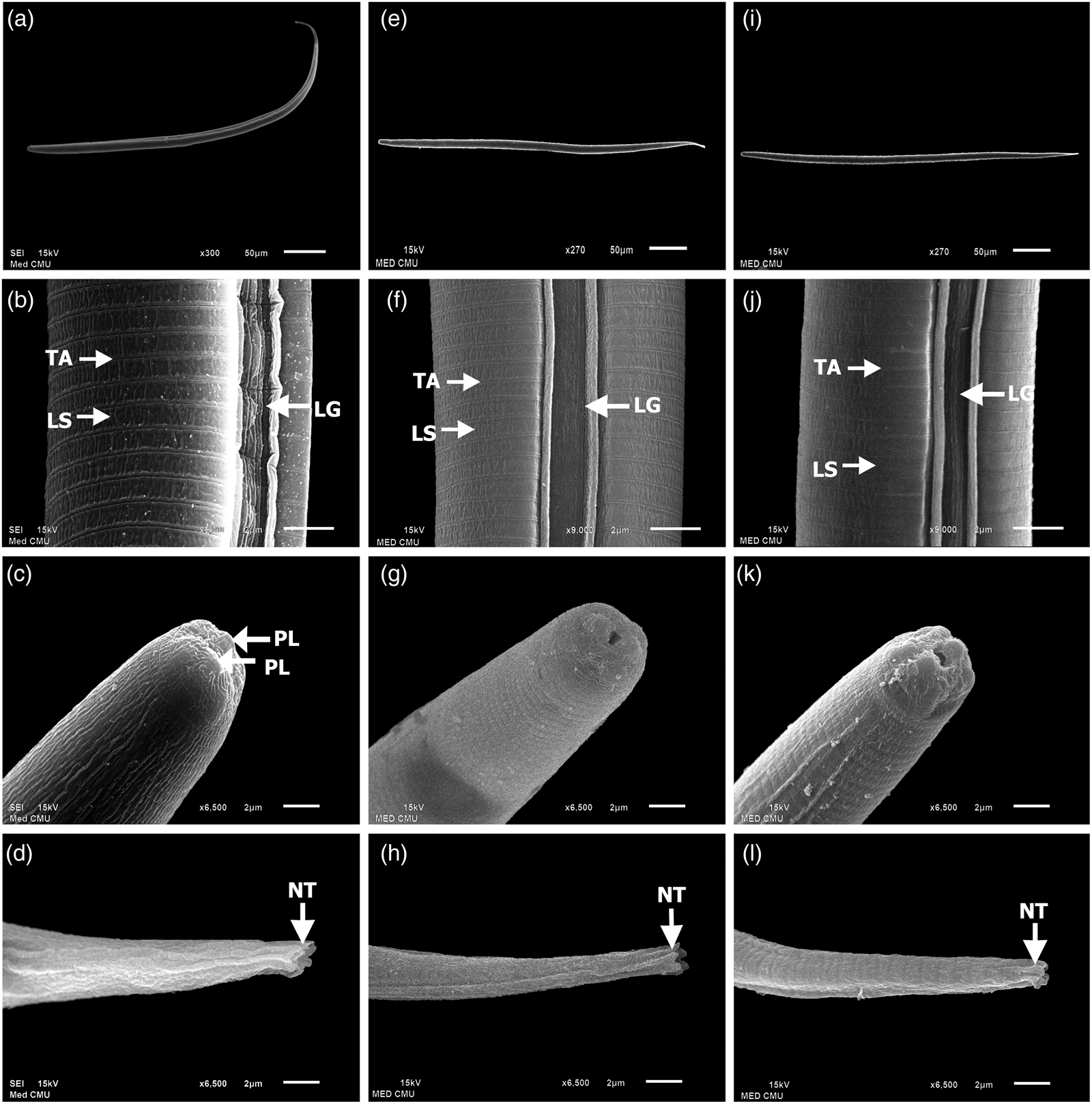
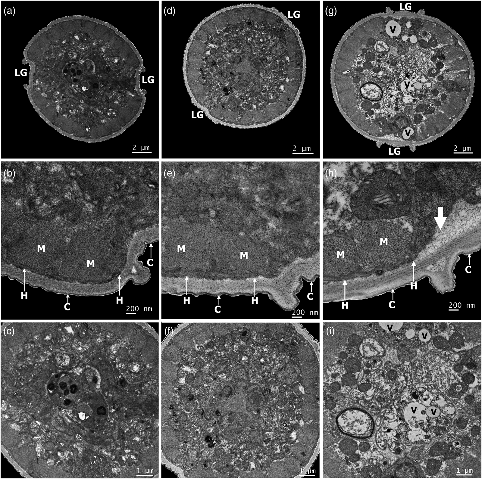
Differences in behavioural responses of the control and treated S. stercoralis L3 were seen evidently under LM. The control larvae were active with normal movement continuously after incubation at 37°C under a 5% CO2 atmosphere, which is in contrast to PHE- and ivermectin-treated ones that moved slowly, became paralysed and eventually died during 24 h of treatment. The larval bodies in the control and treated groups were also different when visualized through a light microscope (fig. 1). While the control larvae appeared as gracefully curved worms with intact internal organs (fig. 1a–c), the ivermectin-treated larvae showed relatively straight bodies with no markedly morphological difference from the control (fig. 1d–f). Furthermore, larval worms treated with PHE exhibited straight bodies infiltrated with numerous unknown vacuoles of variable sizes (fig. 1g–i). The structural architecture of the control, ivermectin- and PHE-treated S. stercoralis L3, as observed under SEM, are shown in fig. 2. The control worm displayed a gracefully curved body (fig. 2a), with two deep and striated longitudinal grooves on the lateral side (fig. 2b). The cuticle appeared as rows of transverse annulation throughout the whole body. Faint longitudinal striations were seen between each transverse annulation (fig. 2b). The anterior part was endowed with two small protruding lips (fig. 2c), whereas the posterior possessed a notch tail (fig. 2d). In contrast, larvae treated with ivermectin (fig. 2e–h) and PHE (fig. 2i–l) showed a similar appearance, but differed relatively from the control. A straight body with swollen cuticle and relatively shallow and smooth lateral longitudinal grooves was detected (fig. 2e–l). In addition, the transverse annulations and longitudinal striations were faded (fig. 2f, j). Such an appearance of both the control and treated larvae was evident in the TEM analysis (fig. 3). The architecture of the control had a normal appearance (fig. 3a–c). Two lateral longitudinal grooves were distinct (fig. 3a). The cuticle appeared as normal, comprising a cuticle, hypodermis and muscular layer with packed muscle cells (fig. 3b). The internal structures were configured normally (fig. 3c). In contrast, morphological aberrations were detected in larvae treated with either ivermectin (fig. 3d–f) or PHE (fig. 3g–i). Larvae in the ivermectin-treated group showed a slightly swollen body, with shallow lateral longitudinal grooves (fig. 3d), and the muscle cells had loose compactness (fig. 3e). The internal body showed no significant difference from that of the control (fig. 3f). More structural aberration was found in the PHE-treated larvae, and apparent swelling of the worm is shown in fig.3g. Lateral longitudinal grooves protruded slightly, but not in a deep groove as in the control. Loose muscle with vacuolation and dissociation between the hypodermis and cuticle were found (fig. 3g–h), and the internal structures showed marked intracellular disorganization with vacuolations (fig. 3i).
Light micrographs of Strongyloides stercoralis L3. (a–c) Control larvae appearing as gracefully curved worms with intact internal organs. (d–f) Ivermectin-treated larvae showing relatively straight bodies with no markedly morphological difference from the control. (g–i) PHE-treated larvae showing straight bodies infiltrated with numerous unknown vacuoles (v) of variable sizes.
Scanning electron micrographs of Strongyloides stercoralis L3. (a–d) Control larva appearing with a gracefully curved shape and normal cuticular sculpturing over the entire body. (b) Intact cuticle with finely transverse annulations (TA) and longitudinal striations (LS), and deep and striated longitudinal grooves (LG) located on the lateral sides. (c) The anterior part endowed with two small protruding lips (PL). (d) The posterior end possessing a notch tail (NT). (e–h) Ivermectin- and (i–l) PHE-treated larvae appearing with straight and swollen bodies. (f–j) Swollen cuticle with faded transverse annulations (TA) and longitudinal striations (LS), and shallow and smooth lateral longitudinal grooves (LG).
Transmission electron micrographs of Strongyloides stercoralis L3 (cross section). (a–c) Control larvae showing normal appearance of the integument and internal structures, with two distinct lateral longitudinal grooves (LG). (b) Intact integument comprising cuticle (C), hypodermis (H) and muscular layer with packed muscle cells (M). (d–e) Ivermectin-treated larvae showing slightly swollen body, with shallow lateral longitudinal grooves (LG) and loose muscle (M). (f) The internal body showing no significant difference from the control. (g–i) PHE-treated larvae showing slightly swollen body, with protruding lateral longitudinal grooves (LG) and vacuolation (V) in the internal structures. (h) Loose muscle (M) with dissociation (white arrow) between the cuticle (C) and hypodermis (H). (i) The internal structures showing marked intracellular disorganization with vacuolations (V).
Discussion
This study carried out the GC–MS technique to show chemical profiles of PHE, the effective nematocide extracted from P. retrofractum fruit. Twenty-six compounds were identified, which constituted for 100% in PHE, with the main components being 4-acetylphenyl (4-benzoylphenoxy) acetate (14.86%) and octyl methoxycinnamate (12.72%), and minor constituents of piperine (9.88%), 7-hydrazino-2H-benzo2,3-G pyridazino [4,5-D, E] quinolin-3-one (8.17%) and 8-heptadecene (7.71%). Identification of the constituents in P. retrofractum fruit extracts was conducted previously with various detection methods based on chemical and spectroscopic evidence. Several compounds such as piperine, piperodione, methyl piperate, oleic acid, glucosides, (2E,12E)-pipertridecadienamide, N-isobutyl-(2E,4E,10Z)-hexadeca-2,4,10-trienamide, N-isobutyleicos-2,14-dienamide, dipiperamides, (E)-N-cinnamoyl-2-methoxypiperidine and (R)-1-(2-oxopyrrolidin-3-yl)-5,6-dihydropyridin-2(1H)-one were recognized from P. retrofractum fruit (Kubo et al., Reference Kubo, Ishii and Ishino2013; Muharini et al., Reference Muharini, Liu, Lin and Proksch2015; Tang et al., Reference Tang, Zhang and Hu2019). It is evident that the chemical constituents identified from various extracts of P. retrofractum fruit, as reported in the present and previous studies, varied qualitatively and quantitatively. In a review of the available literature, few studies have been carried out to elucidate the efficacy of these substances against helminths.
Nematocidal bioassays of the present study revealed the significant lower efficacy of PHE than ivermectin, with the LC50 values of 0.059 mg/ml and 0.021 and μg/ml, respectively, against the L3 of S. stercoralis. This is not surprising because this plant product is the crude extract with complex combinations of either active or inactive constituents, whereas ivermectin is a synthetic single active substance. It is common knowledge that the majority of medicines are derived from natural sources such as plants, which have subsequently been chemically modified to warrant higher potency against human pathogens (Ndjonka et al., Reference Ndjonka, Rapado, Silber, Liebau and Wrenger2013). Despite being less effective than currently available drugs, products of plant origin still have promising potential, due to their compositional complexity of exploiting bioactivity through a wide variety of active mechanisms (Nerio et al., Reference Nerio, Olivero-Verbel and Stashenko2010; Intirach et al., Reference Intirach, Junkum and Lumjuan2016), which may be essential, especially in future drug resistance management regimens. However, intensive research on extractability, toxicology and pharmaceutical production for improving efficacy, safety and user acceptability is needed for practical application of the plant-based products in human helminth therapy.
In addition to determining the mortality/viability of the treated larval worms directly, observations on symptom and structural alterations before and after treatment with test substances is another essential procedure employed for evaluating anthelmintic efficacy. In this research, differences in morphological appearance and behavioural response of control and the PHE-treated S. stercoralis larvae were evidently seen under LM. The determination of ultrastructural changes by means of SEM and TEM were performed previously by several researchers worldwide in order to ascertain the helminthicidal efficacy of synthetic and/or natural substances against nematodes. Of interest, the morphological alterations observed in various studies likely corresponded to those obtained from the in vitro trials presented herein. Abnormalities such as surface damage, grooves on the body cuticle and loss of regular cuticular annulations were detected under SEM in the L3 of nocturnally subperiodic Brugia malayi after culturing with a high concentration (10−4 M) of ivermectin (Tippawangkosol et al., Reference Tippawangkosol, Choochote, Na-Bangchang, Jitpakdi, Pitasawat and Riyong2004). A swollen cuticle, formation of spherical and spike-like projections at the anterior region, and leaf-like expansion on the general cuticle in adult Wuchereria bancrofti treated with a combination of albendazole and diethylcarbamazine were observed in an SEM study by Oliveira-Menezes et al. (Reference Oliveira-Menezes, Lins, Noroes, Dreyer and Lanfredi2007). The SEM observations published by Martinez-Ortiz de Montellano et al. (Reference Martinez-Ortiz de Montellano, Arroyo-Lopez, Fourquaux, Torres-Acosta, Sandoval-Castro and Hoste2013) indicated that adult female Haemonchus contortus, one of the most pathogenic nematodes of ruminants, demonstrated structural alterations after in vitro exposure to tannin-rich (TR) plants: either tzalam (Lysiloma latisiliquum) or sainfoin (Onobrychis viciifolia). The main changes were on the cuticle and buccal capsule, such as transversal and longitudinal thickening/wrinkling of the cuticular ridges and material aggregates at the cephalic region. The authors concluded that the structural changes found in the TR-treated worms might affect their motility and nutrition with possible consequences on their reproduction. Ultrastructural changes in infective L3 of two ruminant nematode species – H. contortus and Trichostrongylus colubriformis – treated with sainfoin (O. viciifolia) extracts were also observed with TEM (Brunet et al., Reference Brunet, Fourquaux and Hoste2011). It was found that the effect of sainfoin on larvae is a non-species-specific process since similar changes, including alteration of the hypodermis, presence of numerous vesicles in the cytoplasm and degeneration and/or death of muscular and intestinal cells, were observed in both H. contortus and T. colubriformis larvae. Furthermore, these observations suggested that the functional changes in the biology of sainfoin-treated larvae were mediated through a direct mode of action – that is, different interactions between the bioactive plant metabolites and nematode structure, depending on the contact route such as cuticle or digestive tract. Evaluation of the ultrastructural changes on the tegument of adult Schistosoma mansoni was carried out using SEM and TEM after in vitro incubation with Mentha x villosa (MVEO) essential oil (Matos-Rocha et al., Reference Matos-Rocha, Cavalcanti and Veras2016). The SEM analysis revealed bubble lesions spreading over the entire body of the worms and loss of tubercles in some regions of the ventral portion. TEM micrographs demonstrated changes in the tegument, presence of vacuoles in the syncytial matrix region and glycogen granules close to the muscle fibres. The authors concluded that MVEO is capable of producing extensive ultrastructural damage to the S. mansoni tegument and this ability correlates with its schistosomicidal effects. Results obtained from these works have documented damage mostly to the worm tegument, which has an important role in many functions of the nematode, including nutrient absorptions, metabolic exchanges and secretion of metabolites, osmoregulation, sensory reception and parasitic defence against the host immune system. Therefore, this structure is an essential organ for worm survival and also an important target for drug action (Faghiri & Skelly, Reference Faghiri and Skelly2009; Lalchhandama et al., Reference Lalchhandama, Roy and Dutta2009; Martinez-Ortiz de Montellano et al., Reference Martinez-Ortiz de Montellano, Arroyo-Lopez, Fourquaux, Torres-Acosta, Sandoval-Castro and Hoste2013).
The LM, SEM and TEM findings in this study and previous trials proved the occurrence of strong interactions between the test substances and worm body system that associate with anthelmintic potential. It is possible to note from the results obtained that associated functional disturbances of the integument and other structural changes observed in this study might result in possible impairments of free movement in S. stercoralis, leading to a significant reduction in activity of the parasite such as slow movement, paralysis and death. In consequence, it is sensible to postulate that abnormalities in both the external and internal structures found in treated S. stercoralis larvae were influenced by ivermectin or PHE, which was most likely absorbed through the integument and into the internal body, thus damaging their structures. However, the toxic phenomenon evidenced herein of either symptom or structural changes in ivermectin- and PHE-treated S. stercoralis was still insufficient for knowledge on specific consequences in worm function. Therefore, the target sites, mechanism of action and cause of larval death remains unknown and warrants more extensive study.
The present work contributes to the scientific knowledge on the nematocidal activity of PHE on morphology and ultrastructure of S. stercoralis. The in vitro nematocidal effects of PHE on S. stercoralis L3, as demonstrated herein, reflect its potential anthelmintic property. To the authors’ knowledge, this study was the first to report P. retrofractum products as an effective anthelmintic agent against S. stercoralis supported by LM, SEM and TEM evidence. The promising helminthicidal activity against S. stercoralis infective larvae proved the potential medicinal value of PHE, which constitutes further development of new herbal drugs for the treatment, prevention and control of strongyloidiasis. Further studies using bioactive ingredients of PHE, either alone or in combination, are needed to evaluate for the in vitro and in vivo anti-Strongyloides activities.
Acknowledgement
The authors thank Miss Wannaree Charoensup, a scientist at the Department of Pharmaceutical Science, Faculty of Pharmacy, CMU, Chiang Mai province, Thailand, for her assistance with the taxonomic identification of plant samples.
Financial support
This work was supported by the Faculty of Medicine Research Fund (PAR-2558-63277, contract number 017/2558), a Diamond Research Grant, Faculty of Medicine, CMU and the Bio and Medical Technology Development Program of the National Research Foundation funded by the Ministry of Science and ICT, South Korea (grant number 2017M3A9E40707).
Conflicts of interest
None.