Case report
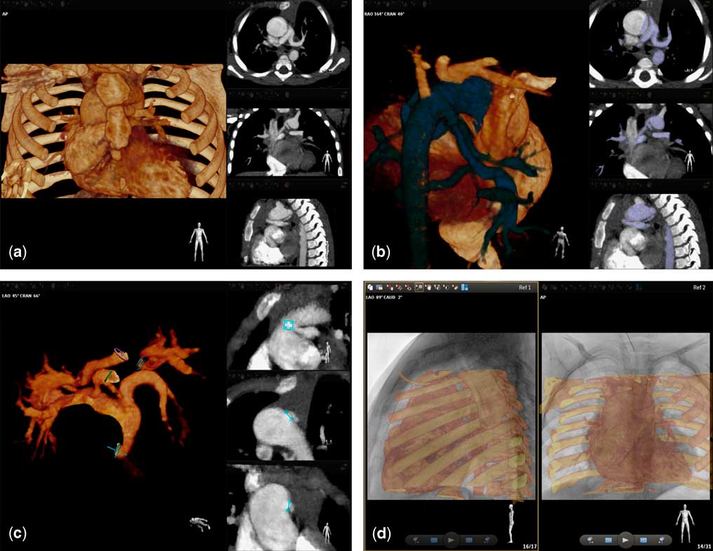
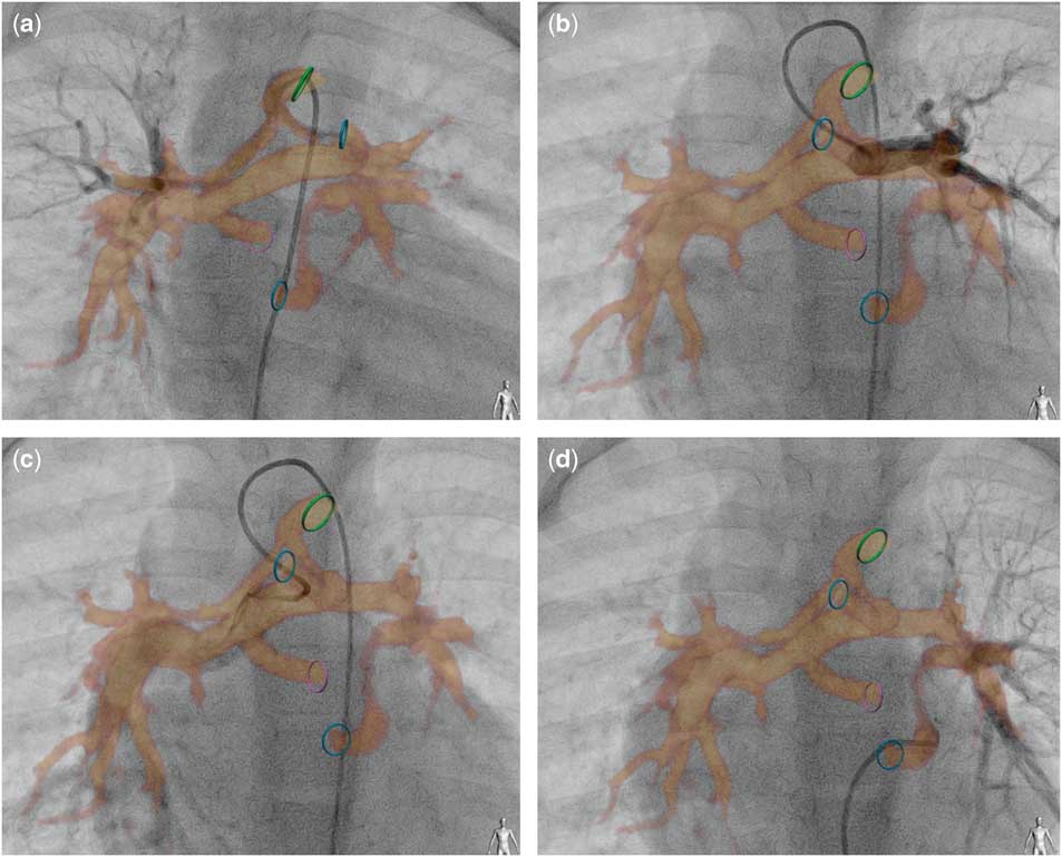
A 3.5-year-old patient (15.5 kg) with tetralogy of Fallot, pulmonary atresia, and major aortopulmonary collaterals was referred for cardiac catheterisation to delineate the current development of pulmonary vasculature with potential closure of collaterals contributing to dual pulmonary blood supply before the next stage of unifocalisation. At the age of 10 months, the patient underwent implantation of a 4-mm central shunt between the ascending aorta and the main pulmonary artery stump, as well as subsequent shunt replacement with a 7-mm tube at 22 months of age. A CT scan (dose length product – 27 mGy cm), performed 20 months earlier, immediately before up-sizing of the central shunt, was used to create a three-dimensional roadmap for catheterisation guidance. The unprocessed data set was uploaded to the dedicated workstation for manipulation with a novel three-dimensional fusion software (VesselNavigator, Philips Healthcare). An automatically created three-dimensional reconstruction (Fig 1a, Supplementary video 1) was manually segmented to expose all vessels providing pulmonary blood supply (Fig 1b). To highlight the origin of the particular vessel from the aorta, colour ring markers were placed on the three-dimensional reconstruction (Fig 1c, left panel), and their proper alignment was corrected in three perpendicular planes (Fig 1c, right panels). In the next stage, spinal bodies and vertebrae of the unprocessed three-dimensional reconstruction were aligned with fluoroscopy images acquired in two perpendicular projections (Fig 1d, Supplementary video 1). After fusion with live fluoroscopy, the movement of a diagnostic catheter within the colour ring markers provided additional reassurance of the proper alignment of the overlay (Supplementary video 2). Next, blood supply to the left and the right lung was explored (Fig 2, Supplementary video 2). Both volume-rendered images (Fig 2) and sole ring markers (Supplementary video 2) were used to monitor the route of a soft coronary guide wire and a diagnostic catheter. As all vessels provided blood supply to separate lung segments, and the consulting surgeon opted for staged unifocalisation of all of them, no embolisation was performed. The fluoroscopy time was 7 minutes and the radiation dose, defined as dose area product, was 810 μGy m2. The remainder of the procedure and hospital stay were uncomplicated.
VesselNavigator-assisted segmentation ( a ), tagging ( b ), and fusion ( d ) of a CT scan ( a ).

VesselNavigator-guided selective angiography to delineate vessels supporting the right ( a and c ) and the left ( b and d ) lung with the overlay of volume-rendered reconstruction. Colour marking rings indicate origins of major vessels from the aorta, which was spared: green – a collateral from the inferior surface of the aortic arch dividing and supporting predominantly the right upper lobe ( a ); top blue – central shunt connecting to the left ( b ) and right ( c ) pulmonary artery; pink – another collateral delivering blood to the right middle and lower lobes; bottom blue – third collateral supporting the left lung ( d ).

The use of three-dimensional imaging for live guidance of diagnostic catheterisations or transcatheter interventions is becoming more popular.Reference Goreczny, Dryzek and Moszura 1 – Reference Goreczny, Morgan and Dryzek 4 Recent advances in fusion software have enabled easy application of pre-intervention imaging, including CT, to create a reliable roadmap for swift manipulation through complex cardiac anatomy.Reference Stangenberg, Shuja and Carelsen 5 In the presented patient, we successfully re-used a CT scan originally performed 20 months earlier and intended for qualification for previous surgery. The overlaid images enhanced selective cannulation of all major vessels originating from the aorta, making aortography unnecessary. Furthers studies are required to explore potential reduction of radiation and contrast exposure with application of this new tool.
Acknowledgements
The authors acknowledge Alexandra Hul (Medical University Lodz, Poland) for editing.
Financial Support
This research received no specific grant from any funding agency, commercial, or not-for-profit sectors.
Conflicts of Interest
None.
Supplementary material
To view supplementary material for this article, please visit http://dx.doi.org/10.1017/S1047951116000895