The generic name Aspidelia was introduced by James Stirton (Reference Stirton1900) for two parmelioid lichens with gall-like tubercles bearing pycnidia, one from New Zealand and the other from the Himalaya. The New Zealand material comprised two specimens collected by Thomas W. N. Beckett (1838–1906) in 1895 and sent to Stirton that year. Correspondence between Beckett and Stirton preserved in Christchurch (CHR), and transcribed by Galloway & Elix (Reference Galloway and Elix1983), revealed that Stirton was immediately impressed by the galls and considered a new genus had to be recognized, proposing the designation ‘Beckettia’. Beckett was quick to point out that a moss he had discovered in New Zealand was about to be given that name by Karl (“Carol”) Müller, and that generic name was published three years later (Müller Reference Müller1898: 77). Stirton (Reference Stirton1900) consequently proposed the generic name Aspidelia instead, but based one of the species epithets on Beckett's surname, A. beckettii.
When introducing his new generic name, Stirton provided separate descriptions of A. beckettii and the species from the Himalaya, A. wattii. Culberson (Reference Culberson1966) argued that Stirton's generic name was not validly published as no separate generic description was provided, which also made the species names invalid (Art. 35.1), and that conclusion has been accepted by subsequent workers on parmelioid lichens (e.g. Galloway & Elix Reference Galloway and Elix1983; Hale Reference Hale1987). Unfortunately, Culberson was incorrect, even under the Code then in operation, as while there was no separate description, Stirton (Reference Stirton1900) did provide a diagnosis of the new genus after the description of A. beckettii: “As I have not seen spermogonia clustered in raised tubercles of a diverse colour from the rest of the thallus, and having spores contained in thecae with thick pellucid walls, I have been constrained to separate this lichen from the Parmelia”. In any case, three subsequent works accepted the genus, and provided separate descriptions or diagnoses of the genus itself (Zahlbruckner Reference Zahlbruckner1908: 216 and Reference Zahlbruckner1926: 238; Räsänen Reference Räsänen1943: 33); these publications appear to have been overlooked by Culberson (Reference Culberson1966) but would have validated the name even if Stirton's original account had been invalid. Interestingly, however, Santesson appears to have accepted Aspidelia as validly published with A. beckettii as type (Farr et al. Reference Farr, Leeusink and Stafleu1979). The name was also accepted by Dodge (Reference Dodge1959), who reported similar excrescences with pycnidia in some African parmelioid lichens. Consequently, had the generic name not been validated by Stirton, it would nevertheless have had to be accepted and attributed to “Stirt. ex Zahlbr.”.
Of the two species names introduced by Stirton (Reference Stirton1900), A. beckettii must be accepted as the type species as “Strn. Gen. nov.” appears after that name, with the diagnosis of the new genus immediately after, and before the description of A. wattii which had just “Strn.” after that name.
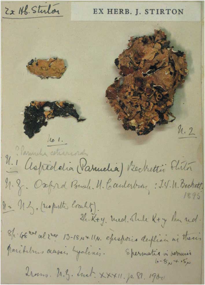
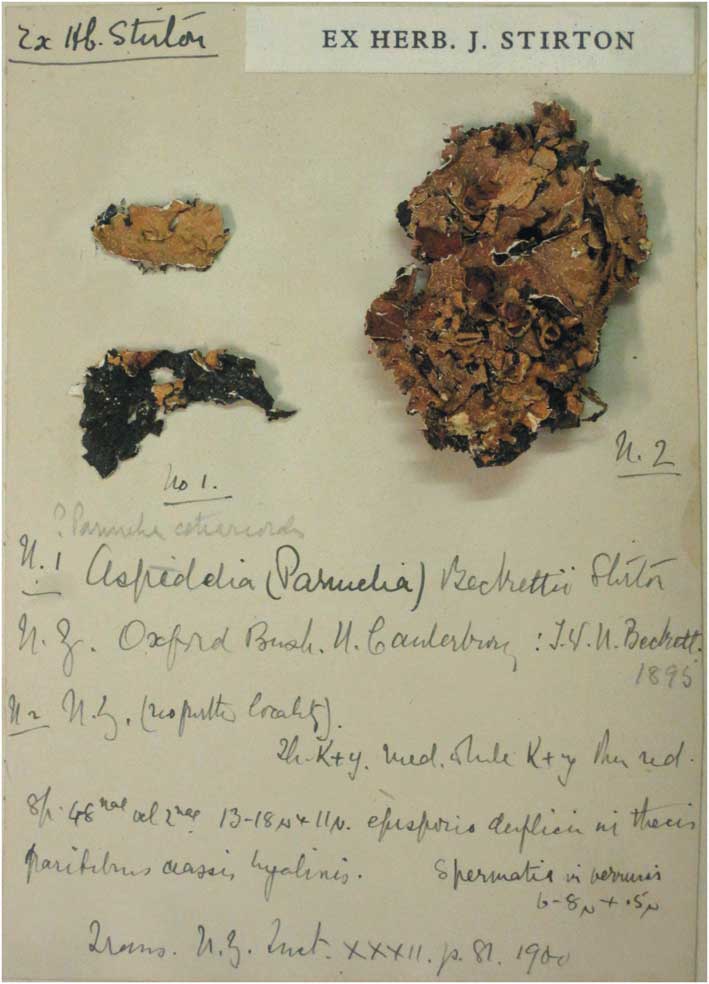
Stirton (Reference Stirton1900) gave the specimen details simply as “Corticola, New Zealand” without any more precise locality information. There are two specimens glued together on the same small sheet in the material from Stirton's herbarium in BM (Fig. 1). The left one is small and comprises two fragments, and has the locality information “No. 1. N.Z. Oxford Bush, U. Canterbury, T.W.N. Beckett 1895”; the right one is a much larger thallus labelled simply as “No. 2. N. Z. (no further locality)”. Two specimens are mentioned in the correspondence between Stirton and Beckett (Galloway & Elix Reference Galloway and Elix1983): “no. 29” with no locality details, and “no. 22” from Oxford Bush; No. 29 seems to have been the smaller collection as Stirton refers to a “scrap of the same growing on No. 22”. As Beckett appears not to have disputed that the two specimens were collected in the same place, and no more precise locality is given in Stirton's published account, the two specimens have to be treated as syntypes from which a lectotype should be selected to fix the application of the species name. Galloway & Elix (Reference Galloway and Elix1983) did not mention either number, but only the locality for the “holotype”, so it is necessary to designate a lectotype and we select the larger of the two specimens here (No. 2; i.e. Beckett's No. 22, see above).
Aspidelia Stirt.
Trans. Proc. N. Z. Inst. 32: 81 (1900) (‘1899’).
Type species: Aspidelia beckettii Stirt.
Aspidelia beckettii Stirt.
Trans. Proc. N. Z. Inst. 32: 81 (1900) (‘1899’); type: New Zealand, (Otago: Canterbury, Oxford Bush?), 1895, T. W. N. Beckett 2 (= No. 22) [BM!—lectotype designated here, MBT 200903; CHR—isolectotype (not seen, illustrated by Galloway & Elix Reference Galloway and Elix1983: 416, fig. 23)].
Aspidelia beckettii (BM—lectotype on right).
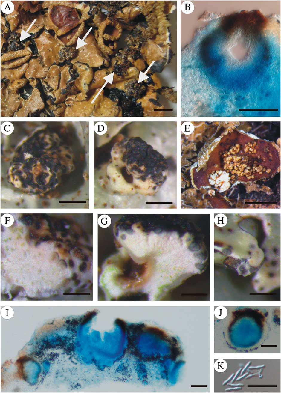
Notoparmelia tenuirima (A & B, BM—lectotype of Aspidelia beckettii; C, D, F–K, MAF Lich-17027; E, O-L-193358). A, thallus portion showing position of galls (arrows); B, vertical section of a gall; C, gall on central part of the thallus; D, gall on margin of an apothecium; E, galls on disc of an apothecium; F, vertical section through a gall on the thallus surface; G, vertical section of a gall on the margin of an apothecium; H, pycnidia on thallus margins, not associated with galls, one cut through; I, vertical section of pycnidia in gall on central part of the thallus; J, vertical section of pycnidium on thallus margin, not associated with galls; K, conidia from a gall on the thallus. All microscope preparations in lactophenol cotton blue. Scales: A = 2·5 cm; B, I & J = 50 μm; C & D = 500 μm; E = 2 mm; F–H = 250 μm; K = 10 μm.
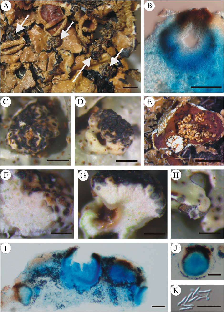
The “raised tubercles” with pycnidia of the lichen were the diagnostic character for the genus, and Stirton described the conidia as cylindrical with the apices “obsolete incrassatulis” and measuring 6–8 × c. 0·5 μm. The reference to the apices suggests these were conidia of the type referred to as “bifusiform” in modern accounts of parmelioid lichens. Galloway & Elix (Reference Galloway and Elix1983) treated Stirton's name as a synonym of Parmelia tenuirima Hook. f. & Taylor 1844, confirming the interpretation of Hale (Reference Hale1987). We found similar tuberculate galls with identical pycnidia and conidia in three more recent collections of Notoparmelia tenuirima, one from Australia and two from New Zealand. Macroscopic and microscopic studies of galls in the lectotype and the more recent collections (Fig. 2C, D, F, G & K) showed that they were identical, and in one case they developed on the disc of an apothecium (Fig. 2E). The pycnidia formed towards the margin of the lobes in MAF Lich-17027 (Fig. 2H) were identical to those found on the galls. We therefore see no reason not to accept Stirton's name as a synonym of N. tenuirima.
Additional specimens of Notoparmelia tenuirima (with galls) examined. Australia: Queensland: dividing range east of Atherton, 15 viii 1970, E. Dahl (O-L-193358).—New Zealand: Southland: Hump Ridge, alt. 500 m, on Nothofagus bark, 2007, A. Knight SPO 23 (MAF Lich-17027=OTA 60607); Lewis Pass Scenic Reserve, Sylkia Flats Picinic Area, just east of Lewis Pass Hwy 7, open silver beech forest, alt. 400 m, 1984, M. E. Hale 65755 (MAF Lich-7663).
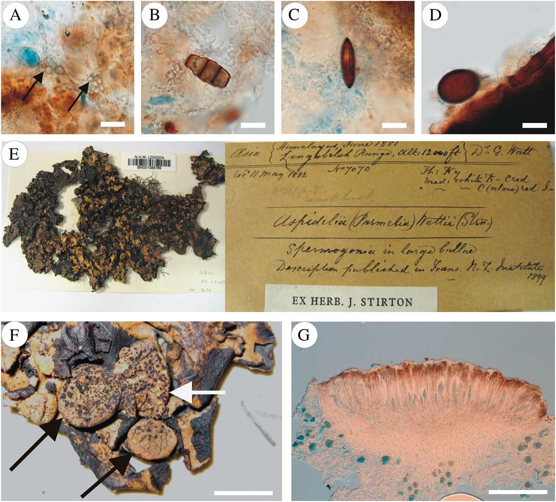
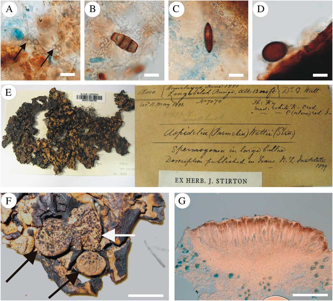
We speculated whether the galls could be caused by a lichenicolous fungus, but could find no evidence of any sporing structures apart from the pycnidia, and nor were there any hyphae distinct from those of the fungal partner of the lichen evident in the sections from the more recent collections. The galls on the lectotype did, however, have a number of other fungal elements associated with its surface (Fig. 3). Amongst these were: hyaline to pale brown chains of swollen cells (Fig. 3A) of uncertain affinity that could represent a growth stage of some lichenicolous fungus, truncated brown septate spores recalling conidia of a Pestalotiopsis that had lost its apical cells and appendages (Fig. 3B), a dark brown fusiform spore with a single vertical germ-slit such as are found in Coniochaetaceae (e.g. Coniochatea) and Xylariaceae (e.g. Rosellinia) (Fig. 3C), and an ovoid dark brown single-celled spore with suggestions of former gelatinous appendages that could be from some member of the Sordariales (e.g. Podospora, Schizothecium) (Fig. 3D). There were also some dark brown, regularly septate hyphae c. 5 μm wide on the surface, which were not constricted at the septa and ladder-like. It seems most probable that the spores were deposited on the specimen during storage or transport over the last 120 years, as no similar structures were found in the other galled collections of Notoparmelia tenuirima studied. The causal agent of the galls consequently remains obscure, though we cannot discount some unidentified lichenicolous fungus in view of the chains of swollen cells discovered.
Notoparmelia tenuirima (BM––lectotype of Aspidelia beckettii) (A–D) and Aspidelia wattii (BM 001096786––lectotype) (E–G). Notoparmelia tenuirima, fungal hyphae and spores on surface and in surface layers of galls: A, hyaline to pale brown chains of swollen cells (arrows); B, truncated brown septate spores (recalling conidia of a Pestalotiopsis); C, dark brown fusiform spore with a single vertical germ-slit (recalling those of Coniochaetaceae or Xylariaceae); D, ovoid dark brown single-celled spore with suggestions of former gelatinous appendages (recalling Sordariales). Aspidelia wattii: E, specimen and its label; F, thallus portion showing galls (arrows) on Cetrelia sanguinea; G, vertical section through the black ascoma of Nesolechia oxyspora on the galls. Scales: A–D = 10 μm; F = 3mm; G = 100 μm.
Accepting that Stirton's name is typified by the lichen-forming element has implications for the recently published name Notoparmelia A. Crespo et al. 2014 (Ferencova et al. Reference Ferencova, Cubas, Divakar, Molina and Crespo2014), which was introduced for 16 species of Australasian parmelioid lichens, including Parmelia tenuirima. Notoparmelia was not, however, a superfluous name and so illegitimate when published (Art. 52.1) as Stirton's specific name was not included in the synonymy when that species epithet was combined into Notoparmelia (Art. 52.2). It is therefore inescapable that Aspidelia provides an earlier legitimate generic name for the genus. This unfortunate oversight means that either 16 new combinations must be made into Aspidelia, or Stirton's name should be proposed for rejection so that Notoparmelia can be retained.
It would also be unfortunate for Aspidelia to be resurrected and come into current use as there are two almost identically spelled generic names: Aspidella E.-J. Gilbert 1941 (Agaricales) in the fungi, and Aspidella Billings 1872, one of the enigmatic Pre-Cambrian Ediacaran fossils which it has been suggested may be lichens (Retallack Reference Retallack1994). The three names have a similar etymology, being based on the Latin diminutive of the Greek “aspis” (a round shield). It could be argued that these names were so similar as to be treated as homonyms under the Code. Although Billings' generic name is earlier than Stirton's, it is currently only formally accepted in zoology so does not render Stirton's name illegitimate; in the eventuality that those fossils were accepted as lichens, another name change for Notoparmelia would be difficult to avoid. This is another reason to avoid Stirton's name being taken up, and is in the spirit of Rec. 54A of the Code which encourages the avoidance of names that exist in zoological nomenclature. More importantly, rejection of Stirton's name would safeguard that of Gilbert, which had generally been treated as a synonym of Amanita but has recently been reinstated for 25 species formally placed in Amanita sect. Lepidella subsect. Vittadiniae and the necessary new combinations made (Vizzini et al. Reference Vizzini, Contu, Ercole and Voyron2012).
In these circumstances, we consider the most appropriate course of action is to propose Stirton's generic name for rejection in order to: 1) avoid having to make new combinations from Notoparmelia; 2) safeguard the generic name of some lichenicolous fungus that may later prove to be the causal agent of the galls; 3) avoid the possibility of confusion with the fossil genus Aspidella; and 4) avoid the need for a new generic name to replace that of Gilbert in Agaricales. A formal proposal for rejection of the name has therefore been made (Ferencova et al. Reference Ferencova, Hawkworth and Crespo2015).
We were also able to locate and re-examine the original material of the second species described in Aspidelia by Stirton, A. wattii from the Himalaya (Himalaya, Lingabelah Range, alt. 1200 ft, June 1881, G. Watt 7070 [ex-hb. Stirton] BM 001096786—holotype) (Fig. 3E). This specimen had been studied by Culberson (Reference Culberson1966) and identified as Cetraria sanguinea Schaer. 1846, and the name was later listed as a synonym of Cetrelia sanguinea (Schaer.) Culb. & C. Culb. 1968 (Culberson & Culberson Reference Culberson and Culberson1968). As Stirton mentioned only a single locality and collector, we presume that his reference to examining two examples refers to the number of galls studied. Stirton gave no details of ascospores or conidia, and neither did Culberson. The specimen has, however, conspicuous tuberculate galls (2–4 mm diam.) with the abundant ascomata of a Nesolechia species (Fig. 3F). This was reported on by Triebel et al. (Reference Triebel, Rambold and Elix1995) who made excellent microtome sections (Fig. 3G) kept with the material in BM and identified the fungus as Phacopsis rufa (Müll. Arg.) Triebel & Rambold 1992. That species was separated from the species now known as Nesolechia oxyspora on the basis of the somewhat curved ascospores, the different host genus, and the distribution. The spore shape, however, evidently comes within the range of N. oxyspora, which is known from 18 other genera in Parmeliaceae (Doré et al. Reference Doré, Cole and Hawksworth2006), so we prefer to treat the name Phacopsis rufa as a synonym of that species pending molecular studies to clarify species concepts in the complex. We decided to include photographs of the fungus and the galls it produces on Cetrelia here as none appear to have been previously published. As both Culberson & Culberson (Reference Culberson and Culberson1968) and Triebel et al. (Reference Triebel, Rambold and Elix1995) applied the name A. wattii to the host and not to the lichenicolous fungus, they were by implication selecting the host element as lectotype so the name can be retained as a synonym of C. sanguinea.
We are indebted to John McNeill for confirming that Stirton's generic name was validly published, to Gintaras Kantvilas for searching for fresh galled material of Notoparmelia tenuirima in Tasmania, to Paul Diederich for advice on possible fungi involved in gall formation, to Holger Thüs for access to collections in BM, to Einar Timdal for the loan of specimens from O, and to Keith Watson for searching for further Stirton material in GLAM. This contribution was completed while DLH was in receipt of funding from the Spanish Ministerio de Ciencia e Innovación project CGL2011-25003.