Bacteria display two distinct life forms during growth and proliferation. In one form, the bacteria appear as single, free-floating planktonic cells, in the other, bacteria are organised into sessile aggregates. The latter form is commonly referred to as the biofilm mode of growth. These two life forms have serious implications for bacterial infections in humans. Acute infections that involve planktonic bacteria are generally treatable with antibiotics. However, in cases where bacteria succeed in forming a biofilm within the human host, the infection might prove to be untreatable and will often develop into a chronic state. The important hallmarks of chronic, biofilm-based infection are an extreme tolerance to antibiotics and conventional antimicrobial agents, and an almost infinite capacity to evade the host defence systems – in particular, the host innate immunity. In order to devise novel intervention principles for biofilm control, we have to understand the molecular mechanisms that underlie biofilm formation and this remarkable tolerance. One important control element that has a role in persistent biofilm infection is cell-to-cell communication, which is also known as quorum sensing (QS).
N-acyl-L-homoserine lactone (AHL)-based signalling systems are the most well studied examples of QS. By 2002, more than 70 Gram-negative bacterial species had been reported to harbor AHL-based systems (Ref. Reference Taga and Bassler1). QS controls a wide range of functions in Gram-negative bacteria, including Ti-plasmid conjugation in Agrobacterium tumefaciens, antibiotic production in Erwinia carotovora (Ref. Reference Taga and Bassler1), virulence gene expression in Vibrio cholerae (Ref. Reference Zhu2), Burkholderia cepacia (Ref. Reference Gilligan3) and Pseudomonas aeruginosa (Ref. Reference Whiteley, Lee and Greenberg4), and surface motility by means of swarming in Serratia liquefaciens (Ref. Reference Eberl5), P. aeruginosa (Ref. Reference Kohler6) and B. cepacia (Ref. Reference Huber7).
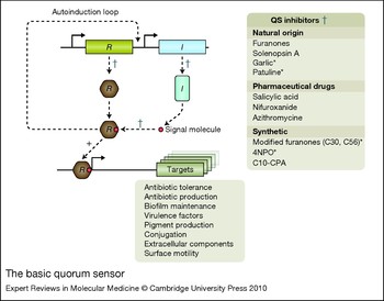
QS systems have also been identified in Gram-positive bacteria. Rather than AHL molecules, Gram-positive bacteria use small peptides, usually 5–17 amino acids in length, as signalling molecules (Ref. Reference Federle and Bassler8). Examples of Gram-positive QS-controlled behavior include: development of genetic competence and sporulation in Bacillus subtilis (Ref. Reference Magnuson, Solomon and Grossman9), virulence expression in Enterococcus faecalis (Ref. Reference Qin10) and Staphylococcus aureus (Ref. Reference Kong, Vuong and Otto11) and the somewhat controversial biofilm regulation in S. aureus (Ref. Reference Boles and Horswill12). The existence of a third type of QS system – the autoinducer-2 (AI-2) system – has been proposed (Ref. Reference Winans and Bassler13); however, this and the Gram-positive QS systems are outside the scope of the present review (see also Fig. 1).
The basic quorum sensor. R represents the regulatory protein and I represents the synthetase protein that generates signal molecules. When the critical cell density is reached, a complex of the R protein and a signal molecule enables autoinduction of the QS sensor and expression of the target genes. A list of QS inhibitors is shown on the right; asterisks indicate QS inhibitors shown to compete with the binding of the signal molecule to the R protein. Crosses indicate putative inhibitory sites. The list below the illustration shows the variety of QS-regulated phenotypes.

Pseudomonas aeruginosa
P. aeruginosa is a well characterised bacterium that is capable of causing chronic infections. It belongs to the genus Pseudomonas, which is in the γ-subclass of Proteobacteria. Characteristics of the genus are simple growth requirements and tolerance to a wide range temperatures (4–42°C). Pseudomonas is one of the most diverse and ecologically significant groups of bacteria known. It can be isolated from most environments, including soil, marshes, coastal marine habitats, plants and mammal tissue (Ref. Reference Stover14). In hospital environments, it grows in moist reservoirs such as food, cut flowers, sinks, toilets, floor mops, dialysis equipment and even in disinfectant solutions (Ref. Reference Murray15). P. aeruginosa is an opportunistic human pathogen, which is not part of the normal human flora, but it can give rise to acute infections in hospitalised patients, in particular immunocompromised individuals, such as patients with AIDS and neutropaenic patients undergoing chemotherapy. P. aeruginosa is a significant source of infections in burn victims and patients having foreign bodies inserted. It causes pneumonia in patients receiving artificial ventilation in intensive care units, as well as keratitis in users of extended-wear soft contact lenses (Refs Reference van Delden and Iglewski16,Reference Lyczak, Cannon and Pier17). It is also the predominant cause of chronic lung infection and mortality in cystic fibrosis (CF) patients (Ref. Reference Koch and Hoiby18). Other susceptible patients include those suffering from venous leg ulcers and diabetic foot ulcers.
Virulence factors
The arsenal of virulence factors produced by P. aeruginosa includes both cell-associated and extracellular products. Among these are rhamnolipids (Ref. Reference Jensen19), lipopolysaccharides (LPSs) (Ref. Reference Tang20), the two ADP-ribosylating enzymes, exotoxin A (Ref. Reference Vasil, Grant and Prince21) and exoenzyme S (Ref. Reference Nicas22), the exoproteases elastase (Ref. Reference Woods23), LasA (Ref. Reference Preston24) and alkaline protease (Ref. Reference Howe and Iglewski25), haemolytic and nonhaemolytic phospholipase C (Ref. Reference Vasil26), the siderophores pyochelin and pyoverdin (Ref. Reference Cox27), the surface-associated polysaccharide alginate (Ref. Reference Pedersen28) and pilus-associated adhesins responsible for binding to specific epithelial cell receptors (Ref. Reference Tang, Kays and Prince29). Pili themselves have on several occasions been shown to be a major antigen capable of inducing an inflammatory response in vitro (Ref. Reference Jendrossek30). These virulence factors are expressed differentially, depending on a number of environmental cues and metabolic factors. To control the production of virulence factors, P. aeruginosa uses an estimated 468 transcriptional regulators, which corresponds to 8.4% of the predicted ORFs (Ref. Reference Stover31). Expression of many of these virulence factors is coordinated by the QS systems (Refs Reference Williams32, Reference van Delden and Iglewski33).
Quorum sensing
The cell-cell communication systems in P. aeruginosa constitute one of the most intensively studied examples of QS in bacteria. The first reports of QS as a controller of virulence surfaced in the mid 1990s (Ref. Reference Latifi34). Later, transcriptomic analysis (Refs Reference Hentzer35, Reference Wagner36, Reference Schuster37) as well as plant (Ref. Reference O'Sullivan and O'Gara38), nematode (Ref. Reference Rasmussen39) and animal infection studies (Refs Reference Hentzer35, Reference Bjarnsholt40, Reference Bjarnsholt41, Reference Christensen42, Reference Jensen43, Reference Rasmussen44, Reference Wu45) have substantiated this. P. aeruginosa QS is based on two systems: a signal generator (I) and receptor (R) encoded by lasI,R and rhlI,R, respectively (Ref. Reference Pesci46). LasI generates 3-oxo-C12-HSL, which is specific for the LasR receptor and RhlI generates C4-HSL, which is specific for the RhlR receptor (see Fig. 1). A second receptor for the LasI-generated signal is the QscR receptor (Ref. Reference Lequette47). The systems encoded by las and rhl are hierarchically ordered, with the las system in control of the rhl system (Ref. Reference Pesci48). The current model is as follows: at low cell densities, the lasI gene, encoding the 3-oxo-C12-HSL synthetase LasI, is constitutively expressed at a low level, resulting in the production of a low concentration of this signal molecule. The lasR gene, encoding the LasR transcriptional regulator, is also expressed at low levels. The majority of the 3-oxo-C12-HSL signal molecules produced are pumped out of the cell by an efflux pump (Ref. Reference Pearson, van Delden and Iglewski49), which is likely to produce an equilibrium of signal molecules inside and outside the cell. When the cell density increases, the concentration of signal molecules increases both inside and outside the cells, and the transcription-inducing complex of LasR and 3-oxo-C12-HSL increases, because of the autoinduction effect (Ref. Reference Seed, Passador and Iglewski50). At a certain concentration of 3-oxo-C12-HSL, a threshold level is reached. At this threshold, binding to the LasR protein is initiated, forming the active transcriptional induction complex, which acts to induce the transcription of target genes including lasI and lasR. The genes regulated by the las system are not induced all at once: they exhibit a temporal expression pattern with increasing cell population density (Ref. Reference Hentzer35). The second receptor for the 3-oxo-C12-HSL, the QscR receptor, controls a regulon comprising a number of specific genes, which overlaps with the two other LasR- and RhlR-controlled regulons (Ref. Reference Lequette47). Since QscR mutants appear hypervirulent, this indicates that one role of QscR is to carefully adjust maximum expression of LasR- and RhlR-controlled virulence. A fourth similar QS regulator, the VqsR, has been described to be involved in siderophore production and antibiotic tolerance (Ref. Reference Juhas51).
The rhl QS system can operate independently of the las system (Ref. Reference Skindersoe52), which itself acts as a positive regulator for the rhl QS system (Ref. Reference de Kievit53). The expression of rhlR is independent of the las system under certain growth conditions, such as phosphate limitation, and is also affected by environmental conditions (Ref. Reference Medina54). Apart from this, the expression of genes regulated by the rhl system follows the same pattern as that described for the las system. However, in contrast to the las signal molecule, the rhl signal molecule C4-HSL diffuses freely in and out of the cells, as a result of its small acyl chain (Ref. Reference Pearson, van Delden and Iglewski55). The active complex of RhlR-C4-HSL acts as a positive transcriptional regulator, inducing its own synthesis and that of a number of other target genes (Ref. Reference Skindersoe52).
A third signal molecule, the Pseudomonas quinolone signal (PQS) is a 4-quinolone encoded by the pqsABCDE operon and pqsH. PQS (2-heptyl-3-hydroxy-4-quinolone) is synthesised via the pqsABCDE gene products, which, through condensation of anthranilate and β-keto dodecanoate generate more than 50 2-alkyl-4-quinolones, among which are HHQ (2-heptyl-4-quinolone), which is converted to PQS by PqsH. HHQ could potentially diffuse between cells and be converted to PQS in the target cell. Of all the synthesised 2-alkyl-4-quinolones HHQ and PQS are the dominant molecules. PQS is produced during logarithmic growth (Ref. Reference Dubern and Diggle56) and through the early stationary phase, after which it peaks in the late stationary phase of growth (Ref. Reference McKnight, Iglewski and Pesci57). The PQS system has been shown to be a part of the P. aeruginosa QS hierarchy. Expression of the pqsABCDE regulon is regulated via PqsR and depends on the ratio between the two AHLs: 3-oxo-C12-HSL induces, whereas C4-HSL reduces, expression of the operon (Ref. Reference McGrath, Wade and Pesci58). It was reported that synthesis of PQS requires a functional LasR protein (Ref. Reference Pesci59). However, later studies showed that a lasR mutant produces PQS in the ‘late’ stationary phase, indicating that only ‘early’ stationary (or transition phase) PQS synthesis requires LasR (Ref. Reference Diggle60). PQS imposes a significant transcription initiation effect on rhlI (Ref. Reference McKnight, Iglewski and Pesci61), and a functional PQS system is required for P. aeruginosa pathogenicity in a nematode model of infection (Ref. Reference Gallagher and Manoil62). In addition, the PQS signal of P. aeruginosa has been detected in the lungs of CF patients (Ref. Reference Collier63).
Pseudomonas aeruginosa biofilms
The various definitions of bacterial biofilm all seem to agree that biofilms consist of numerous bacteria organised in a structured community composed of microcolonies embedded in a self-produced matrix. We believe that attachment to a surface is not an absolute requirement and propose the following definition of a biofilm as: ‘a coherent cluster of bacterial cells embedded in a biopolymer matrix, which compared with planktonic cells, shows increased tolerance to antimicrobials and resists the antimicrobial properties of the host defence’.
The ability of P. aeruginosa to readily form biofilms is likely to contribute to its ecological versatility. Biofilm formation is a survival strategy in hostile environments, which include that of the host organism. Biofilm formation might therefore also be considered to be an important pathogenicity trait. Generally, biofilms consist of bacteria encapsulated in an extrapolymeric substance (EPS) that is either produced by the bacteria or sometimes additionally adapted from the host. Biofilm architecture promotes the development of nutrient and oxygen gradients (Ref. Reference Walters64). The EPS consists of polysaccharides, extracellular DNA and other macromolecular components such as proteins, lipids and biosurfactants. It seems to constitute the scaffolding component of the aggregating bacteria in the biofilm (Refs Reference Nivens65, Reference Allesen-Holm66), acts as scavenger of free oxygen radicals (Ref. Reference Simpson, Smith and Dean67) and binds many antibiotics, including aminoglycosides (Ref. Reference Allison and Matthews68).
Regulation of biofilm formation
Biofilm formation is a developmental progression that involves a number of complex and highly regulated processes (Ref. Reference O'Toole, Kaplan and Kolter69). In vitro, biofilm formation proceeds through an ordered, temporal series of events, which is thought to rely on the controlled expression of a collection of biofilm-specific genes (the biofilm programme) (Refs Reference Sauer70, Reference Petrova and Sauer71). However, the fact that such genes have not yet been identified in P. aeruginosa argues against such a specific genetic biofilm programme in this case (Ref. Reference Hentzer35). Our own confocal-microscopy-based studies reveal variations in the biofilm differentiation of the same strain of P. aeruginosa between experiments, indicating that biofilm formation, and the successive growth and development, proceeds through slightly different but converging paths, each of which might contain chaotic elements. So instead of one particular, surface-contact-triggered predestined biofilm programme, the available data suggest that the biofilm transcriptome reflects an adaptation to the nutritional and environmental conditions (Refs Reference Purevdorj, Costerton and Stoodley72, Reference Reimmann73, Reference Hentzer, Eberl and Givskov74). However, it is possible that with technological improvements in transcriptomic and proteomic analyses, a biofilm-specific genetic programme, which may be expressed in only a subfraction of biofilm cells and therefore is presently overshadowed by the bulk of the cells, might reveal itself. This is in contrast to the classical view of a bacterial culture, where all cells eventually proceed through the various steps of the growth cycle with identical patterns of gene expression. One reason for this difference might lie in the presence of oxygen and nutrient gradients that are characteristic of biofilm growth.
Biofilm dispersal
Biofilms are thought to maintain a ‘steady state’ by means of growth and dispersal. The dispersal occurs either as single cells or as small microcolonies ripped from the biofilm (Refs Reference Stoodley75, Reference Costerton, Stewart and Greenberg76, Reference Webb77). Evidence is accruing that the shift between the biofilm lifestyle and the planktonic lifestyle is regulated via proteins that contain diguanylate cyclase or phosphodiesterase activities and control the intracellular level of the second messenger molecule cyclic diguanosine-5'-monophosphate (c-di-GMP) (Refs Reference Hickman, Tifrea and Harwood78, Reference Meissner79). It appears that in general, high intracellular levels of c-di-GMP upregulate matrix production and biofilm formation, whereas low intracellular levels downregulate matrix production and induce a planktonic lifestyle. Several factors might contribute to dispersal of biofilms, including phage induction (Ref. Reference Webb77), carbon starvation (Refs Reference Gjermansen80, Reference Gjermansen81, Reference Schleheck82) and nitric oxide signalling (Refs Reference Barraud83, Reference Barraud84). The latter two were shown to mediate phosphodiesterase activity in P. aeruginosa, causing decreased intracellular c-di-GMP levels. Recent work shows that P. aeruginosa produces a small organic compound, cis-2-decanoic acid, which induces dispersion of established biofilms (Ref. Reference Davies and Marques85). It was recently shown that this compound, when added exogenously at native concentrations (2.5 nM), was able to induce dispersion in an in vitro P. aeruginosa biofilm. Taken together, these results suggest that dispersal is a programmed process in which cell clusters can slough off as a result of the action of shear forces. These conclusions are based solely on in vitro observations. However, if massive dispersal occurs in medical biofilms, it could have severe implications because it might be a mechanism by which biofilm bacteria disseminate from the infected organ to the entire body, and thereby cause severe systemic infections. However, this would necessitate flow conditions such as those present in blood vessels or in the synovial fluid surrounding a moving artificial limb. However, if the biofilm is located within a discrete tissue or organ, as it is in the CF lung, a chronic skin wound or long-lasting nodules after injection of permanent tissue fillers, then the absence of direct flow limits the dissemination of bacteria, but inflammation might occasionally occur more locally.
Quorum sensing and biofilm formation
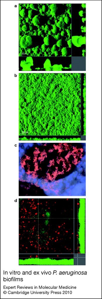
QS involvement in organised bacterial behaviour was first described for S. liquefaciens (Ref. Reference Eberl86), where it controls biosurfactant production engaged in surface conditioning. This, in combination with cell differentiation of a subpopulation enables mass movements over the surface by means of organised group motility. QS has also been implicated in the process of biofilm formation (Ref. Reference Davies87) because P. aeruginosa requires a functional las QS regulon in order to develop differentiated biofilms with mushroom-shaped multicellular structures. However, it appears that in some cases, P. aeruginosa QS mutants are still able to form mushroom-shaped structures in biofilms (Ref. Reference Allesen-Holm66). Because the bacteria in the upper part of P. aeruginosa biofilm have a growth advantage (Ref. Reference Pamp88), there could be a strong selection in QS mutant flow-chamber biofilms for the appearance of secondary mutants that are able to form extended multicellular structures. Recent work has suggested that P. aeruginosa QS mutants can form microcolonies in biofilms, but subsequent formation of mushroom caps is thought to require QS-regulated components, such as rhamnolipid and extracellular DNA (Refs Reference Pamp and Tolker-Nielsen89, Reference Yang90). In addition, there is evidence that chemical inhibition of the PQS system causes P. aeruginosa to form microcolonies on which mushroom caps are not subsequently formed (Refs Reference Yang90, Reference Yang91). When using citrate as a carbon source, the wild type and QS mutant form similar flat biofilms (Ref. Reference Heydorn92). Therefore, the involvement of signalling in the biofilm-forming process appears to depend on the specific in vitro conditions of each laboratory (Ref. Reference Kjelleberg and Molin93). Another contribution to the apparent inconsistency of the involvement of QS in biofilm development is the application of QS mutants with uncertain genetic backgrounds. QS mutants can accumulate secondary mutations (Ref. Reference Beatson94) and secondary QS regulators can also accumulate mutations (Ref. Reference Fox95), both of which could lead to misinterpretation of the effect of QS on biofilm formation. This is supported by observations that P. aeruginosa can form biofilms in a thermal-induced infection of mice independently of QS (Ref. Reference Schaber96)
However, transcriptome analysis unequivocally confirms that QS controls gene expression in biofilms (Ref. Reference Hentzer, Eberl and Givskov74). The majority of QS-regulated genes are expressed 16 hours after inoculation in ‘flow chambers’ and are inducible in a lasI, rhlI background upon the addition of exogenous 3-oxo-C12-HSL and C4-HSL. When using the transcriptome at various stages of the growth cycle of planktonic cells as a comparative background, there appears to be a subclass of QS-regulated biofilm genes that are not expressed in the planktonic mode of growth (Ref. Reference Hentzer, Eberl and Givskov74). A number of genes involved in iron scavenging or metabolism appear to be dependent on a functional QS system for expression in the biofilm mode of growth, which supports the findings that iron availability has a role in the development of biofilm architecture (Ref. Reference Banin, Vasil and Greenberg97). A recent study has provided evidence that, after formation of the initial microcolonies in a P. aeruginosa biofilm, PQS-mediated iron chelation helps to induce genes involved in iron scavenging, which promotes subsequent structural development (Ref. Reference Yang90). In line with this, QS deficiency has been shown to impair the structural stability of the biofilm (Ref. Reference Bjarnsholt40). Microscopic examination revealed that the towers and mushrooms of QS-deficient biofilms were unstable and less robust than their wild-type counterparts. An important link between biofilm stability and QS is the release of extracelluar DNA (eDNA), which is regulated by QS (Ref. Reference Allesen-Holm66). eDNA has recently been shown to be a component of the matrix in P. aeruginosa biofilms (Ref. Reference Whitchurch98), where it helps to stabilise biofilm architecture (Ref. Reference Allesen-Holm66). This might explain why QS-deficient mutants are often reported to be ‘biofilm negative’ in microtitre assays; the wash and staining processes are likely to mechanically remove the less-robust QS-deficient biofilm.
In conclusion, it appears that although QS contributes actively to the regulation of gene expression in the biofilms under in vitro conditions, a functional QS system is not required for P. aeruginosa biofilm formation per se. QS is activated as a consequence of increased cell density, which in turn leads to an increased concentration of QS signal molecules (Ref. Reference MacLehose, Gilbert and Allison99). The spatial build-up of signal molecules is likely to be more pronounced in biofilms than in planktonic cultures, owing to the confined space of the biofilm matrix (Ref. Reference Charlton100).
Antibiotic tolerance
Investigations using a number of bacterial species have fueled the dogma that biofilm bacteria are superior to their planktonic counterparts when it comes to coping with detrimental effects of antimicrobials (Refs Reference Bjarnsholt40, Reference Donlan and Costerton101, Reference Drenkard102, Reference Bjarnsholt103). For many years, the underlying mechanisms were enigmatic. However, recently, a number of molecular mechanisms have been described; these seem to enforce the original hypothesis, which proposed that the biofilm matrix provides extensive shielding in combination with classical resistance mechanisms and physiological adaptations.
Basically, the selection pressure created by antibiotic-mediated killing or growth inhibition strongly promotes development of antibiotic-resistant variants. Bacteria are able to acquire increased resistance by spontaneous mutations, which, in the presence of ample nutrients and challenging antibiotics, enable them to outgrow the susceptible parents. P. aeruginosa produces β-lactamase, which inactivates β-lactam antibiotics upon cleavage (Ref. Reference Bagge104). P. aeruginosa also produces periplasmic glucans, which have been shown to bind directly to the antibiotic tobramycin, preventing it from reaching the site of action by entrapping it in the periplasm (Ref. Reference Mah105). In the presence of hydrogen peroxide, P. aeruginosa uses extracellular catalases (Ref. Reference Elkins106) to degrade hydrogen peroxide (Refs Reference Bjarnsholt40, Reference Stewart107). Biofilm bacteria also show increased tolerance to antimicrobial compounds. Tolerance is a physiological condition that does not involve mutation, but ensures that the bacteria are not killed, although they are not necessarily able to grow in the presence of the antimicrobial compound. Numerous experiments performed over the past 30 years clearly demonstrate that tolerance can, at least in part, be attributed to the physiology of slow growth – a condition observed in large areas of in vitro biofilms. Slow growth is likely to be caused by nutrient-limiting gradients in the biofilm (Ref. Reference Roberts and Stewart108). DNA-array-based analysis has shown that the transcriptome of a biofilm (which shows increased tolerance to antibiotic treatment) resembles the transcriptome of an early stationary phase planktonic culture, particularly with regard to the expression of genes involved in anaerobiosis (Ref. Reference Hoyle and Costerton109). This minimises the effect of most antibiotics because they target biological processes that are predominant in actively growing cells (Refs Reference Donlan and Costerton101, Reference Hoyle and Costerton109).
It has been observed that exponentially growing cells are more tolerant of heavy metals than are stationary phase planktonic cells (Ref. Reference Teitzel and Parsek110). This is in accordance with the finding that P. aeruginosa consists of at least two subpopulations in vitro, one of which grows more actively than the other (Refs Reference Pamp88, Reference Haagensen111). Accordingly, the outer layer, which is most similar to exponentially growing cells is killed by conventional antibiotics, whereas cells in the more central parts tolerate these antibiotics as a result of their slower growth. These cells, however, can be killed by membrane-targeting antibiotics, such as colistin, and are probably also sensitive to heavy metals, which are actively pumped out by growing cells in the outer subpopulation. The microenvironments of the biofilm, such as differences in the pH, pCO2 or pO2, might affect the efficacy of antimicrobial compounds; the action of macrolides and tetracyclines, for example, is compromised at low pH (Ref. Reference Walters112).
Development of antibiotic and biocide tolerance phenotypes appears to be regulated, at least in part, by QS; however, the complete mechanism is not yet known (Refs Reference Hentzer35, Reference Bjarnsholt40, Reference Shih and Huang113, Reference Hentzer114, Reference Hassett115). eDNA can neutralise the activity of many antimicrobials including tobramycin by means of its cation-chelating properties (Ref. Reference Mulcahy, Charron-Mazenod and Lewenza116). Since eDNA release is regulated by QS, this might explain why tobramycin tolerance is partly QS dependent and why treatment with QS inhibitors shows synergistic action with tobramycin (Ref. Reference Bjarnsholt40). The biofilm matrix has also been shown to provide the biofilm-embedded cells with resistance against heavy metals such as zinc, copper, lead (Ref. Reference Teitzel and Parsek110) and silver (Ref. Reference Bjarnsholt103).
It is important to note that the ability of a biofilm to survive otherwise lethal treatments is multifactorial and includes mechanisms for both resistance and tolerance. This protective role of the biofilm in relation to chronic infection means that different guidelines to those used for treatment of acute bacterial infections need to be applied. Hence at Rigshospitalet (Copenhagen University Hospital) the following strategy is used to treat chronically infected patients: (1) the early use of aggressive antibiotics before the biofilm is formed, (2) the use of chronic suppressing antibiotics when the biofilm is established, if it cannot be removed physically.
The role of Pseudomonas aeruginosa in chronic infections
P. aeruginosa biofilms have a key role in many persistent infections, in particular in non-healing leg ulcers (chronic wounds) (Refs Reference Bjarnsholt117, Reference Kirketerp-Moller118) and the chronic lung infection of CF patients (Ref. Reference Barnsholt119). Chronic skin wounds can arise from pressure wounds or cuts in patients with reduced or impaired blood circulation in the lower extremities. The chronic wound remains at an inflammatory stage, which is characterised by a continuing influx of polymorphonuclear neutrophils (PMNs) that release cytotoxic enzymes, free oxygen radicals and inflammatory mediators, causing extensive collateral damage to the host tissue.
CF is a monogenic autosomal recessive, multiorganic disease, although the main cause of morbidity and mortality has its root in the patient's increased susceptibility to lung infections. The genetic cause of CF was identified in 1989 as a defect in the gene encoding the cystic fibrosis transmembrane conductance regulator (CFTR), which is located on chromosome 7 (Refs Reference Kerem120, Reference Riordan121). This causes a dysfunction of a chloride ion channel, which depletes the otherwise normal mucus of water and impairs the mucociliary clearing mechanism of the bronchia. Inhaled microorganisms are therefore able to multiply and an inflammatory response is recruited, leading to highly viscous mucus. The lower respiratory tract in patients with CF is recurrently colonised with a variety of bacteria such as Haemophilus influenzae, S. aureus and P. aeruginosa, before it is chronically infected with P. aeruginosa (80% of adult patients). However, aggressive antibiotic treatment, which delays the onset of chronic infection, remarkably prolong the life expectancy of patients with CF (Ref. Reference Hansen, Pressler and Hoiby122).
There are obvious similarities with respect to the bacterial infection found in patients with CF and those with chronic wounds. First, the conditions remain chronic because of the continuous presence of bacteria (in particular P. aeruginosa). Microscopic inspection of ex vivo samples from wounds and lungs of CF patients reveals that the bacteria are assembled in structures consisting of aggregated cells, which are surrounded but not penetrated by inflammatory cells, in particular phagocytic cells such as PMNs (Fig. 2c). We propose that bacterial persistence, including the extreme tolerance to antibiotics and evasion by the immune system is caused by the bacteria's ability to form these aggregations, which represent bacterial biofilms in vivo. The presence of the matrix component alginate can be demonstrated by using fluorescently labelled antibodies (Refs Reference Bjarnsholt123, Reference Kirketerp-Moller124). Second, both kinds of patients are immunocompromised and suffer from defects in the primary line of defence. The chronic wound consists primarily of granulation tissue composed of a network of collagen fibers, new capillaries and extracellular matrix, together with PMNs, macrophages and fibroblasts. Aggregated microcolonies of bacteria were found embedded in this matrix. This is in accordance with observations that P. aeruginosa is also detected in aggregated microcolonies from the chronic infected CF lung within the lumen of the bronchial airways (Ref. Reference Barnsholt119). Since neither disease causes defects in cellular defence, the PMNs would be expected to eradicate the bacteria. However, similarly to in vitro P. aeruginosa biofilms overlaid with freshly isolated human PMNs (Ref. Reference Jensen125), the bacteria resist phagocytosis by eradicating the PMNs upon contact (Fig. 2c). Recent results demonstrate that P. aeruginosa produces rhamnolipids (Ref. Reference Jensen19), detergents that lead to cellular necrosis and elimination of the PMNs (Refs Reference Jensen19, Reference Alhede126, Reference van Gennip127). Measurements of in vitro biofilms reveal that rhamnolipids are not excreted into the surrounding flow medium but rather stick to the aggregated bacterial biofilm. Accordingly, the rhamnolipids function as a PMN shield (Fig. 2c,d), and the capability of mounting this shield is proposed to significantly contribute to the ability of P. aeruginosa to persist in the lung, as well as in chronic wounds. The result of sustained PMN lysis is that antimicrobials, as well as tissue-devastating compounds, spill out. Examples of such compounds are myeloperoxidase, elastase and matrix metallopeptidase 9 (MMP9); chronic wound fluids are particularly rich in these compounds, in contrast to their lower levels in fluid from acute wounds in humans (Ref. Reference Wysocki, Staiano-Coico and Grinnell128). The same is also true for CF patients compared with patients suffering from acute respiratory failure (Ref. Reference Gaggar129). It is also suggested that the elimination of PMNs facilitates additional colonisation. Antibiotic treatment is likely to reduce the number of planktonic bacteria in the wound and in the lower respiratory areas of the CF lung; however, the biofilm-embedded P. aeruginosa survive, keeping the wound and the bronchial regions chronically infected.
In vitro and ex vivo P. aeruginosa biofilms. (a) In vitro flow cell P. aeruginosa biofilm with glucose as the carbon source, showing characteristic mushroom structures. (b) In vitro flow cell P. aeruginosa biofilm with citrate as the carbon source, showing a flat uniform biofilm. (c) P. aeruginosa biofilm detected in an explanted lung from a cystic fibrosis patient with chronic infection. (d) P. aeruginosa in vitro flow cell biofilm overlaid with human polymorphonuclear neutrophils (PMNs). The PMNs are unable to phagocytose the biofilm bacteria because of rhamnolipid production. Images in a, b and d were obtained using confocal laser-scanning microscopy; c was obtained using epifluorescence microscopy. Bacteria are visualised by GFP staining in a, b and d, and by PNA staining in c. PMNs are visualised by DAPI staining in c, and syto62 in d. (Images in a and b are reprinted from Ref. 170, with permission from John Wiley & Sons)

Control over rhamnolipid synthesis is governed by QS, in particular the PQS and rhl systems (Ref. Reference Fazli130). Rhamnolipid production constitutes a ‘shield’ function to promote necrosis of PMN leukocytes (upon biofilm contact), which subsequently causes collateral tissue damage and promotes development of inflammation. This suggests that quora (which are likely to be represented by the observed bacterial biofilm aggregates) have amassed at certain locations in chronic wounds (Refs Reference Kirketerp-Moller118, Reference Alhede131) or in the CF lung (Ref. Reference Barnsholt119). Such quora are then capable of eliminating PMNs by the production of rhamnolipid (Ref. Reference Morici132). Interestingly, AlgR was shown to reduce expression of RhlR-controlled gene expression in the biofilm mode (Ref. Reference Wu133). Concurrently, the concentration of rhamnolipids was below the detection limit in in vitro biofilms (Ref. Reference Morici132). However, exposure to freshly isolated PMNs superseded AlgR repression and significantly increased expression of rhamnolipids up to a level of 50 µg per g biofilm, which in pure form would causes lysis of PMN within 30 minutes of exposure (Ref. Reference Jensen19). The inducing effect was found to require a functional QS system to induce transcription initiation of the genes regulated by RhlR and PQS, and it is possible that P. aeruginosa, in order to upregulate its PMN shield, receives and responds to signal molecules originating from the host defence cells. Specific effects of cytokines such as interferon gamma (IFN-γ; IFNG) are transmitted through binding of IFN-γ to OprF, resulting in the expression of the QS-dependent virulence determinants PA-I lectin, N-butanoyl-L-homoserine lactone (BHL) and pyocyanin (Ref. Reference Kristiansen134).
Cross-kingdom signalling has a significant role in bacteria–host interactions. Recent evidence also supports the view that QS signal molecules have distinct targets and alter gene expression in mammalian cells (Ref. Reference Skindersoe135). These alterations include modulation of proinflammatory cytokines and induction of apoptosis. In particular, it was observed that P. aeruginosa QS signals decrease the production of IL-12 by murine bone-marrow-derived dendritic cells (BM-DCs) without affecting the release of IL-10. BM-DCs exposed to QS signals during antigen stimulation exhibit a decreased ability to induce specific T-cell proliferation. These results suggest that the P. aeruginosa QS signal molecules impede the T-cell-stimulatory effects of DCs, and function as immunomodulators, removing the host-protecting proinflammatory TH1 from the milieu, thereby enabling the establishment of bacterial infection within the host (Ref. Reference Wu136).
Successful treatment of a nonhealing wound depends on the identification and treatment of the factors that are preventing the healing process. Adaptations from the CF experience might be useful. Early eradication therapy against P. aeruginosa and other bacteria has long been recognised to prevent the onset of the chronic infection (Ref. Reference Hansen, Pressler and Hoiby122). Control of the host response, for example by using growth factors and MMP inhibitors, as well as local modulation of the invading bacteria by the topical application of silver ions, might also promote healing. However, the above-described bacteria–host interactions and shielding open up new treatment scenarios. Lately, several studies have proved the concept of inhibiting QS as a functional and relevant antimicrobial treatment strategy (Refs Reference Hentzer35, Reference Bjarnsholt41, Reference Rasch137, Reference Hodson138). It was recently demonstrated that a single genetic inactivation of rhlA, a gene that is important for rhamnolipid synthesis led to significant attenuation in two animal models of infection (Ref. Reference van Gennip127) The rhlAB operon is responsible for the formation of monorhamnolipids. We propose that QS inhibitor (QSI) compounds conduct functional antimicrobial effects in vivo by preventing the establishment of the PMN shield, and consequently restore proper antimicrobial functions of the PMNs. Since PMN lysis is then likely to be reduced, local inflammation and tissue destruction will be kept to a minimum. We also suggest that release of bacterial eDNA will be obstructed and thereby will reduce the viscocity of the bronchial secretion in CF, which in turn, has been shown to increase and maintain lung function (Ref. Reference Hentzer and Givskov139). Both effects will be expected to tip the balance in favor of bacterial eradication and subsequent healing.
Clinical implications
The previous 15 years of research have shown that QS controls the expression of virulence (but not growth) in a number of pathogens. Despite the fact that QS systems do not control expression of essential genes, QS systems have been shown to constitute new promising antimicrobial drug targets (Refs Reference Bjarnsholt40, Reference Bjarnsholt41, Reference Rasch137, Reference Hodson138, Reference de Nys140). Disruption of the QS signalling cascade might be a particularly valuable strategy in the treatment of chronic biofilm infections. Likewise, it has to be emphasised that the rationale behind this approach is to specifically interfere with inflammation and disease-causing traits, rather than restricting the growth of the bacteria. As production of virulence factors is inhibited, the pathogen can no longer maintain its position in the increasingly hostile host environment and consequently will be cleared by the innate host defences. Given that the mere attenuation of the pathogen does not impose a harsh and direct selective pressure on the cells, it is anticipated that resistant mutants will not arise as readily as in the case of conventional antibiotic treatment. We have denoted this the ‘antipathogenic’ drug principle (Ref. Reference de Nys140)
The first example that nature has already applied QSIs as an antibacterial strategy was demonstrated by the production of halogenated furanones by the Australian macroalga Delisea pulchra (Refs Reference Kjelleberg141, Reference Givskov142, Reference Wu143). These natural brominated furanones do not inhibit the QS of P. aeruginosa. A series of synthetic furanones has since been synthesised. Among these, compounds C-30 (Ref. Reference Hentzer35) and C-56 (Ref. Reference Hentzer114) showed QSI potential in P. aeruginosa. It was shown that biofilms grown with and without furanone C-30 were not significantly different in biofilm formation and biomass (Ref. Reference Hentzer35). However, when treating 3-day-old biofilms with 100 µg/ml tobramycin, grown either with or without furanone C-30, a significant number of dead cells were detected in the furanone C-30-treated biofilm. The antimicrobial properties of both furanone C-30 and furanone C-56 were also demonstrated in a chronic pulmonary mouse model (Refs Reference Hentzer35, Reference Kim144), an implant model (Ref. Reference Christensen42) and a fish model for vibriosis (Ref. Reference Hodson138). The bacteria were cleared significantly faster in mice treated with 1 µg furanone per gramme body weight compared with placebo-treated mice. Other furanone derivatives have been synthesised (Ref. Reference Persson145); however, these furanones also block biofilm formation of P. aeruginosa. This indicates that these furanone derivatives are highly toxic to the bacteria, because QS is not required for biofilm formation. Since the discovery of the halo-furanones, a number of papers have reported the synthesis of QSI compounds by means of organic chemistry methods (Refs Reference Riedel146, Reference Persson, Givskov and Nielsen147, Reference Olsen148, Reference Muh149, Reference Borlee150, Reference Geske, Mattmann and Blackwell151, Reference Geske, O'Neill and Blackwell152, Reference Geske153, Reference Geske154, Reference Mattmann155, Reference Bottomley156). However, a detailed overview of this is outside the scope of the present review.
Many of these compounds have been developed on agonist-based designs and probed for their action by means of specific QS screens (see below). In silico analysis showed an ID50 of 30 nM for the 12-akyl-chain-substituted tetrazol PD12 (Refs Reference Persson, Givskov and Nielsen147, Reference Borlee150). This compound is so far the most active, functional QSI drug ever designed; however, target specificity and functionality have never been validated by transcriptome analysis or animal experiments. It must be emphasised that physiologically relevant concentrations of P. aeruginosa QS signals are likely to be in the range of 1 to 20 µM. Recently, the crystal structure of a LasR peptide bound to OdDHL became available (Ref. Reference Amara157). Thus it is now possible to apply in silico analysis of receptor–ligand interactions (Ref. Reference Yang158). Direct covalent interaction of a reactive isothiocyanate AHL derivative with an amino acid present in the binding site was reported. Based on the three-dimensional structure of the LasR protein (without ligand), computer-based virtual screening has been used to search for putative quorum-sensing inhibitors from a database comprising approved drugs and natural compounds (Ref. Reference Rasmussen159). Three potential QSIs that had effects on P. aeruginosa virulence factor expression were identified.
QSI compounds are found in food, herbal and fungal sources. Several years ago we developed a new tool for high-throughput screening of fresh samples, as well as crude extracts and pure QSI compounds (Refs Reference Rasmussen39, Reference Bjarnsholt41, Reference Bjarnsholt160). The presence of QSI compounds in natural foods is extremely interesting, because, in most cases, vegetables and herbs are nontoxic to humans and are easily available. One of the most potent QSIs ever detected is within extract of garlic (Ref. Reference Bjarnsholt41). The fact that fungi also produce QSIs (Ref. Reference Rasmussen44) supports the view that organisms devoid of functional immune systems rely on chemical defence systems against competing or detrimental bacteria.
The antibiotic azithromycin was also reported to inhibit QS of P. aeruginosa (Ref. Reference Tateda161). This led us to investigate whether QS inhibition is a common feature of antibiotics. Twelve antibiotics were screened for their QS-inhibitory activities using the QSI selector system (Ref. Reference Skindersoe52). Three of the antibiotics tested, azithromycin, ceftazidime and ciprofloxacin, were active in the assay and were further examined for their effects on QS-regulated virulence factor production in P. aeruginosa. Consistent results from the virulence-factor assays, reverse-transcription-PCR, and the DNA microarrays support the finding that these three antibiotics decrease the expression of a range of QS-regulated virulence factors. The data suggest that the underlying mechanism is mediated by changes in membrane permeability, thereby influencing the flux of 3-oxo-C12-HSL (Ref. Reference Skindersoe52). Azithromyzin inhibits or reduces the production of several of the virulence factors of P. aeruginosa, such as elastase and rhamnolipids (Ref. Reference Hoffmann162), and interferes with alginate polymerisation (Ref. Reference Kai163). Recently another group reported that azithromycin inhibits AHL synthesis by blocking unknown pathways involved in regulation of lasI and rhlI (Refs Reference Kai163). It is proposed that azithromycin interferes with one of the precursors of the AHL molecules, indicating that the QSI activity is a secondary effect. Azithromycin does improve the respiratory function in CF patients (Ref. Reference Saiman164, Reference Southern and Barker165).
The principal role of QSIs is to attenuate the virulence of the bacteria, and in our opinion, if the production of the excreted rhamnolipids is attenuated, P. aeruginosa is dramatically weakened. As a result of this, the venom alkaloid solenopsin A is of great interest (Ref. Reference Park166). It is extracted from the fire ant Solenopsis invicta and targets the rhl QS system specifically, thereby inhibiting rhamnolipid production.
Conclusion
Antibiotic resistance is one of the most serious emerging health problems in the world. More and more disease-causing bacteria are resistant to at least one of the currently available antibiotics. Mortality from infectious disease is still a major problem in developed countries and especially in developing countries. As for chronic infections, they often develop in connection with the application of medical devices, implants and artificial organs and joints, but are also seen in connection with development of leg ulcers into non-healing wounds. Such infections are not efficiently treatable with our present assortment of antibiotics; a situation that is developing in parallel with the increase in the elderly population. It has recently been stated by the NIH that: ‘More than 70 percent of the bacteria that cause hospital-acquired infections are resistant to at least one of the drugs most commonly used to treat them’. As implicated in this review, the biofilm mode of growth is important in chronic bacterial infections because biofilms resist or tolerate basically all antibiotics. We would like to emphasise that we do not believe that biofilms are formed by a specific genetic program, and in our hands, biofilm formation can not be inhibited by inhibiting QS.
As is evident from this review, pathogenesis of many bacteria are regulated by QS, which makes QS an obvious new drug target in the control of unwanted microbial activity. Such targets include the production of virulence factors, antibiotic resistance and modulation of the host immune system. We believe that QSI drugs could be beneficial in the fight against biofilm infections, although much more work is needed to develop relevant pharmaceutical molecules.
Acknowledgements and funding
The authors would like to thank the peer reviewers of this manuscript for their useful comments and suggestions. T.B. has received funding from The Carlsberg Foundation and Lundbeck Foundation and M.G. is funded by the German Mukoviszidose e.v. and the Danish Strategical Research Council.