Introduction
Bovine ovaries contain a large number of follicles; however, most of the follicles in ovaries are lost or degenerated, and only a small proportion produce oocytes competent to undergo successful maturation. Many efforts have been made to utilize these oocytes in mice or larger animals (Eppig, Reference Eppig, O'Brien and Wigglesworth1996; Miyano, Reference Miyano2005). One such trial in cows used, as the starting material, growing non-competent oocytes of about 95 μm in diameter that had been isolated from early antral follicles (Harada et al., Reference Harada, Miyano, Matsumura, Osaki, Miyake and Kato1997; Yamamoto et al., Reference Yamamoto, Otoi, Koyama, Horikita, Tachikawa and Miyano1999). Hirao et al. (Reference Hirao, Itoh, Shimizu, Iga, Aoyagi, Kobayashi, Kacchi, Hoshi and Takenouchi2004) reported that such growing oocytes had grown to an average of 113 μm in diameter and had acquired the competence for maturation during a 14-day culture with surrounding granulosa cells in a medium containing polyvinylpyrrolidone (PVP) and 17β-estradiol. Using this system, a live calf was later obtained (Hirao et al., Reference Hirao, Itoh, Shimizu, Iga, Aoyagi, Kobayashi, Kacchi, Hoshi and Takenouchi2004); however, the percentage of oocytes competent to reach metaphase II is low in this system.
In the present study, we were interested in supplementing the culture medium with androstenedione in order to grow oocytes. One major reason for our interest is that recent studies have revealed the contribution of androgens to female reproductive function (Walters et al., Reference Walters, Allan and Handelsman2008; Sen & Hammes, Reference Sen and Hammes2010). Another important reason is that theca cells are not involved in the culture system used in this study. In the ovary, oocytes are enclosed by layers of granulosa cells, which in turn are enclosed by layers of theca cells. These somatic cells synthesize and secrete different steroid hormones in a concerted action. 17β-Estradiol is a primary hormone transformed from androstenedione by the aromatase in granulosa cells (Fortune & Armstrong, Reference Fortune and Armstrong1978; Liu & Hsueh, Reference Liu and Hsueh1986) and is involved in the proliferation and differentiation of granulosa cells. Androstenedione is synthesized by the theca cells. It has been reported that androgens enhance follicular development and granulosa cell proliferation (Daniel & Armstrong, Reference Daniel and Armstrong1980), and that the androgen receptor is present in granulosa cells (Schreiber & Ross, Reference Schreiber and Ross1976); however, there is a lack of data on the effect of androstenedione on bovine oocytes undergoing growth in vitro.
In the present study, we investigated the effects of androstenedione on the growth of bovine oocytes collected from early antral follicles. In addition, the meiotic competence of the oocytes after growth culture was assessed.
Materials and methods
Collection of oocyte–granulosa cell complexes
Bovine ovaries were obtained from an abattoir and transported to the laboratory in calcium- and magnesium-free Dulbecco's phosphate-buffered saline (Nissui Pharmaceutical) supplemented with 2 mM dimethylthiourea (Iwata et al., Reference Iwata, Ohota, Hashimoto and Nagai2003). Collection of complexes consisting of an oocyte and cumulus/granulosa cells was based on the method described by Harada et al. (Reference Harada, Miyano, Matsumura, Osaki, Miyake and Kato1997). Briefly, early antral follicles measuring 0.4–0.7 mm in diameter were isolated from the surface of the ovaries. The follicles were opened using forceps and a blade to dissect out the oocyte–granulosa cell complexes associated with a group of mural granulosa cells, hereafter referred to as oocyte–granulosa cell complexes or simply complexes. Each collected complex was transferred to a microdrop of the collection medium, HEPES-buffered modified Minimum Essential Medium (pH 7.2, Gibco-BRL) prepared with Earle's balanced salt solution, 20 mM HEPES (Gibco-BRL), 5 mM sodium bicarbonate, 4% (w/v) polyvinylpyrrolidone (PVP; Sigma, molecular weight 360,000), 1 mM sodium pyruvate, and 0.08 mg/ml kanamycin sulfate (Sigma). Only oocytes with a healthy appearance were used.
Growth culture of oocytes
The culture medium used for oocyte growth was tissue culture medium (TCM)-199 (Gibco-BRL) supplemented with 5% (v/v) fetal bovine serum (FBS; HyClone), 50 μg/ml ascorbic acid 2-glucoside (Hayashibara Biochemical Laboratories), 4% (w/v) PVP (molecular weight 360,000), 1 mM sodium pyruvate, 0.08 mg/ml kanamycin sulfate, 4 mM hypoxanthine, 55 μg/ml cysteine and 0.05 μM dexamethasone (Sigma). The medium was also supplemented with either 0, 10 or 100 ng/ml 17β-estradiol, or 0, 10 or 100 ng/ml androstenedione (Sigma). Each complex was transferred into culture dishes (96-well culture plates; Biocoat Collagen I Cellware, Becton Dickinson Falcon), which were coated with a collagen mixture consisting of 0.3% (w/v) collagen solution (Cellmatrix Type I; Nitta Gelatin) prepared according to the manufacturer's instructions. The day of complex isolation was designated day 0. The complexes were cultured in 0.2 ml medium at 38.5 °C under an atmosphere of 5% O2:5% CO2:90% N2 from day 0 to day 4, and an atmosphere of 5% CO2 in air from day 4 to day 14. On days 1, 4, 6, 7, 8, 10, 12 and 14, oocyte–granulosa cell complexes that showed degenerative signs, such as cytoplasmic degeneration of oocytes and/or complete detachment of granulosa cells from oocytes resulting in degradation of the complex structure, were classified as degenerative complexes and excluded from further examination. The diameter of each oocyte, excluding the zona pellucida, was measured to the nearest 0.5 μm with an ocular micrometer (Olympus) on days 1, 7 and 14. Half of the medium was changed every other day. After the size measurement on day 14, the oocytes and surrounding granulosa cells were picked up from a 96-well culture plate and transferred to microdrops prepared for the induction of oocyte maturation as described below. Oocytes that had at least some firmly attached granulosa cells and that showed no sign of degeneration were considered as the surviving oocytes and were subjected to in vitro maturation.
Maturation culture of oocytes
After the 14-day culture period, each of the surviving oocytes was transferred into a 10 μl microdrop of the maturation medium, which consisted of TCM-199 supplemented with 0.1 mg/ml sodium pyruvate, 0.08 mg/ml kanamycin sulfate, 5% (v/v) FBS and 100 ng/ml follicle stimulating hormone (FSH; NIDDK). The oocytes were cultured at 38.5 °C for 22–24 h under an atmosphere of 5% CO2 in air. At the end of the culture period, the oocytes were denuded mechanically of granulosa cells using a small-bore pipette with the help of hyaluronidase (Sigma). The oocytes were then mounted onto a slide and fixed with acetic alcohol. The following day, the chromatin was stained with acetic orcein and examined under an inverted microscope. Oocytes collected from large antral follicles (4–6 mm) were cultured and examined in the same manner as for the in vivo control.
Data presentation and statistical analysis
The experiments were repeated at least four times. Statistical differences in the diameters of the oocytes were analysed by the Steel–Dwass test (multiple comparisons). Other values were analysed by Fisher's exact test with Holm correction (multiple comparisons). A probability of less than 0.05 was considered statistically significant.
Results
Morphology of complexes and oocyte growth
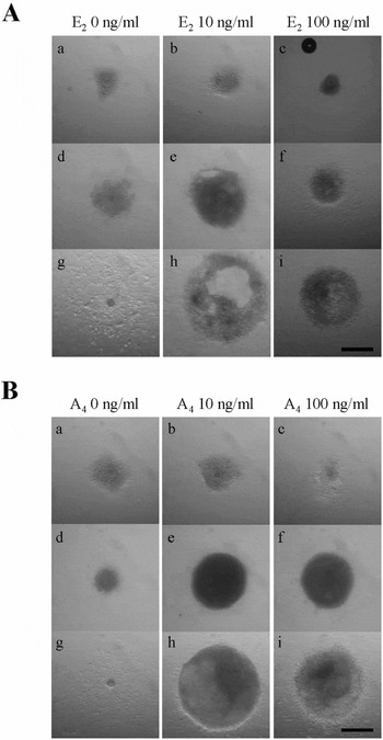
Figure 1 shows the typical morphology of oocyte–granulosa cell complexes in the medium supplemented with 0, 10 or 100 ng/ml 17β-estradiol or androstenedione during culture. As a common morphology on day 1, each complex had an oocyte at the centre, around which the piled granulosa cell populations spread outward to form a thin cell layer on the periphery of the complex (Fig. 1Aa–Ac,Ba–Bc). Over the next few days, some complexes gradually developed a small cavity (or cavities) in a dense area of granulosa cells, and then, within another few days, a well developed cavity was seen in a proportion of the complexes in medium supplemented with the steroid hormones. Without the steroids, the complexes usually possessed no cavity on day 7 (Fig. 1Ad,Bd). In contrast, more than half of the complexes in the medium containing 10 or 100 ng/ml of 17β-estradiol or 10 or 100 ng/ml androstenedione developed a small dome-like structure (Fig. 1Ae,Af,Be,Bf). By day 14, in the medium without 17β-estradiol or androstenedione, the oocytes had became gradually denuded (Fig. 1Ag,Bg), while a proportion of the complexes in the medium supplemented with the steroid hormones had developed and retained the domelike structure (Fig. 1Ah,Ai,Bh,Bi).
Typical morphology of oocyte–granulosa cell complexes in the medium supplemented with 0, 10 or 100 ng/ml 17β-estradiol (A) or androstenedione (B) on day 1 (a–c), day 7 (d–f) and day 14 (g–i). The complexes in the medium supplemented with 17β-estradiol (Ah, Ai) and androstenedione (Bh, Bi) exhibited a dome-like structure on day 14. E2: 17β-estradiol; A4: androstenedione. Scale bars represent 300 μm.

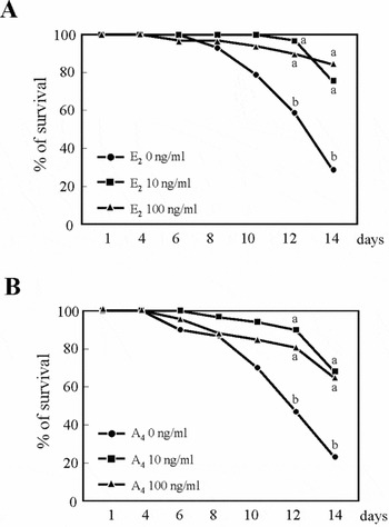
Figure 2 shows the viability of oocyte–granulosa cell complexes over the culture period. A sharp and steady decrease of the survival rate was found in the groups without 17β-estradiol or androstenedione from day 6 to day 14. In the medium supplemented with steroid hormones, the viability of complexes was better sustained over almost the whole culture period. High rates of surviving complexes on day 14 were found in the cultures with 10 and 100 ng/ml 17β-estradiol (Fig. 2A; 75% and 84%, respectively). In the medium supplemented with 10 and 100 ng/ml androstenedione, 68% and 65% were recovered as viable, respectively (Fig. 2B).
The percentage of oocyte–granulosa cell complexes surviving (without degenerative signs) in the medium supplemented with 0, 10 or 100 ng/ml 17β-estradiol (A) or androstenedione (B) for 14 days. E2: 17β-estradiol; A4: androstenedione. a, bValues with different superscripts at the same culture period differ significantly (Fisher's exact test with Holm correction, p < 0.05).

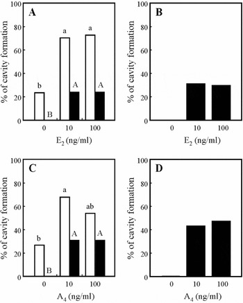
Figure 3 shows the percentages of oocytes accompanied by one or more cavities. Regardless of the concentration, the addition of 17β-estradiol promoted the formation of cavities; over 70% of the complexes had developed a cavity during the culture period (Fig. 3A). However, the percentage of complexes retaining the cavity was only about 24% on day 14 in both groups. In the medium without 17β-estradiol, 24% of the complexes developed a small cavity, although by day 14 this cavity had completely disappeared (Fig. 3A). Of the oocytes surviving until day 14 in the presence of 17β-estradiol, about 30% of the complexes had sustained the cavity in both groups (Fig. 3B). Essentially the same pattern was seen in the results of the androstenedione-supplementation experiments (Fig. 3C,D). Although 100 ng/ml androstenedione appeared slightly less effective than 10 ng/ml androstenedione in promoting the overall cavity formation during the culture, the percentages eventually became equivalent between the two concentration groups (Fig. 3C,D).
Effect of the addition of 17β-estradiol (A,B) or androstenedione (C,D) on the formation of a cavity in the complexes. The white columns in (A) and (C) represent the proportion of the complexes (to the total number of complexes used for culture) that had developed a cavity at some time point during the 14-day culture period. The black columns in (A) and (C) represent the proportion of the complexes which had retained the cavity until the end of the culture period. The columns in (B) and (D) represent the proportion of the complexes having the cavity on day 14 to the number of oocytes that were viable on day 14. Values with different superscripts (a, b or A, B) in the same frame differ significantly (Fisher's exact test with Holm correction, p < 0.05).

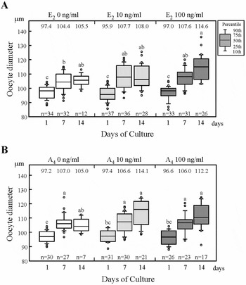
Figure 4 shows the growth of oocytes during the culture period. The box plots represent the distribution of the oocyte sizes. In all groups the mean diameter (shown at the top of each frame) of oocytes surviving on days 7 and 14 had increased from those measured on day 1. The mean diameter of oocytes measured after seeding on the culture substrate was 96.9 μm (n = 191). The mean diameters of the oocytes on day 14 in the medium supplemented with 10 and 100 ng/ml 17β-estradiol increased significantly to 108.0 ± 1.5 μm and 114.6 ± 1.7 μm (Fig. 4A), compared with day 1 (95.9 ± 0.8 μm and 97.0 ± 0.9 μm, respectively). Similarly, in the medium supplemented with 10 and 100 ng/ml androstenedione, the mean diameters on day 14 increased significantly to 114.1 ± 1.9 μm and 112.2 ± 2.3 μm (Fig. 4B), compared with day 1 (97.4 ± 0.7 μm and 96.6 ± 1.0 μm, respectively).
The size distribution of oocytes cultured in the medium supplemented with 0, 10 or 100 ng/ml 17β-estradiol (A) or androstenedione (B) for 14 days. The box plots represent the size of the viable oocytes on day 1, day 7 and day 14 of culture. The numbers above the box plots indicate the mean diameters (μm), and the numbers of oocytes used are shown at the bottom of each frame. E2: 17β-estradiol; A4: androstenedione. a–cValues with different superscripts in the same frame differ significantly (Steel–Dwass test, p < 0.05).

Meiotic competence
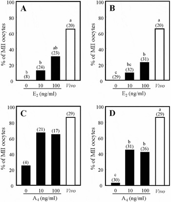
Before culture, oocytes collected from early antral follicles did not mature to metaphase II (0/38). After 14 days of the growth culture, the oocytes enclosed by granulosa cells were further cultured for maturation to assess their maturational competence (Fig. 5). Among the surviving oocytes grown in the medium supplemented with 100 ng/ml 17β-estradiol, 30% reached metaphase II (Fig. 5A). The percentage of matured oocytes after the growth using 10 ng/ml 17β-estradiol was intermediate between the two groups (13%). When the results were expressed as a percentage of the oocytes initially used for culture, a significant difference was found between the groups with 0 ng/ml and 100 ng/ml 17β-estradiol supplementation (Fig. 5B). Conversely, 67% and 65% of oocytes recovered from the medium supplemented with 10 and 100 ng/ml androstenedione, respectively, matured to metaphase II (Fig. 5C), which was comparable with the percentages of oocytes grown in vivo (86%; oocyte diameter 123.7 ± 0.8 μm). Only one-quarter of oocytes that were recovered from the medium without androstenedione supplementation progressed to metaphase II, but no significant difference was found between the other groups, due to the paucity of surviving oocytes in this group. As Fig. 5D indicates, when a comparison was made on the basis of the total number of oocytes, the difference in the number of oocytes between the medium with and the medium without androstenedione was remarkable.
Meiotic competence of the oocytes that were cultured for growth in the medium supplemented with 17β-estradiol (A,B) or androstenedione (C,D). The proportions of metaphase II oocytes to the number of oocytes on day 14 are shown in (A) and (C). The proportions of metaphase II oocytes to the number of oocytes initially introduced in the growth culture are shown in (B) and (D). E2: 17β-estradiol; A4: androstenedione. The numbers of oocytes used for calculation are shown in the parentheses. a–cValues with different superscripts differ significantly (Fisher's exact test with Holm correction, p < 0.05).

Discussion
In the present study, the androstenedione-supplemented medium supported the growth of more than 65% of bovine oocyte–granulosa cell complexes without showing any degeneration of oocytes throughout the 14-day-culture period. The addition of either 17β-estradiol or androstenedione was absolutely necessary to maintain the organization of oocyte–granulosa cell complexes under the conditions used in this study. Our findings also indicated that it may be possible to completely replace 17β-estradiol with androstenedione in the culture medium. The effects of these steroid hormones on the oocyte–granulosa cell complexes were similar in the present study, but some detailed but important differences were noticed. It was of particular interest that androstenedione appeared to show a more advantageous effect than 17β-estradiol, with respect to the meiotic competence of the resultant oocytes.
It has been well established that oocytes acquire meiotic competence during the final stage of oocyte growth (Sato et al., Reference Sato, Matsuo and Miyamoto1990; Fair et al., Reference Fair, Hyttel and Greve1995). An oocyte size of more than 110 μm in diameter seems to be a prerequisite for the progression to metaphase II. This parallel between oocyte size and meiotic competence was also evident in the present study. The oocyte groups with a mean diameter that exceeded 110 μm exhibited good competence of oocyte maturation, while the oocyte groups with a diameter of only 105 μm failed to progress to metaphase II. A crucial observation worthy of consideration is that the addition of 10 ng/ml androstenedione was not less effective than the addition of 100 ng/ml 17β-estradiol in terms of oocyte meiotic competence. This finding suggests not only that androstenedione is useful to promote the acquisition of meiotic competence of oocytes, but also that the effects of androstenedione are not exerted entirely through the conversion to 17β-estradiol. The promotion of meiotic competence in the medium that contained androstenedione can be attributed, in part, to androstenedione itself. Furthermore, this result indicates that exogenous factor(s), in this case androstenedione, may affect the acquisition of meiotic competence of oocytes separately from their growth.
The results in the present study suggest that both 17β-estradiol and androstenedione have a beneficial effect on the association between oocyte and granulosa cells that is crucial for oocyte growth (Anderson & Albertini, Reference Anderson and Albertini1976; Eppig & Schroeder, Reference Eppig and Schroeder1989). However, the percentage of viable oocytes decreased towards the end of the culture period except in the group supplemented with 100 ng/ml 17β-estradiol. The problem in many instances was denudation of the oocytes. It appeared that one of the basic conditions responsible for such denudation was the use of culture medium without FSH. It seems possible that androstenedione requires a more normal function of the granulosa cells in order to exert its effects than does 17β-estradiol, which exhibits more straightforward mitogenic activity. Conversely, we have not investigated the effects of the growth factors produced and excreted from theca cells, which were absent from our culture system. Although androstenedione may be potent enough to promote extensive growth of healthy oocyte–granulosa cell complexes, androstenedione alone may not be sufficient to sustain the structure of enlarged complexes that have almost full-grown oocytes.
The dome-like structure has been documented in cultured oocyte–granulosa cell complexes from preantral or early antral follicles in several mammals (Cain et al., Reference Cain, Chatterjee and Collins1995; Demeestere et al., Reference Demeestere, Delbaere, Gervy, Van Den Bergh, Devreker and Englert2002; Hashimoto et al., Reference Hashimoto, Ohsumi, Tsuji, Harauma, Miyata, Fukuda, Hosoi, Iritani and Morimoto2007). In the present study, on day 14, none of the complexes had cavities in the medium without the steroid hormones. It is important to note, however, that about 25% of the complexes had formed a small cavity by the middle stage of the culture, although they eventually lost these cavities. Considering that no FSH was added to the medium, a possible explanation for this finding is that the oocytes produced factors both promoting granulosa cell proliferation (Gilchrist et al., Reference Gilchrist, Ritter and Armstrong2001) and directing cavity formation (Shen et al., Reference Shen, Miyano and Kato1998) during the first week, but that the effect did not last for 14 days. Conversely, even in the presence of steroid hormones, the dome-like structure was lost in half or more of the complexes. Again, it was suggested that some crucial factors may be missing from the culture system, and that these factors should be identified and added to the medium in order to further improve the culture system.
In conclusion, the present study indicates that the effects we expected from 17β-estradiol on the growth of bovine oocyte–granulosa cell complexes can be mostly achieved by the addition of androstenedione to the culture system. Androstenedione at a low dose even promotes the acquisition of meiotic competence. Other observations, however, indicated that androstenedione alone is not sufficient to sustain the continued growth of enlarged complexes at the later time period. It will be important to determine the conditions that ‘strike a balance’ between the use of androstenedione and the maximization of oocyte–granulosa cell complex viability.
Acknowledgements
The authors would like to thank the staff of the National Agricultural Research Center for Tohoku Region for supplying bovine ovaries. This work is supported in part by the Grant-in-Aid for Scientific Research of the Japan Society for Promotion of Science to Y.H. and T.M.