I. INTRODUCTION
The usual approach of tissue engineering is to seed cells on scaffolds to create biological tissue equivalents.Reference Mooney and Mikos1 In the past decade, an array of techniques such as weaving, knitting, electrospinning, wet-spinning, microfluidics, and bioprinting has been developed to produce micro/nanofibers that mimic biochemical, mechanical, and structural details of a cell niche.Reference McCullen, Autefage, Callanan, Gentleman and Stevens2–Reference Liu, Thomopoulos and Xia6 The fibers were then seeded with cells to create constructs that offered a high surface area for cell migration as well as enabled efficient mass transport.Reference Jun, Kang, Chae and Lee5–Reference Freeman, Woods, Cromer, Wright and Laurencin7
Advanced fiber fabrication processes have also achieved in situ incorporations of cells; for instance, electrospinning was explored for directly drawing fibers with cells using a coaxial needle configuration to generate simple scaffolds, cell sheet, and complex architectures.Reference Jayasinghe8,Reference Lopez-Rubio, Sanchez, Sanz and Lagaron9 However, the encapsulation of live cells in electrospun fibers faces many challenges as the diameter of the nanofiber is typically smaller than a single cell.Reference Lopez-Rubio, Sanchez, Sanz and Lagaron9 Wet-spinning is a relatively simple technique to produce cell-laden microfibers; however, these fibers mostly explored the gelation chemistry of calcium and alginate.Reference Ram Lee, Lee, Kang, Kim and Lee10,Reference Shin, Park, Lee, Park, Park, Lee, Whang and Lee11 Prolonged exposure to these chemicals can be injurious to the cells. Thus, further cell viability studies must be performed. Microfluidic spinning is similar to wet-spinning, with the exception of that the crosslinking agent is supplied by the coaxial flow instead of a bath.Reference Tamayol, Akbari, Annabi, Paul, Khademhosseini and Juncker4,Reference Jun, Kang, Chae and Lee5,Reference Liu, Zhou, Chai, Zhang, Wu, Huang, Su and Jiang12 Though microfluidic spinning is a versatile method of fabricating cell-laden fibers, however, due to the relatively slow fiber production process, fabricating 3D structures is time-consuming.Reference Jun, Kang, Chae and Lee5 Bioprinting techniques allow fabrication of fiber-based scaffolds containing live cells with high resolution, controlled geometry, pore size distribution, and microstructure.Reference Hsieh, Lin and Hsu13,Reference Levato, Visser, Planell, Engel, Malda and Mateos-Timoneda14 However, shear stress associated with bioprinting may affect cell viability and detailed post fabrication physiological/genetic alteration studies of cells remain to be carried out. Thus, all the present technologies of cell-laden fiber production suffer from certain limitations and need further improvement in the technology.
In our previous study, chitosan microfibers were produced in the citric acid (CA) bath for tissue engineering applications.Reference Ghosh, Rameshbabu, Das, Francis, Pawar, Subramanian, Pal and Dhara15 The carboxyl anions of the CA bath, pH 7.4 participated in ionotropic complexation with the glucosamine moieties of chitosan solution forming an instantaneous gel that was drawn steadily to produce mechanically strong fibers. Moreover, CA is a nontoxic metabolic product of the body (Krebs cycle), it is also readily available, and is inexpensive.
In the present study, the ionic gelation property of chitosan solution in the CA bath is further explored to produce cell-laden fibers. Amniotic mesenchymal stem cells (AM-MSC) were conditioned in different ratios of CA/media solution and then analyzed for adherence, viability, and proliferation. A nontoxic and cytocompatible CA/media ratio was selected from the abovementioned studies and used to study gelation kinetics of chitosan solution using a rheometer. The chitosan gels were chemically characterized to confirm cross-linking of chitosan solution in CA/media. Subsequently, chitosan fibers were produced in the CA/media bath containing cells under sterile conditions at physiological pH and room temperature. The morphology of the cells adhered on the fibers was examined at various time periods using microscopy. The cell-laden fibers (living fibers) produced under benign conditions can provide a platform technology for creating viable 3D building blocks for tissue engineering applications.
II. MATERIALS AND METHODS
A. MSC isolation and culture
All the tissue samples were obtained after informed consent according to the policy approved by the ethical committee (Midnapore Medical College, Paschim Medinipur, India). Term placentas obtained from cesarean sections were collected in phosphate-buffered saline (PBS) containing penicillin and streptomycin (200 U/mL penicillin, 200 µg/mL streptomycin) and transferred to the laboratory under stringent sterile conditions. AM-MSCs were isolated as reported previously.Reference Alviano, Fossati, Marchionni, Arpinati, Bonsi, Franchina, Lanzoni, Cantoni, Cavallini, Bianchi, Tazzari, Pasquinelli, Foroni, Ventura, Grossi and Bagnara16,Reference Rameshbabu, Ghosh, Subramani, Bankoti, Kapat, Datta, Maity, Subramanian, Roy, Chaudhury and Dhara17 Briefly, amniotic membrane (AM) was separated from chorion through blunt dissection. AM was washed several times with Hank’s balanced salt solution to remove blood clots. AM was chopped into small pieces and digested with 0.25% trypsin–EDTA solution for 30 min and the supernatant was discarded. The tissue was subjected to second digestion with 1 mg/mL collagenase IV solution in DMEM (all purchased from GIBCO) for 50 min at 37 °C. The supernatant was transferred to a fresh tube and spun at 1500 rpm for 10 min. The pellet was suspended in 5 mL of culture medium containing: DMEM, 20% FBS, penicillin 100 U/mL and streptomycin 100 µg/mL. MSC cultures were grown at 37 °C in 5% CO2 and the media (with 10% of FBS) was subsequently changed every four days. When the culture reached 90% confluence, cells were recovered using 0.25% trypsin–EDTA for expansion or other studies.
The MSCs were characterized by immunofluorescence. Briefly, the cells were cultured on chamber slides till confluence was reached, fixed with 4% formaldehyde solution in PBS, permeabilized with 0.1% Triton X-100 solution in PBS, unspecific sites blocked with 1% bovine serum albumin (BSA; Sigma, St. Louis, Missouri) in PBS, incubated with antibodies, and imaged by means of fluorescent microscopy (Zeiss Axiovision, Jena, Germany). The antibodies used were FITC/PE-conjugated CD44, CD105, and CD166 (Biolegend, Cat: 338803, Cat: 323203, and Cat: 343903 respectively).
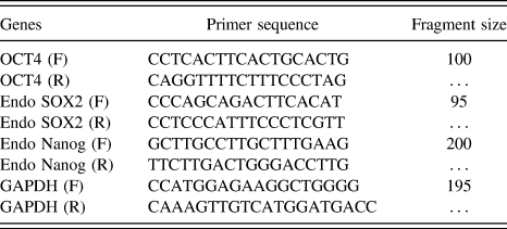
Gene expression studies were further carried out to confirm stem cell characters of the isolated cells. Total RNA was extracted from 5 × 10⁶ MSCs using TRIzol reagent (Invitrogen, Carlsbad, California) following the standard protocol. The isolated RNA was then treated with RNase-free DNase to remove contaminated DNA if any. Concentration and purity of RNA were determined using a nanodrop spectrophotometer (ND-1000, Nanodrop Technologies, Wilmington, Delaware). Approximately 1 μg of total RNA was used for cDNA synthesis using MMLV reverse transcriptase 1st strand cDNA synthesis kit following manufacturer’s protocol (Epicentre Biotechnologies, Madison, Wisconsin). Semiquantitative reverse transcriptase PCR (sqRT-PCR) was carried out with the gene-specific primers (Table I), which were designed to amplify a small region (100–600 bp) from the 3′ end of the respective genes. PCR amplification was performed on a DNA thermal cycler (Applied Biosystems, Foster City, California) using a typical reaction mixture of 50 µL. The reaction was carried out for a cycle of 94 °C for 2 min, followed by 30–32 cycles at 94 °C for 30 s, 55 °C for 30 s, and 72 °C for 30 s, followed by a final extension at 72 °C for 5 min. Each PCR product was run on 1% agarose gels, stained with ethidium bromide (10 µg/mL), and imaged on top of a UV light box (GEL DOC, BIORAD). For quantification, the pixel intensity of each band was determined using ImageJ (NIH Image, Bethesda, MD). The means (±standard deviations) of three replicates of RT-PCR values were presented as a relative intensity. GAPDH was used as an internal control for normalization of variations in reaction efficiency from sample to sample and the initial variations in sample concentration.
Primer sequences of targeted genes and their reaction profiles.

B. Cytocompatibility of MSCs in CA/media
CA (Merck, India) was dissolved in MilliQ water (resistivity 18.2 MΩ cm) to form a final concentration of 30 mM and adjusted to pH 7.4. Furthermore, CA was mixed with media in different ratios (CA/media; 0:100, 25:75, 50:50, 75:25, and 100:1). MSCs were trypsinized from culture flasks, centrifuged, and exposed to different CA/media for 10 min. The cells were then centrifuged, washed with PBS, seeded on coverslips, and cultured in media for 3 days. The actin and nuclei of the cells were stained using rhodamine-phalloidin (catalog no. R415, Invitrogen) and DAPI (4′,6-diamidino-2-phenylindole, catalog no. D1306, Invitrogen, Carlsbad, California). The cells were fixed, permeabilized using PBS containing 0.1% Triton X-100, stained using fluorescent dyes, and imaged using fluorescence microscopy (Zeiss Axiovision, Jena, Germany). To perform viability test, the cells were stained with live and dead assay kits (LIVE/DEAD Cell Viability Assays, Invitrogen Life Technologies, Carlsbad, California) following manufacturer’s protocol. The proliferation ability of the cells conditioned in different CA/media concentrations was assessed by proliferating cell nuclear antigen (PCNA; Thermo Scientific, Waltham, Massachusetts) staining kit using manufacturer’s instructions. Apoptotic cells were determined by the DeadEnd colorimetric terminal deoxynucleotidyltransferase-mediated dUTP nick-end labeling (TUNEL) system (Promega Corp., Madison, Wisconsin). Ten random microscopic regions of interest were evaluated per tissue slide for determining the number of apoptotic cells.
C. Production of living fiber
1. Gelation kinetics of chitosan in citrate/media
4 wt% Chitosan (MW 710,000; >90% deacetylated, Marine Chemicals, Cochin, India) stock solution was prepared similarly as provided in our previous studies.Reference Ghosh, Rameshbabu, Dogra and Dhara18–Reference Ghosh, Rameshbabu and Dhara20 Gelling behavior of chitosan in CA/media in the ratio of 25:75, pH 7.4 was evaluated using a rheometer (Malvern Instruments, Malvern, U.K.) with a measurement gap of 150 μm. A solvent trap was used to prevent evaporation of water during measurements. Gelation kinetics was evaluated by change in elastic and viscous modulus as a function of time at constant strain (0.01) and frequency (1 Hz), at 25 °C. Strain sweep was carried out on the formed chitosan gels at a constant frequency of 1 Hz to determine the linear viscoelastic region by shearing them until structure breakdown occurred.
2. Chemical characterization
Solid state 13C NMR spectra of the chitosan gels formed in the presence of CA/media were recorded at 500 MHz on a Bruker Advance II-500NMR spectrophotometer (Billerica, Massachusetts).
3. Preparation of crosslinked fibers in the cell-citrate bath
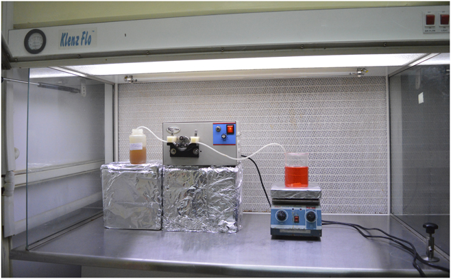
Chitosan solution was extruded using a single channel peristaltic pump (Riviera Glass Pvt. Ltd., Mumbai, Maharashtra, India) in a citrate coagulation bath containing 2 × 10⁶ cells/mL under sterile conditions as provided in Fig. 1 and movie (Supplementary Material). The fibers were retained in the CA/media for 10 min for completing the gelation process. The CA/media was then replaced with media and the cell-laden fibers were cultured in the incubator. The fibers were named living fibers. To examine the presence of cells on the fibers, MSCs were prelabeled with CellTracker™ Green CMFDA (5-chloromethyl fluorescein diacetate; Invitrogen, Carlsbad, California) following manufacturer’s protocol. The cells in the living fibers were imaged immediately using florescent microscopy. After that, the cells were cultured in complete media and were imaged again after 36 h. To investigate the maintenance of the cell phenotype, cells at 7th day of culture were released from fibers for morphology analysis using 0.25% trypsin–EDTA. The cell pellets were recovered after centrifugation, plated on lysine coated cover slips, and allowed to grow till confluence. The morphology of the cells was determined by florescent microscopy after staining them with rhodamine-phalloidin and DAPI.
Preparation of living fibers under a sterile condition, at physiological pH and room temperature.

D. Statistical analysis
All data in this study were expressed as the mean ± standard deviation. Statistical analysis was performed using t-test in OriginPro 8 software (Northampton, Massachusetts) for TUNEL assay. Significance was determined at P < 0.05.
III. RESULTS
A. MSC isolation and culture
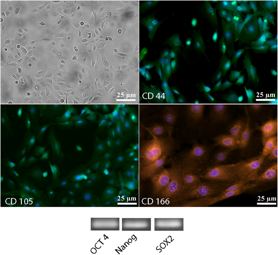
AM-MSC adhesion occurred within 24 h on tissue culture flasks. Initially, the cells were round in shape but eventually acquired a typical fibroblast-like morphology (Fig. 2). This is in agreement with earlier reports.Reference Díaz-Prado, Muiños-López, Hermida-Gómez, Rendal-Vázquez, Fuentes-Boquete, de Toro and Blanco21 Isolated AM-MSCs were characterized by immunofluorescence and reverse transcriptase PCR studies (Fig. 2). The cells expressed characteristic stem cell surface markers: CD44, CD105, and CD166. Furthermore, they expressed octamer-binding transcription factor (OCT)4, the homeobox transcription factor NANOG, and sex-determining region Y-box 2 (SOX2), the genes known to be required for self-renewal and pluripotency.Reference Niwa, Masui, Chambers, Smith and Miyazaki22,Reference Chambers, Colby and Robertson23
Characterization of isolated amniotic MSCs. the phase contrast image of MSCs on day 5 at passage 2; Immunofluorescence-based detection of expression of characteristic stem cell markers namely CD44, CD105, and CD166; sqRT-PCR-based analysis for RNA expression of self-renewal and pluripotency-related different genes like OCT4, Nanog, and SOX2 in the isolated amniotic MSCs.

B. Cytocompatibility of MSCs in CA/media
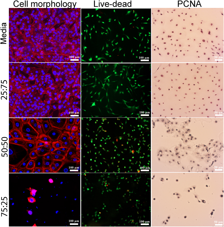
The adherence and morphology of the AM-MSCs conditioned with different CA/media ratios were investigated using rhodamine-phalloidin and DAPI staining (Fig. 3). The cells cultured in media and 25:75 CA/media had well spread actin filaments with round and prominent nucleus. Higher concentration of CA in media affected the shape of cytoskeleton considerably. After treating cells with 50:50 and 75:25 CA/media, change in the morphology of the cells was observed. The cells conditioned in 75:25 CA/media could not adhere well and were characterized by damaged cytoskeleton and deformed nuclei. The cells conditioned in 100% CA could not adhere and were excluded from the studies.
Viability of the amniotic MSCs after conditioning in different ratios of citric acid solution and media after 3 days. The morphology of the cells was investigated with rhodamine-phalloidin and DAPI assay. Cell viability was assessed by using a live–dead assay kit. Cell division was evaluated by PCNA staining. (CA, citric acid; the ratios indicate citric acid/media).

Viability assessment using live-dead staining kit indicated minimal cell death of the MSCs conditioned with CA/media concentrations of 0:100 (media only), 25:75, 50:50, and 75:25 (Fig. 3). However, the cells had proper morphology when cultured in media and 25:75 CA/media concentration. At higher CA concentrations, the cells were round in shape. PCNA staining of the cells treated with media and 25:75 CA/media demonstrated the presence of proliferating cells, indicating their normal progression through the cell cycle (Fig. 3). This established that 25:75 CA/media facilitated effective cell growth. Apparently from the microscopic studies, a lesser number of cells retained their proliferation potential when conditioned in 50:50 and 75:25 CA/media.
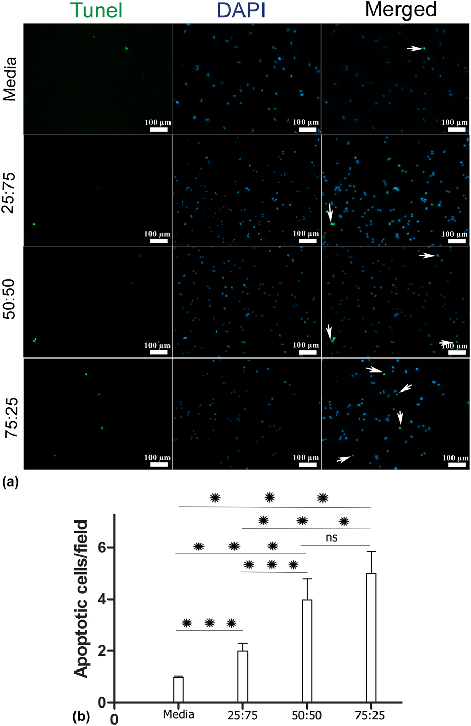
The change in the metabolism of the cells at different CA concentrations was examined with TUNEL assay [Figs. 4(a) and 4(b)]. At higher concentration of CA, more apoptotic cells were observed (marked with arrows). This was further confirmed by the quantitative data for the TUNEL assay. It is to be noted that the cells conditioned at 25:75 CA/media were used for living fiber formation.
(a) Representative images of apoptosis through the TUNEL assay (scale bar, 100 μm). (b) Quantitative data for the TUNEL assay. Values represent mean ± SD (n = 5). P < 0.05. (CA, citric acid; the ratios indicate CA/media; arrows represent apoptotic cells).

C. Production of living fiber
1. Gelation kinetics of chitosan in CA/media
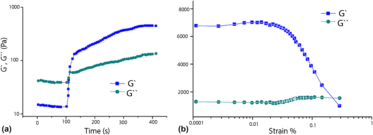
To evaluate gelation kinetics of chitosan solution in CA/media, the in situ cross-linking progression (on the rheometer) was monitored via dynamic time sweep analysis [Fig. 5(a)]. Initially, the viscous modulus (G″) was higher than the elastic modulus (G′) which signifies the solid state of the material. The addition of CA/media at 100 s increased the G′ sharply, indicating instantaneous gelation. However, the G′ obtained in the present study is significantly lower than the G′ obtained during gelation kinetics of chitosan with CA reported earlier.Reference Ghosh, Rameshbabu and Dhara20 This may be because of the lower viscosity of the polymer used and dilution of the cross-linker, CA with media leading to less availability of the citrate anions for gelation with glucosamine moieties of chitosan. Furthermore, strain sweep was measured to evaluate the breaking point of the gel at a constant frequency of 2 Hz [Fig. 5(b)]. Initially, G′ was independent of shear strain; however, after a critical strain value, the G′ started decreasing dramatically, marking the transition from a linear to the nonlinear range.
Rheological assessment of chitosan with CA/media in the ratio 25:75 at pH 7.4, 25 °C. (a) G′ and G″ as a function of time at constant strain and frequency of 0.01 and 1 Hz, respectively. (b) G′ and G″ of citrate–chitosan gel on strain sweep. G′, elastic modulus; G″, viscous modulus.

2. Chemical characterization
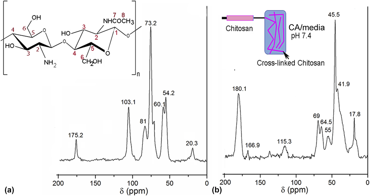
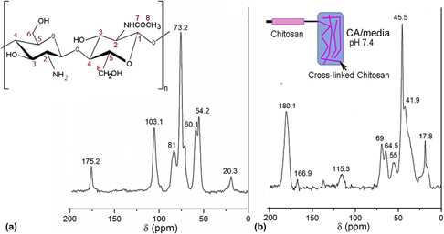
The NMR spectra of chitosan and cross-linked gel with CA/media are shown in Fig. 6. Chitosan showed four characteristic peaks between 60.1 and 103.1 ppm [Fig. 6(a)] due to the presence of carbon atoms (C1, C3, C4, C5, and C6) in the six-membered rings. The peaks at 54.2 and 20.3 ppm are associated with the carbon atom (C2) attached to primary amine group and the methyl carbon (i.e., C8), respectively. The peak at 175.2 ppm is assigned to carbonyl carbon (i.e., C7) of the amide group [Fig. 6(a)]. In case of cross-linked fibers, all the characteristic peaks of chitosan are present, but the peaks of amide carbon shifted to 166.9 ppm [Fig. 6(b)]. The new peaks at 180.1 and 41.9 ppm could be attributed to the presence of carbonyl carbon of the carboxylate group and the –CH2 group of the CA. The presence of these additional peaks confirms the cross-linking between the glucosamine moiety of chitosan and the citrate ions.
13C NMR spectra of (a) chitosan and (b) the product obtained via cross-linking of chitosan with CA/media in the ratio of 25:75, pH 7.4 (CA, citric acid).

3. Production of cross-linked fibers in cell-citrate bath
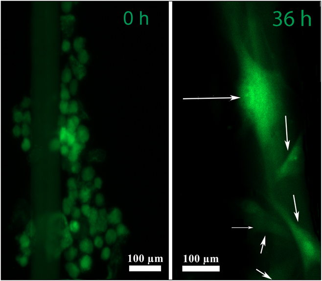
Among natural polymers, chitosan has been extensively used for fiber fabrication because of its intrinsic shear thinning behavior.Reference Terada, Kobayashi, Zhang, Tiwari, Yoshikawa and Hanagata24 This property enables the polymer to be extruded through narrow needles for fiber production under desired bath conditions. In this study, chitosan solution was extruded in CA/media bath containing cells to produce cell-laden fibers. To study cell adhesion on the fibers, the MSCs were prelabeled with a cell tracker. It was observed that the cells were round in shape immediately after the formation of the fibers (Fig. 7). After 36 h, the cells on the fibers assumed well spread morphology.
Living fiber formation. The cells were labeled with cell tracker and visualized immediately and after 36 h of fabrication of living fibers. The arrows represent cells in the fibers.

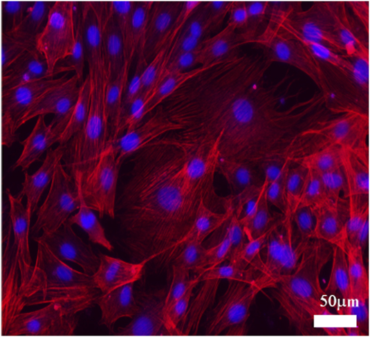
The cells’ phenotype might also be affected during generation of cell-laden fibers and prolonged cell culture.Reference Murphy and Sambanis25 To address the question, the amniotic MSCs were retrieved from the fibers after 7 days and plated on lysine-coated coverslips. The cells were able to attach and demonstrated the normal morphology in 2D (Fig. 8). Thus, the cell functionality was not compromised during the formation of cell-laden fibers.
AM-MSCs retrieved from fibers after 7 days plated on cover-slips. Fluorescent imaging by rhodamine-phalloidin and DAPI staining shows cell–cell contact and retention of cell morphology.

IV. DISCUSSION
Chitosan [poly-(β-1/4)-2-amino-2-deoxy-D-glucopyranose] is a linear cationic polysaccharide obtained from chitin, found in the shells of shrimp, lobsters, and crabs.Reference Maciel, Yoshida and Franco26 It is a copolymer made of D-glucosamine and N-acetyl-D-glucosamine bonds, in which glucosamine is the predominant repeating unit in its structure. It is biocompatible, biodegradable, bioactive, nontoxic, and nonimmunogenic, with antibacterial capability.Reference Oryan and Sahvieh27,Reference Manivasagan and Oh28 Therefore, this natural polysaccharide has widely been used as a wound dressing and in bone and cartilage regeneration.Reference Jiang, Deng, James, Nair and Laurencin29 However, chitosan only dissolves in acidic condition as the protonation of amino groups of chitosan occurs in acidic solution (pK a value ∼6.5–6.8); which compromises cell viability significantly. Mihaila et al., reported the addition of weak base to chitosan solution to increase the pH value of chitosan without precipitation till the pK a value for forming cell-laden fibers.Reference Mihaila, Popa, Reis, Marques and Gomes30 However, with an increase in the pH of chitosan, deionization of amino groups occurred which weakened the binding affinity to the oppositely charged ions, deteriorating the mechanical stability of the fibers.
To circumvent the problem, instead of increasing the pH of chitosan solution, the polymer in this study was extruded in CA/media bath, pH 7.4 to produce fibers. Prior to that, the morphology of the cells conditioned in different concentrations of CA/media was evaluated by rhodamine-DAPI am staining. It was found that the cells exhibited a favorable phenotype with well-spread actin filaments when exposed in 25:75 CA/media, by contrast, the cells showed the irregular morphology when conditioned in higher concentrations of CA. Also, concentrations of CA/media higher than 25:75 were detrimental to the viability of the cells as revealed by live–dead and PCNA staining. Thus, CA/media in the ratio of 25:75 were found to be optimal for cell adhesion and viability and could be a potential coagulation bath for production of cell-laden fibers. Consequently, we performed rheological investigations of chitosan solution with CA/media (25:75) to assess fiber forming ability. Rheological data revealed that elastic modulus of chitosan solution increased immediately upon addition of CA/media (25:75) resulting in the formation of a chitosan gel. NMR studies of the chitosan gel confirmed the cross-linking between the glucosamine moiety of chitosan and the citrate ions. The instantaneous gel-forming ability of chitosan in CA/media was successfully used for fiber production by extruding chitosan solution at a constant speed in 25:75 CA/media, pH 7.4 containing an adequate quantity of cells at room temperature in a sterile condition. The cells were prelabeled using a cell tracker and their attachment to the fiber immediately after fabrication was confirmed by microscopy. The viability of the cells immediately after the fiber fabrication process provides evidence of the formation of living fibers.
This approach can provide multiple flexibilities in terms of gelation layer thickness, the diameter of the fibers, mechanical strength, and amendable biodegradability that could be varied by adjusting the viscosity of the polymer solution, the concentration of the cross-linker (i.e., CA/media), and the inner diameter of the tip used in extrusion. Although in the present study, AM-MSCs were used for the production of living fibers, other cell types can also be used and standardized according to the end applications. The accomplishment in the fabrication of living fibers indicates the possibility toward biofabrication of living structures via bioprinting to mimic topology, architecture, and functionality of the desired tissues that will enable clinical solutions for tissue engineering applications. Our future studies will investigate the production of 3D scaffolds of appropriate mechanical properties using the living fibers for tissue engineering applications.
V. CONCLUSION
The present study explored various concentrations of CA/media ratio permissible for the viability of the mesenchymal stem cells (MSCs). The optimum concentration of CA/media that was nontoxic to cells was selected and used to evaluate gelation kinetics with chitosan solution. Subsequently, chitosan solution was extruded in CA/media containing cells at benign conditions for production of the living fibers. The MSCs were shown to be distributed along the fibers. The living fibers can be used to create viable 3D building blocks for tissue engineering applications.
Supplementary Material
To view supplementary material for this article, please visit https://doi.org/10.1557/jmr.2018.143.
ACKNOWLEDGMENTS
Fellowship from Council of Scientific and Industrial Research (CSIR) and Department of Science and Technology (DST), Govt. of India to P.G. and A.P.R. is acknowledged. Financial support from CSIR, DST, Department of Biotechnology, and Defense Research and Development Organization, (Govt. of India) are acknowledged. Dr. Asit Baran Panda is acknowledged for his help in NMR studies.