Pulseless cardiac arrest is typically defined as the cessation of cardiac mechanical activity, determined by unresponsiveness, apneoa, and the absence of a palpable central pulse. Such pulseless arrest accounts for around one-twentieth of admissions to paediatric intensive care units, be they medical or exclusively cardiac.Reference Slonim, Patel, Ruttimann and Pollack1–Reference Suominen, Palo, Sairanen, Olkkola and Rasanen3 Though the incidence of cardiac arrest is higher in children admitted to a cardiac as opposed to a paediatric intensive care unit, the outcome of these patients is better, with just over two-fifths surviving when treated in the cardiac intensive care unit, versus between one-sixth and one-quarter of those admitted to paediatric intensive care units. Possible explanations for the disparity in rates of survival rates are that the aetiologies of the arrest, along with the site of arrest and the availability of better monitoring and easy access to interventions and management subsequent to the arrest, are inherently different.
In a large retrospective national registry of cardiac arrest occurring in hospital, children who received chest compressions for bradycardia with pulses had a significantly higher rate of survival to discharge, at 60%, than did those presenting with pulseless cardiac arrest, with only 27% surviving to discharge.Reference Nadkarni, Larkin and Peberdy4 This suggests that early resuscitation before the patient becomes pulseless, along with early recognition and intervention, are likely to improve outcomes.
Cardiac arrest is the end-result of diverse aetiologies and pathophysiologic mechanisms, ultimately leading to electrical or mechanical arrest due to progressive hypoxic-ischaemic events or metabolic disturbances. The child without cardiac disease typically presents as a result of global asphyxia secondary to evolving respiratory failure. The patient with cardiac problems, in contrast, may require resuscitation due to a persistent low cardiac output state, but is also likely to experience an acute decompensation, such as a respiratory event, an arrhythmia, a vagal episode, occlusion of an aortopulmonary shunt, pulmonary hypertensive crisis, or a coronary arterial event.
Recently published reports of in-hospital cardiac arrests in children can be derived from the multi-centric National Registry of Cardiopulmonary Resuscitation provided by the American Heart Association.Reference Nadkarni, Larkin and Peberdy4 This multi-centric observational registry characterized the process and outcome of cardiopulmonary resuscitation of children in hospital. The population is heterogeneous, but in a recent summary, most arrests occurred in children with progressive respiratory insufficiency and/or progressive circulatory shock. Before the arrest, almost three-fifths of these children were mechanically ventilated, just under two-fifths were receiving continuous infusions of vasopressors, and three-tenths had continuous invasive monitoring of arterial blood pressure. Medical and surgical cardiac illness accounted for just over one-third of the 880 events. Few achieved complete recovery. Just over half attained sustained return of spontaneous circulation, one-third survived for 24 hours, and only just over one-quarter survived to be discharged from hospital. Of the survivors, two-thirds had good neurological outcomes.Reference Nadkarni, Larkin and Peberdy4 These statistics excluded one-fifth of the patients reported to the database who received chest compressions whilst retaining pulses.
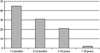
During the past 4 years at the Children’s Hospital of Philadelphia, 3.1% of the average 1000 annual admissions to the cardiac intensive care unit have received cardiopulmonary resuscitation while in the unit.5 A similar incidence of cardiac arrest is also observed in the cardiac intensive care unit at Children’s Hospital, Boston.6 Infants less than 2 months of age represented 45% of these patients (Fig. 1). Overall survival of those receiving cardiopulmonary resuscitation was 46%. Survival was better for those receiving cardiopulmonary resuscitation after cardiac surgery, at 53%, compared with survival of 33% for pre-operative or non-surgical patients undergoing resuscitation. The latter group included patients not felt to be candidates for either surgical intervention or mechanical circulatory support. Clearly there is room for improvement in outcomes from cardiac resuscitation in children with cardiac disease. In this review, therefore, we summarize the newest developments in paediatric resuscitation, with an expanded focus upon the unique challenges and importance of anticipatory care in infants and children with cardiac disease.
Ages of patients receiving cardiopulmonary resuscitation during their admission to the cardiac intensive care unit at Children’s Hospital of Philadelphia from January, 2003, through April, 2007.

Phases of cardiac arrest and cardiopulmonary resuscitation
Interventions to improve outcomes for all children suffering cardiac arrest should be targeted to optimize therapies according to the aetiology, timing, duration, intensity and phase of resuscitation. There are at least four phases of cardiac arrest, namely the phase prior to the arrest, the phase of absent flow, or cardiac arrest without compressions, the phase of low flow with compressions, and the phase of recovery subsequent to resuscitation.
The pre-arrest phase
The phase prior to the arrest, offering the opportunity for anticipation and protection, represents the greatest opportunity to improve survival by preventing a pulseless arrest. Interventions during this phase focus on prevention. Suitable anticipation and prevention of cardiac arrest, focusing on the mobilization of the medical response teams to address rapid restoration of oxygenation, ventilation, and circulation, should be anticipated to improve outcomes. This is particularly important for patients with known cardiac problems. Those exhibiting the pre-arrest phase may present in low cardiac output syndrome following intra-operative myocardial ischaemia, arrhythmias following cardiac surgery, or in the context of myocarditis or dilated cardiomyopathy, pulmonary hypertensive crisis with right ventricular failure, or inadequate flow of blood to the lungs during occlusion of a shunt, cyanotic spells, or ductal closure. Early and thoughtful circulatory and respiratory support can most commonly prevent those in this phase proceeding to cardiac arrest. Close surveillance, with anticipatory intervention, are the keys to successful outcomes in these patients.
Low cardiac output syndrome
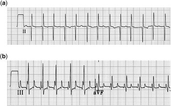
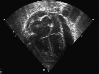
The post-operative syndrome of low cardiac output is well described. Following cardiopulmonary bypass and aortic cross clamping, neonates and infants have a predictable decrease in cardiac index over the first 6 to 18 hours after removal of the aortic cross clamp.Reference Wernovsky, Wypij and Jonas7 In the worst case, myocardial dysfunction can progress to hypotension, inadequate coronary arterial perfusion, and cardiac arrest. The best outcomes occur with early recognition, monitoring, pharmaceutical intervention, or mechanical circulatory support to avoid the progression to pulseless cardiac arrest. If the patient has an intracardiac shunt, thermodilution is not accurate for measuring cardiac output. Clinical surrogates include trends in vital signs and physical examinations, erosion of an excess of blood base, levels of lactate in the serum, transcutaneous mixed saturations of oxygen as measured by near-infrared spectroscopy, and invasive monitoring of arterial blood and central venous pressures. Patients with functionally univentricular physiology may demonstrate systemic hypoxaemia due to inadequate delivery of oxygen, resulting in inadequate mixing and low mixed venous saturations. Patients with low cardiac output syndrome must be evaluated for arrhythmia (Fig. 2), pericardial effusion (Fig. 3), residual anatomical lesions, and in those with functionally univentricular physiology an excessive ratio of pulmonary to systemic flows. Milrinone has been shown to reduce the incidence of low cardiac output syndrome in infants and children following biventricular repairs.Reference Hoffman, Wernovsky and Atz8 Positive pressure ventilation will help reduce the afterload on the systemic ventricle. Initiation of positive pressure ventilation, however, may precipitate a cardiac arrest in a patient with pericardial tamponade, pulmonary hypertension with right ventricular failure, or post-operative right ventricular dysfunction such as after a neonatal repair of Tetralogy of Fallot.
Use of temporary epicardial atrial pacing wires to diagnose atrial flutter. (a) Surface electrocardiogram where p waves are difficult to visualize. (b) Temporary epicardial pacing wires demonstrating atrial flutter.

Echocardiographic demonstration of a significant pericardial effusion.

Arrhythmia
Symptomatic post-operative arrhythmias include complete heart block, particularly in the patients with functionally univentricular circulations, and rapid junctional ectopic tachycardia. Symptomatic complete heart block is most effectively treated with both atrial and ventricular temporary epicardial pacing wires, permitting preservation of atrial-ventricular synchrony. In the absence of atrial pacing wires, a transoesophageal lead will usually sense the atrium. In our experience, post-operative complete heart block in patients with hypoplastic left heart syndrome carries a 67% mortality,9 as it most likely represents myocardial metabolic derangements or iscahemia, and there should be a low threshold for initiating extracorporeal membrane oxygenation in this setting.
Junctional ectopic tachycardia can be associated with complete heart block. When symptomatic, the tachycardia may be slowed by avoiding hyperthermia, minimizing inotropic agents, and by cautious administration of antiarrhythmic drugs, including amiodarone or procainamide. In the best case, atrial overdrive pacing will regain atrial-ventricular synchrony; whereas in the worst case, extracorporeal membrane oxygenation may be necessary.
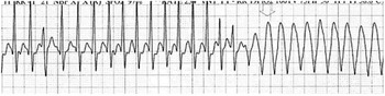
Patients with acute compromise to coronary arterial flow due to air embolism, mechanical or anatomic compromise, and/or inadequate perfusion pressure, can present with ventricular arrhythmia or ventricular fibrillation (Fig. 4). For these patients, electric cardioversion or defibrillation, phenylephrine and lidocaine are often a successful combination, followed immediately by diagnostic evaluation. The Guidelines for Pediatric Advanced Life Support provided by the American Heart Association recommend the use of epinephrine in this setting. Epinephrine, however, may precipitate further arrhythmia. The guidelines note that there is insufficient data to recommend for or against the use of vasopressin in these circumstances, but it may increase the pressure of coronary arterial perfusion.
Electrocardiogram of an infant undergoing attempted decannulation from extracorporeal membrane oxygenation following surgical palliation for an anomalous left coronary artery from the pulmonary trunk. The trace is consistent with inadequate coronary arterial perfusion, and the patient remained on support for several more days before successful separation from bypass.

Acute fulminant myocarditis
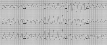
This disease is characterized by an abnormal electrocardiogram (Fig. 5), arrhythmia, a normal cardiac silhouette on the chest radiograph, and normal left ventricular dimensions on echocardiography. Affected patients progress rapidly to cardiac shock. Pharmaceutical interventions should be employed whilst activating the team required to establish extracorporeal membrane oxygenation. Amiodarone in this setting often precipitates cardiac arrest. Although positive pressure ventilation reduces the systemic ventricular afterload, the process of endotracheal intubation may precipitate a cardiac arrest, and should ideally be performed with extracorporeal membrane oxygenation standby.
Electrocardiogram of a patient with acute fulminant myocarditis.

Pulmonary hypertensive crisis
Patients with persistent pulmonary hypertension and associated right ventricular failure are at high risk for cardiac arrest. The presence of an intra-atrial communication may help maintain systemic cardiac output at the expense of hypoxaemia. The right ventricle should be supported by maintaining preload, and providing additional inotropic support, such as with dopamine. Isoproterenol is an old, but often effective, agent with which to manage an acute pulmonary hypertensive crisis because of its pulmonary vasodilative and chronotropic properties. Bradycardia or ventricular arrhythmias are ominous signs of impending arrest. Positive pressure ventilation can result in unacceptable right ventricular afterload and resultant cardiac arrest. Delivering a high concentration of inspired oxygen, inducing an acute metabolic alkalosis with sodium bicarbonate, or a respiratory alkalosis with hyperventilation, and inducing pulmonary vasodilation with inspired nitric oxide remain the mainstays of resuscitation. Heart-lung transplantation subsequent to extracorporeal membrane oxygenation is nearly uniformly fatal,Reference Kirshbom, Bridges, Myung, Gaynor, Clark and Spray10 thus mechanical support is not a beneficial option during cardiovascular collapse.
Inadequate flow of blood to the lungs
Hypoxemia due to inadequate pulmonary blood flow can occur in the pre-operative state with premature ductal closure or a cyanotic spell, or in the post-operative state with thrombosis of a shunt. Premature ductal closure can occur prior to post-natal diagnosis or if there is an interruption to the delivery of prostaglandins. Re-initiation of prostaglandins is almost uniformly therapeutic. In the current era, routine repair of tetralogy of Fallot occurs within the first six months of life, and cyanotic spells have become a rare occurrence. Should such spells occur, treatment includes provision of oxygen, calming or sedation, increasing intravascular volume, correction of acidosis, avoiding systemic vasodilation, and increasing systemic vascular resistance with phenylephrine.
Persistent hypoxaemia in the shunted patient with a functionally univentricular heart raises the spectre of evolving thrombosis of the shunt. If suspected, the treatment includes systemic heparinization to prevent ongoing thrombosis, increasing systemic vascular resistance with boluses of fluid and phenylephrine to promote increased antegrade flow across the shunt, and an emergent cardiac catheterization for definitive diagnosis and intervention. Over a period of 4 years, from 2003 through 2006, at the Children’s Hospital of Philadelphia, transcatheter or surgical intervention was needed in 30 of 324 patients with flow of blood to the lungs dependent upon a shunt. Although many interventions were undertaken on an urgent or emergent basis, only 5 shunts occluded acutely requiring cardiopulmonary resuscitation and or extracorporeal membrane oxygenation for rapid resuscitation.11 The use of extracorporeal oxygenation to maintain cardio-respiratory function during a cardiac arrest in a patient with a functionally univentricular heart and acute obstruction of a shunt, who fails to respond to conventional techniques of resuscitation, is associated with a high rate of survival.Reference Allan, Thiagarajan, del Nido, Roth, Almodovar and Laussen12
Respiratory support
Respiratory insufficiency is a common precipitating event for cardiac arrests in children. The goal of assisted ventilation is to ensure adequate delivery of oxygen to meet metabolic demand and removal of carbon dioxide. Effective bag-mask ventilation remains the cornerstone of providing effective emergency ventilation. An advanced airway can minimize both aspiration of gastric contents into the airway, and respiratory embarrassment due to gastric distention during bag-mask ventilation. Initiation of positive pressure ventilation can be the inciting event of cardiopulmonary arrest in tenuous patients with cardiac problems. Patients with right ventricular dysfunction can develop acute right heart failure due to increases in right ventricular afterload from positive pressure ventilation. Increased intrathoracic pressure and increased lung volumes with positive pressure ventilation can further exacerbate the physiologic derangements in cardiac tamponade. A spontaneously breathing patient with acute heart failure can increase minute ventilation to compensate for metabolic acidosis. Mechanical ventilation must match the inherent minute ventilation to avoid worsening acidosis leading to further haemodynamic compromise and resultant arrest.
Open sternum
The option exists to reopen the sternum in patients after cardiac surgery. Clear indications would include pericardial tamponade and unstable haemodynamics. During periods of low cardiac output syndrome, oxygenation and blood pressure can improve by opening the sternum. Cannulation for extracorporeal membrane oxygenation can be achieved percutaneously, usually through the vessels on the right side of the neck, or transthoracically. Effective chest compressions in the neonate are more difficult with an open sternum.
Mechanical circulatory support
Extracorporeal membrane oxygenation is commonly used for circulatory support in children with refractory low cardiac output after cardiac surgery, persistent hypoxaemia, arrhythmias, cardiac arrest, or failure to wean from cardiopulmonary bypass.11–Reference Delmo Walter, Stiller and Hetzer17 Some centres report using the technique to support neonates early after surgical palliation for hypoplastic left heart syndrome to avoid the hypoxaemia and potential cardiac arrest during the low cardiac output syndrome.Reference Ungerleider, Shen and Yeh18 Survival to hospital discharge in the data for 2004 reported to the Extracorporeal Life Support Organization was 41%, albeit that outcomes for individual centers show considerable variability because the populations treated differ in anatomic diagnosis, surgical procedures, and indications for extracorporeal membrane oxygenation.Reference Kolovos, Bratton and Moler14 In particular, survival to hospital discharge for shunted patients with functionally univentricular circulations supported with extracorporeal membrane oxygenation ranged from 39 to 64%.9, Reference Allan, Thiagarajan, del Nido, Roth, Almodovar and Laussen12, Reference Ravishankar, Dominguez and Kreutzer20 Even higher survival discharge from hospital, at 81%, is reported in those patients when they were supported for hypoxaemia,11 at 64% when extracorporeal membrane oxygenation was initiated greater than 24 hours after surgery,Reference Aharon, Drinkwater and Churchwell19, Reference Ravishankar, Dominguez and Kreutzer20 and at 80% when supported for acute thrombosis of a shunt.Reference Ravishankar, Dominguez and Kreutzer20
Patients with failing dilated cardiomyopathy or fulminant myocarditis may also benefit from institution of mechanical circulatory support prior to an arrest. The introduction of the Berlin HeartReference Hetzer, Potapov and Stiller21, Reference Blume, Naftel, Bastardi, Duncan, Kirklin and Webber22 now enables the provision of left ventricular assistance in patients as small as 10 kg.
The phase of no-flow
Interventions during the pulseless phase of untreated cardiac arrest focus on early recognition of the arrest, and the initiation of basic and advanced life support. In a critically ill patient, when the invasive arterial trace dampens or the blood pressure cuff fails to register a blood pressure, it is necessary to prove there is not a problem with the patient before focussing on inadequacies in the monitoring. The traditional “ABC” teaching of cardiopulmonary resuscitation is based on evaluating and managing the airway, the breathing, and the circulation. In children with known cardiac problems, the circulation should be considered as the more common aetiology, and the clinician should be asking himself or herself several important questions:
• Is there a pulseless arrhythmia for which defibrillation is the initial intervention?
• Is an intervention needed simultaneously with the establishment of a secure airway, such as drainage of pericardial tamponade, or management of acute occlusion of a systemic-to-pulmonary arterial shunt?
• Should epicardial or transcutaneous pacing be initiated?
• Should the extracorporeal membrane oxygenation team be activated?
It goes without saying that it is also essential to secure the airway so as to provide effective oxygenation and ventilation. Effective compression of the chest is also needed whenever there is insufficient delivery of oxygen to the brain or heart.
The phase of low flow
Circulation, oxygenation, and ventilation
The goal during this resuscitative phase is to achieve effective compressions of the chest so as to optimize the pressures of coronary arterial perfusion and the flow of blood to critical organs. Continuous effective chest compressions are generally the best way to provide circulation during cardiac arrest. The current recommendations are to “push hard” and “push fast”. Because the flow of blood will stop without chest compressions, it is important to minimize their interruption. Full recoil should be permitted, and over-ventilation avoided in order to allow adequate venous return during the decompression phase of external cardiac massage. Children in the early post-operative phase after cardiac surgery can present additional challenges for resuscitation. Aggressive compressions of the chest can result in disruption of fresh suture lines and haemorrhage, resulting in significant neurological injury. An open sternum may provide access for direct cardiac massage in an older patient, but clearly limits effective compressions in a neonate. Hepatic compressions, or a combination of hepatic and chest compressions, may help cardiac output in some patients.
Higher pressures of coronary arterial perfusion, improved values of exhaled carbon dioxide, and good cardiac output during the phase of cardiopulmonary resuscitation associated with low flow are all consistently associated with an improved return of spontaneous circulation, and improved short- and long-term outcomes. Patients with congenitally malformed hearts may pose considerable limitations for compression of the chest so as to maintain an effective cardiac output and delivery of oxygen during resuscitation. This is particularly true in patients with regurgitation across the arterial or atriventricular valves, patients with a shunted functionally univentricular circulation, or those with a cavo-pulmonary connection. In this latter group, effective flow of blood to the lungs is difficult to maintain during chest compressions, thereby affecting oxygenation and preload to the systemic ventricle, and the elevated central venous pressure further compromises cerebral blood flow, contributing to a high incidence of neurological injury.Reference Booth, Roth, Thiagarajan, Almodovar, delNido and Laussen23
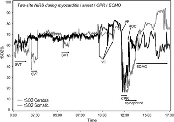
In the absence of an intracardiac shunt, monitoring of end-tidal values of carbon dioxide can be reliable in judging the effectiveness of cardiac output generated by cardiopulmonary resuscitation. In patients with intracardiac shunting, pulmonary hypertension, or flow of blood to the lungs through a shunt, such values may be less reliable. Invasive monitoring of arterial blood pressure, transcutaneous estimation of saturations of oxygen, and intermittent point-of-care measurement of arterial blood gases are often a more accurate assessment of effective cardiopulmonary resuscitation. Assessment of delivery of oxygen to the organs during all phases of resuscitation may be possible with non-invasive near-infrared spectroscopy. Current implementations of this technique permit continuous monitoring of regional venous-weighted saturations of oxyhaemoglobin in a tissue volume about two centimetres below the site of the probe, and can be used to assess both cerebral and somatic organ oxygenation.Reference Hoffman, Stuth and Jaquiss24–Reference Hoffman26 The signal obtained is related to the regional relationship between supply and demand of oxygen. This can help guide the vigour of resuscitation, administration of vasoactive medications, specific manipulation of cerebrovascular resistance and perfusion pressures, and the decision to institute extracorporeal circulatory support. At the Children’s Hospital of Wisconsin, near-infrared spectroscopy is routinely used to monitor critically ill patients (Fig. 6).
The near infrared spectroscopy tracings (cerebral, forehead; and somatic, flank) from a patient with acute myocarditis. A low cardiac output state was characterized by a pattern of circulation with somatic rSO2 less than the cerebral rSO2. Episodes of supraventricular tachycardia (SVT), ventricular tachycardia (VT) and ventricular fibrillation (VF) are noted. Circulatory arrest requiring resuscitation was marked by a profound drop in both cerebral and somatic oxygenation. The patient achieved return of circulation (ROC) before being placed on extracorporeal membrane oxygenation (ECMO). End-organ function was maintained and the patient achieved a complete recovery.

During cardiopulmonary resuscitation, cardiac output and pulmonary blood flow are between one-tenth and one-quarter of the values achieved during normal sinus rhythm. Consequently, much less ventilation is necessary for adequate exchange of gases from the blood traversing the pulmonary circulation. Experimental and clinical data from experience in adults indicate that over-ventilation during cardiopulmonary resuscitation is common, and can substantially compromise venous return and cardiac output. Over-ventilation and increased intrathoracic pressure can significantly compromise pulmonary arterial flow in the patient with a functionally univentricular heart palliated with a cavopulmonary anastomosis or systemic-pulmonary shunt.
Extracorporeal membrane oxygenation
Several series and retrospective reviews have reported the use of extracorporeal membrane oxygenation as a rescue therapy for children suffering cardiac arrest.Reference de Mos, van Litsenburg, McCrindle, Bohn and Parshuram2, Reference Allan, Thiagarajan, del Nido, Roth, Almodovar and Laussen12, Reference Kolovos, Bratton and Moler14 In the experience at the Children’s Hospital of Philadelphia and the Children’s Hospital Boston, approximately half of patients suffering cardiac arrest on the cardiac intensive care unit require extracorporeal membrane oxygenation to re-establish circulation during cardiopulmonary resuscitation. At the Children’s Hospital of Philadelphia, following the first stage of palliation for hypoplastic left heart syndrome over the period 2002 to 2006, 33 of 269 patients (13%) required cardiopulmonary resuscitation, and of these patients, 21 (63%) were stabilized with extracorporeal membrane oxygenation. Outcome for these patients depends primarily upon the aetiology of the arrest, with early survival reported to vary between 35% and 55%.11–Reference Kolovos, Bratton and Moler14, Reference Ravishankar, Dominguez and Kreutzer20 Extracorporeal membrane oxygenation during cardiopulmonary resuscitation, therefore, is a potentially life-saving intervention in an otherwise fatal situation.
Pharmacologic interventions
A complete review of cardiovascular medications are beyond the scope of this paper, but have been discussed elsewhere.Reference Tabbutt, Helfaer and Nicols27 Although animal studies indicate that epinephrine can improve initial resuscitation success after cardiac arrests, no single medication has been shown to improve survival in children. There are, nonetheless, some special considerations in patients with cardiac disease when first-line drug therapy may differ from standard guidelines for advanced life support in children.
Neonates have reduced intracellular stores of calcium, and are particularly responsive to administration of calcium, particularly if suffering the post-operative low cardiac output syndrome. Immediate improvement in myocardial function can be observed by echocardiography during administration of calcium, with a simultaneous increase in blood pressure. Rapid intracardiac delivery can result in reflex bradycardia. This differs from children with non-cardiac problems treated in intensive care, where multiple doses of calcium during cardiac arrest was independently associated with hospital mortality.Reference de Mos, van Litsenburg, McCrindle, Bohn and Parshuram2
Patients with progressive hypotension due to low cardiac output syndrome or myocardial dysfunction will likely benefit from judicious administration of epinephrine. The α-adrenergic action of this agent increases systemic vascular resistance, increasing diastolic blood pressure, which in turn increases the pressure of coronary arterial and blood flow, and increases the likelihood of return of spontaneous circulation. Epinephrine also increases cerebral blood flow during cardiopulmonary resuscitation because peripheral vasoconstriction directs a greater proportion of flow to the cerebral circulation. The β-adrenergic effect increases myocardial contractility and heart rate, and relaxes smooth muscle in the skeletal muscle vascular bed and bronchuses. Epinephrine also increases the vigour and intensity of ventricular fibrillation, increasing the likelihood of successful defibrillation. Epinephrine, however, can cause a compromised myocardium to fibrillate, so titration to effect with small doses is particularly important, especially in patients whose central venous access results in intracardiac delivery. Although epinephrine may be life-saving, special consideration should be give to using multiple or large doses in patients with shunted functionally univentricular physiology. In such patients, where systemic blood flow is dependent on the balance of systemic and pulmonary vascular resistances, the increased systemic vascular resistance that accompanies the administration of epinephrine may decrease systemic perfusion and increase pulmonary blood flow, resulting in inadequate tissue perfusion. In these patients, we consider the absence of response to 1 or 2 doses of epinephrine to be an indication to support the circulation with extracorporeal membrane oxygenation. While the circuit is being prepared, systemic blood flow should be maintained with good chest compressions rather than repeated administration of intravenous epinephrine. Vasopressin has also been reported to be useful in low cardiac output states associated with sepsis syndrome and organ recovery in children. While vasopressin will not likely replace epinephrine as a first line agent in children suffering cardiac arrest, there is preliminary data to suggest that its use in conjunction with epinephrine deserves further investigation in this setting.
Phenylephrine is very helpful in restoring coronary arterial perfusion in patients with adequate cardiac function and low systemic blood pressure. Although phenylephrine is not recommended in the guidelines for Pediatric Advanced Life Support as provided by the American Heart Association, common scenes for its use include patients with hypertrophic cardiomyopathy, William’s syndrome, aortic stenosis, or systemic vasodilation due to sedative or analgesic medications. Phenylephrine has no beta-1 activity and is therefore less likely to initiate ventricular arrhythmias.
Patients with pulmonary hypertension and relative bradycardia respond well to isoproterenol. Similarly, in post-operative patients with sinus nodal dysfunction or subsequent to transplantation, isoproterenol is an alternative to atrial pacing.
The compromised myocardium is sensitive to acidosis. Arrhythmias are common, myocardial function is worsened and pacing thresholds are increased. Early correction of acidosis with appropriate ventilation, effective chest compressions and base replacement may help return of spontaneous circulation.
Ventricular arrhythmias may be primary with reasonable underlying myocardial function or may reflect a myocardium compromised either in the post-operative period, during end-stage cardiomyopathy or with fulminate myocarditis. Lidocaine is generally the first line medication. More recently, amiodarone has been introduced. In the absence of significant ventricular dysfunction, amiodarone is low risk. There are no published comparisons of anti-arrhythmic medications for refractory ventricular fibrillation in children. Adult experience with amiodarone cannot be extrapolated to infants and children, as the mechanism of ventricular fibrillation in children is rarely local myocardial ischaemia, as it is in adults. In children, ventricular arrhythmias arise from a global myocardial dysfunction, and rapid administration of amiodarone to compromised myocardium in the pre-arrest phase often results in cardiac arrest. There is inadequate data on administration of amiodarone during cardiac arrest to comment. Administration of anti-arrhythmic medications should not delay defibrillation for a patient with ventricular fibrillation. After unsuccessful attempts at electrical defibrillation, medications to increase the effectiveness of defibrillation should be administered, with the first drug being epinephrine. If epinephrine with or without vasopressin and a subsequent repeat attempt to defibrillate are unsuccessful, the anti-arrhythmic agents amiodarone or lidocaine should be considered. Once defibrillated, the use of epinephrine with an irritable myocardium may promote further episodes of ventricular fibrillation.
Cerebral protection
Compromised neurological outcome is an unfortunate complication of extended cardiopulmonary resuscitation. Animal models demonstrate that cerebral hypothermia during and following warm ischaemic arrest can improve neurologic outcome. Many current practices for cerebral protection are derived from experience in the cardiac operating room. Application of ice to the head, particularly in neonates, may be preventive, but has not been studied. Conditions that optimize cerebral flow of blood, such as optimizing the pH and determining the preferred pharmacologic manipulation of cerebral pressures of perfusion, have yet to be established.
Post-resuscitation phase
The phase of recovery is a period of high risk for cerebral injury, ventricular arrhythmias, and other reperfusion injuries. Thoughtful attention to management of temperature, glucose, blood pressures, coagulation, acid-base state, oxygenation, and optimal ventilation may be particularly important to improve functional outcome. The timing, intensity, duration and focus of interventions is likely very important. Emerging data suggest that interventions that can improve short-term outcome during one phase may be deleterious during another
Temperature
Mild induced hypothermia is currently the most celebrated goal-directed post-resuscitation therapy for adults.28, Reference Bernard, Gray and Buist29 Extrapolation of these studies to children is difficult. Emerging neonatal trials of selective brain cooling and systemic cooling show promise in neonatal hypoxic-ischemic encephalopathy, suggesting that induced hypothermia may improve outcomes.Reference Gluckman, Wyatt and Azzopardi30, Reference Shankaran, Laptook and Wright31 Fever following cardiac arrest, brain trauma, stroke and other ischemic conditions is associated with poor neurological outcome. Hyperthermia following cardiac arrest is common in children.Reference Hickey, Kochanek, Ferimer, Graham and Safar32 At a minimum, it is advisable to avoid even mild hyperthermia in children following cardiopulmonary resuscitation. Scheduled administration of anti-pyretic medications, as well as the use of external cooling devices, are often necessary to avoid hyperthermia in this population.
Myocardial support
Post-arrest myocardial stunning occurs commonly after successful resuscitation in adults and children. The pathophysiologly is thought to be similar to sepsis-related myocardial dysfunction and post-cardiopulmonary bypass myocardial dysfunction, including increases in inflammatory mediator and nitric oxide production. Post-arrest myocardial stunning is worse after a more prolonged untreated cardiac arrest, after more prolonged cardiopulmonary resuscitation, after defibrillation with higher energy shocks and after greater number of shocks.Reference Tang, Weil and Sun33 Optimal treatment of post-arrest myocardial dysfunction has not been established. Myocardial dysfunction has been treated with various continuous inotropic/vasoactive agents, including dopamine, dobutamine, and epinephrine. In addition, milrinone improves the haemodynamic state of children with post-cardiopulmonary bypass myocardial dysfunctionReference Hoffman, Wernovsky and Atz8 and septic shock.Reference Liet, Jacqueline, Orsonneau, Gras-Leguen, Potel and Roze34 The new inotropic agent levosimenden has also been effective in treating myocardial stunning in adultsReference Garcia Gonzalez and Dominguez Rodriguez35 and children with low cardiac output.Reference Egan, Clarke and Williams36
Blood pressure
Cerebral autoregulation is lost in children and infants during deep hypothermic cardiopulmonary bypass and is impaired with exposure to circulatory arrest.Reference Greeley, Ungerleider, Smith and Reves37 Hence, they may not maintain cerebral perfusion pressure in the face of systemic hypotension, and likewise may not be able to protect the brain from acutely increased blood flow in the face of systemic hypertension. It is rational to attempt to minimize blood pressure variability as much as possible following resuscitation from cardiac arrest. It seems reasonable to aggressively treat and prevent hypotension.
Extracorporeal membrane oxygenation
Potential advantages of extracorporeal membrane oxygenation during the post-resuscitative phase come from its ability to maintain tight control of physiologic parameters. Rates of flow of blood, oxygenation, ventilation, anti-coagulation, and body temperature can all be manipulated precisely through the circuit for extracorporeal membrane oxygenation. As we learn more about the processes of secondary injury following cardiac arrest, extracorporeal membrane oxygenation might enable controlled perfusion and temperature management to minimize reperfusion injury and maximize cellular recovery.
Nutrition
The optimal nutrition or blood glucose for the post-resuscitation phase has not been determined. The timing of introduction of enteral feeds to the reperfused bowel has also not been determined. Maximizing enteral nutrition, maintaining normal blood glucose levels and early introduction of trophic feeds makes intuitive sense.
Conclusions
There is a growing interest in improving outcomes for the resuscitation of children suffering cardiac arrest. Registries, clinical studies, and research from animal models and basic science have added to the wealth of information. Evolving understanding of the pathophysiology of events, and titration of the interventions to the timing, aetiology, duration, and intensity of the cardiac arrest can improve the outcomes for resuscitation. Infants and children with congenitally malformed hearts and compromised myocardial function require special consideration. Current guidelines for advanced life support must be modified for children. Once cardiac arrest occurs, return of spontaneous circulation requires extracorporeal membrane oxygenation in approximately half of the patients suffering arrest in the cardiac intensive care unit. Thus, the pre-arrest phase is of utmost importance. Close surveillance, anticipation and diagnosis, and specific intervention are the keys to improved outcomes. Low cardiac output syndrome, functionally univentricular physiology, flow of blood to the lungs dependent on shunts, pulmonary hypertension, coronary arterial insufficiency, primary arrhythmia, fragile suture lines, tamponade physiology, and open sternums all require individualized interventions. Management to minimize reperfusion injury after resuscitation is in need of clinical and scientific data, and is critically important to improve the outcomes after resuscitation.Reference Sunde, Pytte and Jacobsen38 Current areas of clinical focus, such as control of glucose, mild hypothermia, and mechanical provision of circulatory support, are just the beginning.
Exciting new epidemiological studies, such as the National Registry of Cardiopulmonary Resuscitation for in-hospital cardiac arrests, and the large-scale, multi-centric Resuscitation Outcome Consortium funded by National Heart Lung Blood Institute, are providing new data to guide our practices for resuscitation, and generate hypotheses for new approaches to improve outcomes. Despite evidence-based guidelines and extensive provision of training and credentialing in resuscitation medicine, slow compression rates, inadequate depth of compression, and substantial pauses are common. Innovative technical advances, such as directive and corrective real-time feedback, and dynamic training and debriefing of the team, can substantially improve self-efficacy and operational performance. In the past, recommendations for treatment of cardiac arrests in children were based on data extrapolated from animal studies and experiences in adults. These sub-optimal approaches are no longer acceptable. Clinical trials are necessary to provide appropriate evidence-based recommendations. By strategically focusing therapies to specific phases of cardiac arrest and resuscitation, and to the evolving pathophysiology, there is great promise that cardiac critical care interventions will lead the way to more successful cardiopulmonary and cerebral resuscitation in children. Emerging technology, interfaced with evolving teams and systems of post-resuscitative care, will likely facilitate high quality interventions and ensure optimal odds for survival.