A male infant with 5 kg and no relevant medical history, asymptomatic and apparently healthy until 3 months of age, was referred to pediatric cardiology evaluation for a continuous heart murmur diagnosed in a routine paediatric consultation.
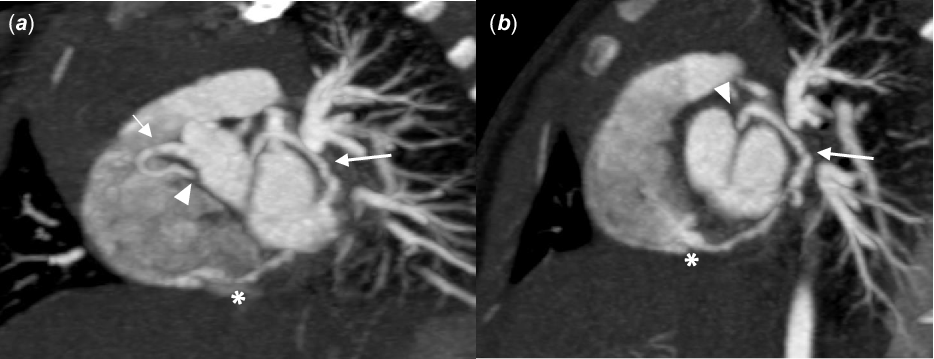
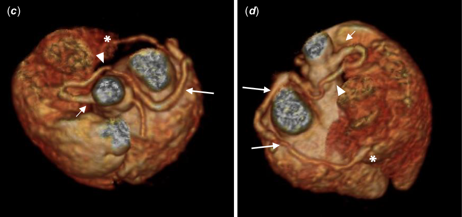
The echocardiogram revealed a globular left ventricle with preserved systolic function and a dilated right coronary artery at the origin, with a tortuous appearance and an anomalous course; there were no other structural anomalies in the echocardiogram. A computed tomography (CT) angiography was performed and confirmed the diagnostic hypothesis of a fistula from the right coronary artery. CT showed a dilated right coronary artery at the origin in the right coronary sinus with a retroaortic course, continuing in the left atrioventricular groove and reaching the left atrium on its posteroinferior wall, then extending to the right, parallel to coronary sinus, and draining into the right ventricle (Figs 1 and 2). There was no evidence of ischemic lesions on the electrocardiogram. Percutaneous therapeutic approach has been proposed. It will be closed when the patient reaches 10 kg for less risk of venous access injury.
CT angiography reconstructed images (maximum intensity projection), coronal (a) and sagittal (b) oblique planes. Images depict the dilated right coronary artery at the origin in the right coronary sinus (short arrows) with retroaortic course (arrowheads), continuing in the left atrioventricular groove (long arrows) and entering the right ventricle (*).

CT angiography 3D reconstructed images of the right coronary fistula to right ventricle: anteroposterior (c) and posteroanterior (d) views. Right coronary artery originated of the in the right coronary sinus (short arrows); retroaortic course (arrowheads) followed by a left atrioventricular groove course (long arrows); fistula in the right ventricle (*).

Coronary fistula is a rare condition, comprising of 0.2–0.4% of all congenital heart diseases. 1 It can be associated with other congenital anomalies in 30% of cases. Depending on the magnitude of the left-to-right shunt, the clinical presentation can range from asymptomatic to signs of heart failure or myocardial ischemia. Early therapeutic intervention is important to minimize overload of the right atrium and ventricle and to avoid other potential complications.
Acknowledgements
We thank R.R., M.A., G.S. and C.S. for their contribution.
Financial support
This research received no specific grant from any funding agency, commercial or not-for-profit sectors.
Conflicts of interest
None.
Ethical standards
The authors assert that all procedures contributing to this work not involve human and/or animal experimentation.