Introduction
There are many reports of a link between cardiac lymphatic system abnormalities and pathologies of the heart. Reference Jones and Min1–Reference Cui, Urschel and Petrelli4 Abnormalities of the cardiac lymphatics have been shown to be associated with ventricular fibrillation, Reference Bansal5,Reference Ishikawa, Akishima-Fukasawa and Ito6 fibroblastic proliferation, Reference Miller3,Reference KLINE, MILLER, PICK and KATZ7 heart failure Reference Miller3 and interestingly, congenital heart disease (CHD). Reference Richards and Garg8–Reference Clark10 According to the Centers for Disease Control and Prevention, heart defects are the most common type of birth defect in the United States, affecting nearly 1% of births each year. 11 Although there is a plethora of information on the risk factors that can influence the development of CHD, there is limited information on the involved mechanism. It has been proposed that abnormal development or obstruction in the cardiac lymphatic system is responsible for heart defects including coarctation of the aorta, hypoplastic left heart and bicuspid aortic valves. Reference Loscalzo, Van and Ho9,Reference Clark10 It has also been suggested that the development of the cardiac lymphatic system and the major vessels of the heart may share the same genetic origin. Reference Irrthum, Devriendt and Chitayat12,Reference Fang, Dagenais and Erickson13 As a result, a mutation in the gene or genes responsible can result in simultaneous defects in both the great vessels of the heart and the cardiac lymphatic vessels. Furthermore, the close anatomic relationship between the cardiac lymphatic vessels and the great vessels of the heart may also suggest a potential “cause and effect” relationship whereby an abnormality in one vessel can encroach on the other vessel resulting in a defect. Knowledge of this potential relationship is very important as it can broaden our understanding of a potential cause of congenital heart defects, which can in turn influence the development of better ways of diagnosing and preventing them. This systematic review explores evidence suggesting a possible connection between cystic hygroma, nuchal translucency and the genetic link underlying cardiac lymphatic abnormalities and congenital heart disease
Materials and methods
In this systematic review, the authors adhered to the Preferred Reporting Items for Systematic Reviews and Meta-Analyses (PRISMA) guidelines. The researchers conducted a literature search on the PubMed database using the search string: (“cardiac” OR “heart”) AND “lymphatics” AND “congenital” up to July 10, 2024. Backwards citation of references from retrieved articles was also performed to locate additional articles for inclusion in the current analysis.
Selection criteria
Articles were selected based on the following inclusion criteria: 1) Discussion of abnormalities in the lymphatic system; 2) discussion of congenital or acquired heart diseases; 3) studies that used animal subjects as human models; 4) text available in the English language. The exclusion criteria for articles consisted of 1) discussion of abnormalities in the lymphatic system without mentioning congenital or acquired heart diseases; 2) discussion of congenital or acquired heart diseases without mentioning abnormalities in the lymphatic system; 3) animal studies relevant to veterinary medicine; 4) letters, editorials, expert opinions and other descriptive reports.
Results
The initial search of the PubMed database yielded 1125 articles, with another 42 identified through backwards citation searching. After applying inclusion and exclusion criteria to the 1167 articles, 78 articles were sought for retrieval and 76 articles were retrieved. After retrieval, 39 reports were further excluded after review of the entire text thereby leaving 37 articles for final inclusion in this review. ( Figure 1).
The PRISMA flow chart.

Evidence with neck webbing
Eight studies examined the increased incidence of congenital heart defects in fetuses with web neck anomaly. Reference Loscalzo, Van and Ho9,Reference Clark10,Reference Chervenak, Isaacson and Blakemore14–Reference Byrne, Blanc, Warburton and Wigger19 Four of these studies involved autopsies of fetuses with cystic hygroma. Reference Lacro, Jones and Benirschke16–Reference Byrne, Blanc, Warburton and Wigger19 Across all eight studies, most fetuses exhibited chromosomal abnormalities, with Turner syndrome being the most common. Reference Loscalzo, Van and Ho9,Reference Clark10,Reference Chervenak, Isaacson and Blakemore14–Reference Byrne, Blanc, Warburton and Wigger19 Frequently observed congenital heart defects included coarctation of the aorta, Reference Loscalzo, Van and Ho9,Reference Clark10,Reference Berdahl, Wenstrom and Hanson15,Reference Lacro, Jones and Benirschke16,Reference Byrne, Blanc, Warburton and Wigger19 bicuspid aortic valve, Reference Loscalzo, Van and Ho9,Reference Miyabara, Nakayama, Suzumori, Yonemitsu and Sugihara17 and atrioventricular canal defects. Reference Berdahl, Wenstrom and Hanson15
Evidence of a link with nuchal translucency
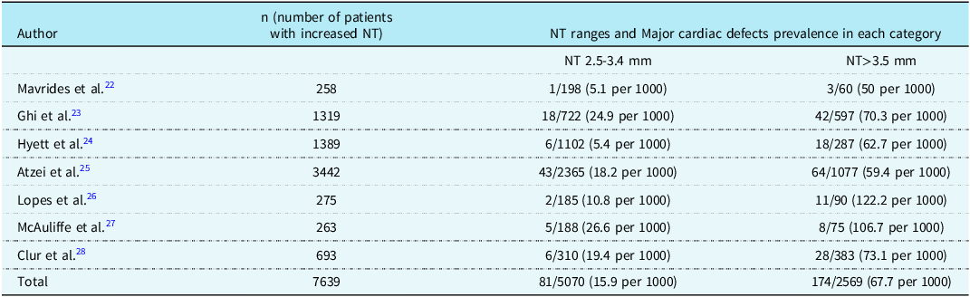
Ten studies investigated the association between increased nuchal translucency and the incidence of congenital heart defects. Reference Hyett, Moscoso, Papapanagiotou, Perdu and Nicolaides20–Reference Hyett, Moscoso and Nicolaides29 Seven of these studies specifically focused on the association between increased nuchal translucency and major cardiac defects in chromosomally normal fetuses. Reference Mavrides, Cobian-Sanchez and Tekay22–Reference Clur, Mathijssen and Pajkrt28 The most common defects reported were; atrioventricular septal defect, Reference Mavrides, Cobian-Sanchez and Tekay22–Reference Clur, Mathijssen and Pajkrt28 ventricular septal defect, Reference Ghi, Huggon, Zosmer and Nicolaides23,Reference Hyett, Perdu, Sharland, Snijders and Nicolaides24,Reference Lopes, Brizot, Lopes, Ayello, Schultz and Zugaib26,Reference Clur, Mathijssen and Pajkrt28 pulmonary atresia/stenosis, Reference Mavrides, Cobian-Sanchez and Tekay22,Reference Ghi, Huggon, Zosmer and Nicolaides23,Reference Atzei, Gajewska, Huggon, Allan and Nicolaides25,Reference McAuliffe, Hornberger, Winsor, Chitayat, Chong and Johnson27,Reference Clur, Mathijssen and Pajkrt28 transposition of great arteries, Reference Mavrides, Cobian-Sanchez and Tekay22,Reference Atzei, Gajewska, Huggon, Allan and Nicolaides25,Reference Lopes, Brizot, Lopes, Ayello, Schultz and Zugaib26 coarctation of the aorta, Reference Mavrides, Cobian-Sanchez and Tekay22,Reference Ghi, Huggon, Zosmer and Nicolaides23,Reference Atzei, Gajewska, Huggon, Allan and Nicolaides25 hypoplastic left heart syndrome Reference Mavrides, Cobian-Sanchez and Tekay22–Reference Atzei, Gajewska, Huggon, Allan and Nicolaides25,Reference McAuliffe, Hornberger, Winsor, Chitayat, Chong and Johnson27,Reference Clur, Mathijssen and Pajkrt28 and tetralogy of Fallot. Reference Mavrides, Cobian-Sanchez and Tekay22,Reference Ghi, Huggon, Zosmer and Nicolaides23,Reference Atzei, Gajewska, Huggon, Allan and Nicolaides25 In these seven studies examining chromosomally normal fetuses with increased nuchal translucency, the prevalence of major cardiac defects ranged from 5.1 to 26.6 per 1,000 cases for NT measurements of 2.5–3.4 mm, and from 50 to 122.2 per 1,000 cases for NT above 3.4 mm. Reference Mavrides, Cobian-Sanchez and Tekay22– Reference Clur, Mathijssen and Pajkrt28 Among 7,639 cases across these studies, the overall prevalence of cardiac defects was 15.9 per 1,000 for NT 2.5–3.4 mm, and 67.7 per 1,000 for NT > 3.5 mm. Reference Mavrides, Cobian-Sanchez and Tekay22–Reference Clur, Mathijssen and Pajkrt28 Further description of values by study are listed in Table 1. One study suggested that the increased nuchal translucency could be due to a rise in the number of fetal jugular lymphatic sacs. Reference Bekker, Haak, Rekoert-Hollander, Twisk and Van Vugt21
Studies investigating the prevalence of major cardiac defects in chromosomally normal fetuses with increased nuchal translucency

NT = Nuchal Translucency.
A potential genetic linkage
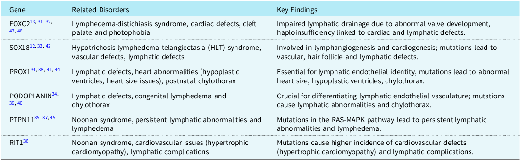
Nineteen studies investigated the genes involved in the embryonic development of both the lymphatic and cardiovascular systems. Reference Irrthum, Devriendt and Chitayat12,Reference Fang, Dagenais and Erickson13,Reference Burger, Bekker, de Groot, Christoffels and Haak30–Reference Mellor, Tate and Stanton46 Of these, nine studies used mice as human models. Reference Burger, Bekker, de Groot, Christoffels and Haak30,Reference Kume, Jiang, Topczewska and Hogan32,Reference Sakamoto, Hara and Kanai-Azuma33,Reference Mishima, Watabe and Saito38,Reference Schacht, Ramirez and Hong40–Reference Johnson, Dillard and Baluk44 Five papers focused on the FOXC2 gene, which is linked to Lymphedema-distichiasis syndrome. Reference Fang, Dagenais and Erickson13,Reference Brice, Mansour and Bell31,Reference Kume, Jiang, Topczewska and Hogan32,Reference Petrova, Karpanen and Norrmén43,Reference Mellor, Tate and Stanton46 Three studies examined the SOX18 transcription factor gene, associated with hypotrichosis-lymphedema-telangiectasia syndrome (HLTS). Reference Irrthum, Devriendt and Chitayat12,Reference Sakamoto, Hara and Kanai-Azuma33,Reference François, Caprini and Hosking42 The PROX1 gene’s role in lymphatic and cardiac development was highlighted in four studies, Reference Shakoor, Wu and Muley34,Reference Mishima, Watabe and Saito38,Reference Wigle and Oliver41,Reference Johnson, Dillard and Baluk44 while three papers implicated the PODOPLANIN gene . Reference Shakoor, Wu and Muley34,Reference Breiteneder-Geleff, Soleiman and Kowalski39,Reference Schacht, Ramirez and Hong40 Additionally, four studies discussed Noonan syndrome, which is associated with mutations in both the PTPN11 gene Reference Joyce, Gordon and Brice35,Reference Sarkozy, Conti and Seripa37,Reference Smpokou, Tworog-Dube, Kucherlapati and Roberts45 and the RIT1 gene. Reference Kouz, Lissewski and Spranger36 These Studies are summarized in Table 2.
Highlights genes involved in both cardiovascular and cardiac lymphatic development, which may explain the link between lymphatic abnormalities and congenital heart defects

Discussion
Evidence with neck webbing
Nuchal translucency (NT) refers to the fluid-filled space at the back of a fetus’s neck, visible during an ultrasound scan. Increased NT, due to a more significant fluid buildup, is referred to as nuchal edema, which is visible as swelling at the back of the fetus’s neck. Cystic hygroma is a significantly more severe form of increased nuchal translucency, often presenting as a multi-loculated fluid collection. Reference Chervenak, Isaacson and Blakemore14,Reference Bekker, Haak, Rekoert-Hollander, Twisk and Van Vugt21,Reference Castelli, Todros, Mattutino, Torre and Panattoni50 Neck webbing (pterygium colli) is the result of excess skin folds in the neck area, which develop due to the resolution of a nuchal cystic hygroma formed in utero. Nuchal cystic hygroma occurs when there is an obstruction of the jugular lymphatic vessel, causing lymph to accumulate in the nuchal region’s lymphatic sacs. Reference Chervenak, Isaacson and Blakemore14 The optimal window for nuchal translucency screening is between 11 and 14 weeks of gestation. Reference Nicolaides47 Cardiovascular defects, such as bicuspid aortic valves and aortic coarctation, are common in individuals with Turner syndrome and other conditions involving neck webbing. About one-third of females with Turner syndrome have congenital heart defects. Reference Richards and Garg8 Aside from genetic factors, it has been suggested that another proposed cause for these defects is lymphatic obstruction extending into the mediastinum. This can lead to significant fetal lymphedema that compresses the heart and major vessels, altering the blood flow and potentially causing defects like aortic coarctation, left ventricular hypoplasia and aortic valve anomalies . Reference Loscalzo, Van and Ho9,Reference Chervenak, Isaacson and Blakemore14
Putte (1977) explored the embryology of the lymphatic system to explain the link between webbed neck and congenital heart defects. Normally, jugular lymphatic sacs, which form lateral to the internal jugular vein, drain upper body lymphatic fluid into the venous system through the internal jugular vein. In Turner syndrome, these sacs fail to connect properly to the veins, causing lymph fluid to accumulate and the sacs to dilate. This elevated lymphatic pressure may also cause cardiac lymphatics to expand, potentially compressing the ascending aorta and altering intracardiac blood flow, which can lead to complex congenital heart defects. Reference Putte48 Observations showed that lymphatic vessels converge at the heart’s base, with lymphatic ducts running alongside the aorta to the jugular lymphatic sac. Reference Clark10 Clark (1984) suggested a similar theory where increased hydrostatic pressure in the obstructed jugular lymphatic sac might lead to the expansion of cardiac lymphatics at the base of the heart, compressing the developing ascending aorta and causing abnormalities in the left heart outflow tract. Reference Clark10 Lacro (1988) further examined Clark’s theory in a study of 12 fetuses with Turner syndrome and nuchal cystic hygroma, aborted between 16- and 26-weeks’ gestation. In 9 of these fetuses, distended lymphatics were observed at the heart’s base, supporting the idea that increased pressure in the jugular lymphatic sac can extend to the cardiac lymphatics. Reference Lacro, Jones and Benirschke16
Studies on embryos with a 45X genotype and cystic hygroma, accompanied by dilated lymphatics around the heart and great vessels, have reported various left-sided congenital defects, such as hypoplastic left heart, aortic valve defects, aortic hypoplasia and coarctation. Reference Lacro, Jones and Benirschke16,Reference Miyabara, Nakayama, Suzumori, Yonemitsu and Sugihara17 In a review of 193 patients with a 45X karyotype, Clark found that 30% of those with webbed necks had congenital heart defects, while 9% had normal hearts. Reference Clark10 Coarctation of the aorta was observed in 25% of patients with neck webbing, but only in 3% of those with a normal neck. Reference Stevenson and Hall49 In the study by Lacro et al. (1988), 75% of fetuses with cystic hygroma and hydrops fetalis had left-sided heart defects. Reference Lacro, Jones and Benirschke16 While the characteristic features of Turner syndrome are often attributed to the abnormal 45X karyotype, in a prospective study conducted by Loscalzo et al. (2005) of 134 females with Turner syndrome, they found a strong association between central lymphedema (indicated by neck webbing) and both bicuspid aortic valve and aortic coarctation, regardless of karyotype. Reference Loscalzo, Van and Ho9 The findings from Byrne et al. (1984) studies on female fetuses with cystic hygromas also suggest that nuchal cystic hygroma is a better indicator of cardiac abnormalities than the 45 X karyotype. Reference Byrne, Blanc, Warburton and Wigger19 Berdahl et al. (1995) provided supporting evidence by analyzing data from the Iowa Birth Defects Registry, identifying 120 fetuses with webbed neck or excessive nuchal skin born between 1983 and 1988. Of these, 66% had blood flow defects. They also compared the incidence of congenital heart defects and flow-related abnormalities in patients with Down, Ullrich-Turner, and Noonan syndromes, with and without a webbed neck. The study found a higher incidence of flow-related defects in those with a webbed neck compared to those without. Reference Berdahl, Wenstrom and Hanson15
Research on fetuses with evidence of lymphatic abnormalities such as neck webbing indicates a strong link between abnormalities in the cardiac lymphatic system and congenital heart disease. Neck webbing, resulting from jugular lymphatic obstruction, is particularly associated with congenital heart defects, especially coarctation of the aorta. Reference Clark10 These studies show a high prevalence of cardiac defects in individuals with neck webbing. Therefore, the presence of webbed neck or nuchal cystic hygroma on prenatal ultrasound or newborn examination should prompt thorough screening for congenital heart disease.
Evidence of a link with nuchal translucency
The nuchal translucency (NT) test is a common screening method used to identify fetuses at risk for chromosomal abnormalities such Down syndrome. This test uses ultrasound to measure the fluid at the back of the fetus’s neck, typically between 10 and 14 weeks of gestation. Bekker et al. (2005) suggested that an increase in the number of fetal jugular lymphatic sacs could be a cause of increased nuchal translucency, noting that 22 of 26 fetuses with increased nuchal translucency had a greater number of jugular lymphatic sacs, most likely due to a delay in the development of the lymphatic system. Reference Bekker, Haak, Rekoert-Hollander, Twisk and Van Vugt21,Reference Castelli, Todros, Mattutino, Torre and Panattoni50
Initially, heart defects in fetuses with increased NT were thought to result from chromosomal abnormalities. However, studies in euploid fetuses revealed that the incidence of cardiovascular defects increased with higher NT and that the degree of NT reflected the risk of congenital heart disease. Reference Hyett, Moscoso, Papapanagiotou, Perdu and Nicolaides20,Reference Carvalho51 In a study by Hyett et al. (1996), 19 out of 21 cases of chromosomally normal fetuses with increased NT showed cardiac abnormalities, with aortic isthmus stenosis being the most common. Reference Hyett, Moscoso, Papapanagiotou, Perdu and Nicolaides20 Carvalho (2005) suggested that nuchal translucency could serve as a predictor for heart defects, although it cannot be linked to a specify the type of cardiac abnormality. Reference Carvalho51
Nuchal edema and congenital heart defects both involve abnormal differentiation during embryogenesis. Our findings confirm a strong association between increased nuchal translucency (NT) in the first trimester and major cardiac defects in fetuses with normal karyotypes, with the risk of such defects rising as NT measurements increase. Given the significant relationship between NT and congenital heart disease, it can be used as a screening tool for congenital heart defects. According to Bansal (2013), one-third of fetuses with severe CHD can be identified through nuchal screening based on this association. Reference Bansal5 Thus, echocardiography is recommended for all fetuses with increased NT, particularly those with measurements above the 99th percentile.
A potential genetic linkage
Recent advances in comparative molecular analysis have shown that vascular and lymphatic endothelial cells share a common lineage . Reference Lampejo, Ghavimi, Hägerling, Agarwal and Murfee52,Reference Hong, Shin and Detmar53 This connection is supported by findings in lymphedema-distichiasis syndrome, an autosomal dominant disorder caused by a mutation in the FOXC2 gene and characterized by limb lymphedema, double rows of eyelashes, cardiac defects, cleft palate, extradural cysts and photophobia. Reference Fang, Dagenais and Erickson13,Reference Brice, Mansour and Bell31,Reference Mellor, Tate and Stanton46,Reference Lampejo, Ghavimi, Hägerling, Agarwal and Murfee52 The FOXC2 gene is involved in the development of both the heart and the cardiac lymphatic system. Reference Kume, Jiang, Topczewska and Hogan32,Reference Jiang, Tian, Nicolls and Rockson54 Mutations in FOXC2 result in normal-looking vessels with impaired lymphatic drainage due to abnormal valve development and defective mural cell coating in the collecting vessels. Reference Petrova, Karpanen and Norrmén43 Therefore, haploinsufficiency of the FOXC2 gene can give rise to both cardiac defects and lymphatic abnormalities. Reference Fang, Dagenais and Erickson13,Reference Brice, Mansour and Bell31
In a systematic review by Burger et al. (2015), 15 genes were found to be essential for both cardiac and lymphatic vascular development in mouse embryos. Fourteen of these genes, when altered, resulted in both cardiac defects and NT. These genes primarily were involved in encoding components of the endothelium, emphasizing a link to endothelial differentiation, which is crucial for forming the heart, blood vessels and lymphatic vessels. Reference Burger, Bekker, de Groot, Christoffels and Haak30 These findings suggest that alterations in genes critical to both lymphatic and cardiovascular development may explain the strong association between the two, with abnormal endothelial differentiation as the underlying pathophysiology.
The SOX18 transcription factor is another gene involved in both cardiovascular and cardiac lymphatic development . Reference Irrthum, Devriendt and Chitayat12 SOX18 plays a crucial role in lymphangiogenesis Reference Jones and Min1,Reference Csányi and Singla55 and cardiogenesis. Reference Wegner56 In hypotrichosis–lymphedema–telangiectasia (HLT) syndrome, mutations in SOX18 lead to vascular, hair follicle and lymphatic defects. Reference Irrthum, Devriendt and Chitayat12 Francois et al. (2008) found that mice with SOX18 mutations showed abnormalities in both the lymphatic and blood vascular systems. Reference François, Caprini and Hosking42 Additionally, SOX18, along with SOX7 and SOX17 (collectively known as SOX F genes), are involved in the development of the heart, blood vessels, and lymphatic vessels, guiding endothelial cell differentiation during these processes. Mice with SOX17/SOX18 mutations exhibited deformities in anterior blood vessels, reduced endocardial cell differentiation, and abnormal endocardial fusion, which disrupts the formation of heart septa and valves, potentially giving rise to defects like atrioventricular septal defects. Reference François, Caprini and Hosking42,Reference Francois, Koopman and Beltrame57 However, Sakamoto et al. (2007) did not find significant anomalies with SOX17/18 mutations, though weak SOX7 expression was linked to malformations in the anterior dorsal aorta and head/cervical vasculature, suggesting a regional effect. Reference Sakamoto, Hara and Kanai-Azuma33 Despite these findings, the role of SOX17/18 in cardiac lymphatics and CHD remains uncertain and requires further research.
The PROX1 gene is essential for the development of both lymphatic vessels and the heart. It plays a key role in establishing and maintaining lymphatic endothelial cell identity by determining the specification of these cells from their precursors. Reference Wigle and Oliver41,Reference Jiang, Tian, Nicolls and Rockson54 Deleting PROX1 can disrupt lymphatic development including cardiac lymphatics, as shown by Johnson et al. (2008), where PROX1 knockout mice developed blood-filled lymphatics and irregular lymphatic vasculature. Reference Johnson, Dillard and Baluk44 Shakoor (2021) reported that PROX1 knockout mice had structural heart abnormalities, such as reduced heart size, hypoplastic ventricles, and a disorganized interventricular septum. Total PROX1 loss led to significant lymphatic defects in the aorta, myocardium, atrioventricular cushions and coronary arteries, resulting in a failure to separate blood and lymphatic vessels, which led to the development of postnatal chylothorax in some mice. Reference Shakoor, Wu and Muley34 . According to Csányi (2019), PROX1 knockout mouse embryos did not survive beyond mid-gestation due to liver and lymphatic defects. Reference Wigle and Oliver41 Additionally, a missense mutation in integrin alpha-9, whose expression is controlled by PROX1, was linked to severe chylothorax in human fetuses. Reference Mishima, Watabe and Saito38,Reference Brouillard, Boon and Vikkula58
The gene PODOPLANIN is also believed to play a role in the association between lymphatic system abnormalities and cardiovascular defects. Shakoor et al. (2021) found that PODOPLANIN-deficient mice had issues with separating blood and lymphatic vessels and some developed postnatal chylothorax. These knockout mice exhibited congenital lymphedema, impaired lymphatic transport and absent lymphatic capillaries. Reference Shakoor, Wu and Muley34,Reference Schacht, Ramirez and Hong40 Thus, PODOPLANIN is crucial for differentiating lymphatic endothelial vasculature from embryonic. Reference Breiteneder-Geleff, Soleiman and Kowalski39,Reference Vuorio, Tirronen and Ylä-Herttuala59
Noonan syndrome is an autosomal dominant disorder marked by abnormal development in various parts of the body. Noonan syndrome occurs due to mutations in genes of the RAS-MAPK pathway, and the lymphatic defects in these syndromes have been demonstrated to have a genetic basis. Reference Brouillard, Boon and Vikkula58 Joyce et al. (2016) found that all 11 patients with Noonan syndrome in their study had persistent lymphatic abnormalities, with nine patients having genital edema and eight patients having chylous reflux with lymphorrhea. The PTPN11 gene, which encodes SHP2, is commonly mutated in Noonan syndrome, appearing in 6 of the 11 patients studied. Reference Joyce, Gordon and Brice35 PTPN11 mutation causes Noonan syndrome in about 50% of cases, with half of these patients also experiencing lymphedema. Reference Smpokou, Tworog-Dube, Kucherlapati and Roberts45 Other mutations, such as a missense RIT1 variant, have also been associated with the syndrome. Kouz et al. (2016) noted that the phenotype associated with RIT1 mutations was particularly linked to a higher incidence of cardiovascular issues, such as hypertrophic cardiomyopathy and lymphatic complications. Reference Kouz, Lissewski and Spranger36
These studies suggest that mutations in shared genes may explain the possible association between lymphatic abnormalities and congenital heart defects. This supports the hypothesis of a shared molecular marker linking cardiac lymphatics and CHD. The genes discussed are highlighted in Table 2 and represent key candidates for future research into the underlying causes and potential treatments of CHD.
Conclusion
There seems to be significant evidence to support a potential association between abnormalities of the cardiac lymphatic system and the development of CHD. The proximity of the cardiac lymphatics to the great vessels of the heart may suggest a “cause and effect” relationship. As a result, finding a cystic hygroma or webbed neck on ultrasound should prompt further investigation for heart defects. Increased nuchal thickening should also stimulate further search for cardiac defects. There also seems to be a possible genetic relationship between cardiac lymphatic abnormalities and congenital heart disease, but further research needs to be done to confirm this. As demonstrated in some of the included studies, even in fetuses with a normal karyotype, the presence of in-utero nuchal cystic hygroma was associated with a higher incidence of cardiac defects, such as bicuspid aortic valve or aortic coarctation. This conflicts with the currently prevalent belief that the characteristic phenotypic features seen in Turner syndrome can be solely attributed to the abnormal 45X karyotype. In these studies, the karyotype seemed to be independent of these defects and was therefore less reliable than cystic hygroma or webbed neck in indicating the presence of cardiac abnormalities. Understanding this potential association would be of great clinical significance as it would pave the way for new diagnostic approaches and treatment options.
Acknowledgements
None
Financial support
This research received no specific grant from any funding agency, commercial or not-for-profit sectors.
Competing interests
The author(s) declare none