Case report
We report an unusual case of thrombotic complication in a 6-year-old boy with hypoplastic left heart syndrome and dextrocardia after a fenestrated total cavopulmonary connection, using an extracardiac conduit (16 mm). The total cavopulmonary connection with fenestration (due to a borderline size of the left pulmonary artery) was performed at the age of 5 years, followed by Warfarin anticoagulation therapy. The blood oxygen saturation increased from 80–84% to 85–91% after completion of the total cavopulmonary connection. The mean pressure in the Fontan system was 13–14 mmHg. There were no significant residual lesions after surgery. Echocardiography revealed good contractility of the systemic right ventricle, trivial to mild tricuspid regurgitation, and a mean fenestration gradient of 5 mmHg.
The patient’s overall condition was satisfactory at his first two outpatient visits – 1 month and 4 months after the operation. There were no signs of failure of the Fontan circulation, and the child was active, with no activity limitation or dyspnoea. The echocardiographic findings remained unchanged. However, 1 year after surgery the patient showed significant deterioration with shortness of breath during exercise, increased fatigue during daily activities, and a newly acquired hepatomegaly (4 cm below the costal margin). Echocardiography revealed an increase of the mean fenestration gradient to 10 mmHg. Also, a new restriction of flow through the atrial communication was found. This had been non-restrictive immediately after the total cavopulmonary connection completion, and at patient’s outpatient visits at 1 and 4 months after surgery. The sum of gradients suggested a mean pressure of 25 mmHg in the Fontan system. Therefore, the patient was indicated for a cardiac CT scan and subsequent surgical intervention.
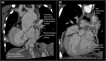
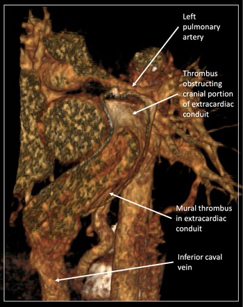
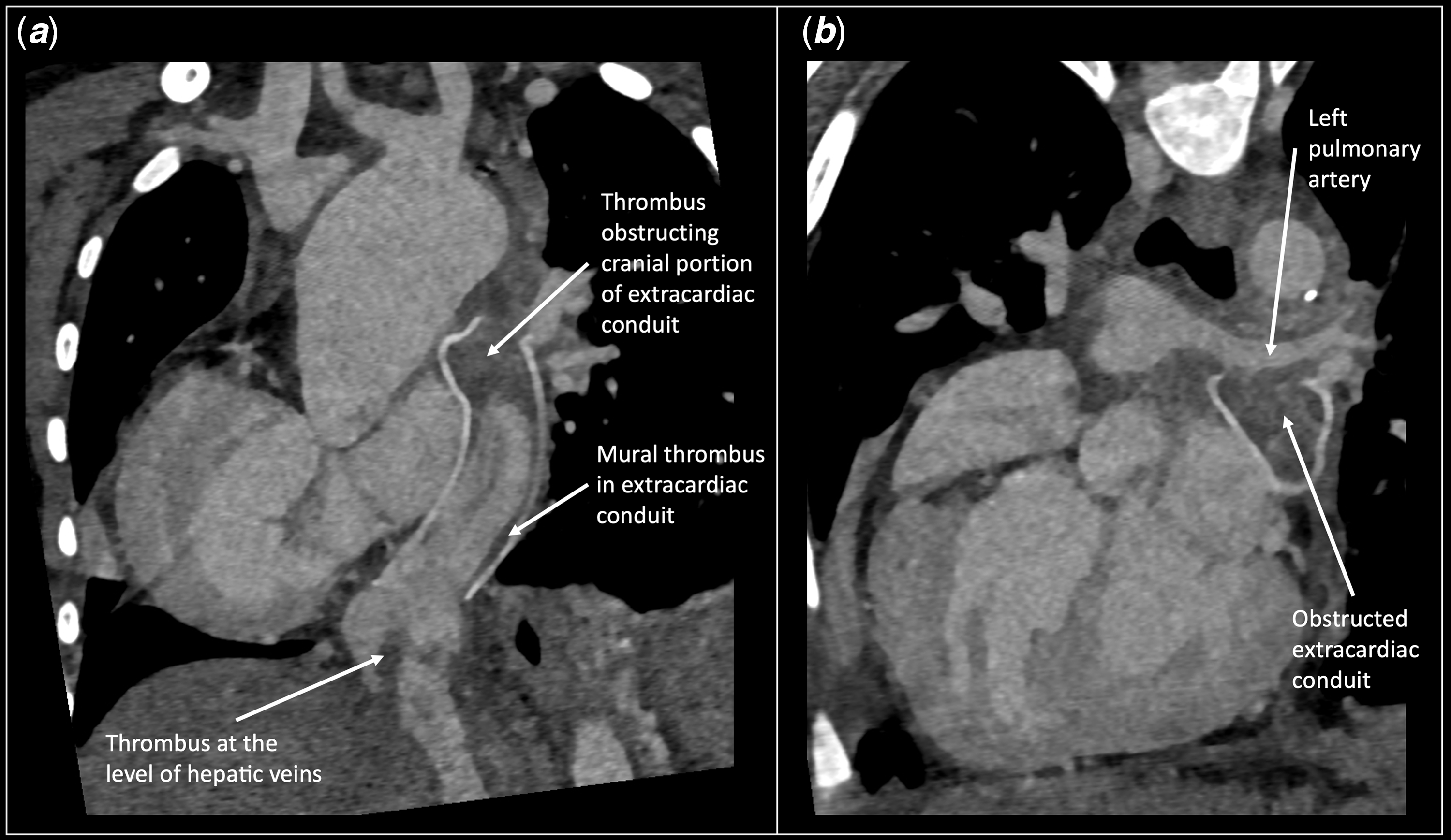
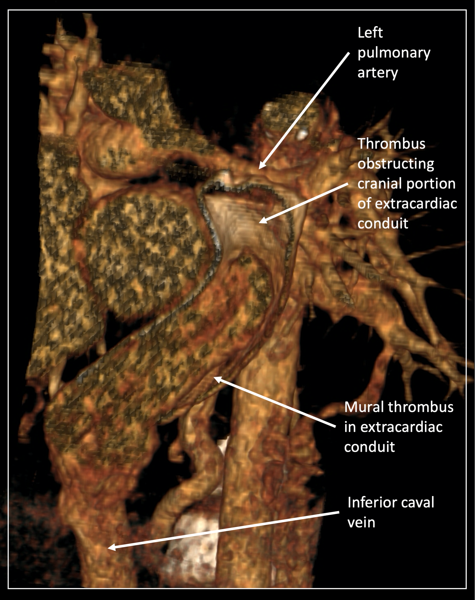
The CT scan revealed multiple thrombi in the total cavopulmonary connection. The most significant were one thrombus that completely obstructed the flow through the cranial portion of the extracardiac conduit, another extensive mural thrombus that narrowed the remaining inferior portion of the extracardiac conduit, and a third thrombus that partially obstructed the inferior caval vein at the level of the hepatic veins (Figures 1 and 2). Given the fact that the patient was kept on adequate anticoagulation (the international normalised ratio range 2.5–2.9) from completion of the total cavopulmonary connection, the formation of this many thrombi seemed rather unlikely without any other comorbidity. Screening of thrombophilic mutations was negative. Routine blood tests and a physical examination performed at admission did not reveal any potential cause of the thrombosis. Haematocrit was 0.44, haemoglobin was 14 g/dl, and the international normalised ratio was 2.7. The boy had no signs of haemoconcentration.
Contrast CT angiography; ( a ) frontal view of the thrombi in the extracardiac conduit; ( b ) backward tilted frontal view of total obstruction between the extracardiac conduit and left pulmonary artery.
Three-dimensional CT angiography reconstruction of the extracardiac conduit.
Because COVID-19 and subsequent generalised disorders, such as paediatric inflammatory multi-system syndrome temporally associated with SARS-CoV-2 (PIMS-TS), are known to cause a hypercoagulable state,Reference Connors and Levy1 the patient was tested for a previous or ongoing COVID-19 infection. The polymerase chain reaction from a nasopharyngeal swab was negative, but the presence of circulating IgG antibodies against SARS-CoV-2 was confirmed by micro blot array. These were most likely anamnestic and therefore suggest that the patient had gone through a clinically inapparent COVID-19 infection several weeks or months previously.
The patient underwent a thrombectomy, left pulmonary artery plasty, and atrial communication enlargement. The intraoperative course was unfortunately complicated by cerebral ischaemia due to low cardiac output before going on bypass. There were no thrombotic complications.
Discussion
Thrombotic complications are not uncommon in patients after a total cavopulmonary connection,Reference Singh, Jerrom, DClinP and Saxena2–Reference Attard, Huang, Monagle and Ignjatovic4 especially in the first year after surgery, and the risk factors are not well understood. In this case, multiple factors may have played a role in the development of almost complete thrombotic occlusion of the total cavopulmonary connection circuit while the patient was on effective anticoagulation therapy.
Although we are not able to determine exactly when the patient went through COVID-19 infection, we believe that a recent presence of a high-risk prothrombogenic state such as SARS-CoV-2 in combination with newly developed restrictive atrial communication led to the formation of significant thrombi in the total cavopulmonary connection, thus severely impairing the patient’s haemodynamics associated with significant clinical worsening. The time of formation of the thrombi was unclear, but clinical symptoms suggested an interval of several weeks before the 1-year follow-up visit.
Thrombotic complications are most often described in severe cases of COVID-19 infection or associated inflammatory multi-system syndrome, but cases of thromboembolic events in non-severe patients,Reference Saleh, Aboelghar and Salem5 or as the first or only symptom of the infection,Reference Fan, Umapathi and Chua6,Reference del Nonno, Colombo, Nardacci and Falasca7 have also been reported.
COVID-19 infection may have a significant impact on Fontan patients and should be handled carefully even if inapparent.
Acknowledgements
None.
Financial support
This work was supported by MH CZ – DRO, Motol University Hospital, Prague, Czech Republic 00064203.
Conflicts of interest
None.