




No CrossRef data available.
Article contents
An institutional review of the value of computed tomographic angiography in the diagnosis of congenital cardiac malformations
Published online by Cambridge University Press: 14 April 2005
Abstract
The ultra-fast, thin-cut computerised tomographic angiogram is an efficient method to diagnose extracardiac lesions associated with congenital cardiac disease. For the purposes of this review, we evaluated various facets of the technique as used in 30 patients who were referred for diagnosis of congenital cardiac disease. The technique had high diagnostic accuracy, with a sensitivity of 93 percent in 15 of these patients referred for either interventional catheterisation or surgery. There were no immediate side-effects associated with the scanning procedure. The scan was also found to be more cost-effective as compared to an alternative noninvasive modality for imaging modality, namely magnetic resonance imaging. The angiographic technique, however, does expose the child to between 2 and 2.5 rems of radiation, despite the short period of scanning, of 10 plus or minus 2 seconds.
Information
- Type
- Original Article
- Information
- Copyright
- 2005 Cambridge University Press
The diagnostic imaging of congenital cardiac malformations has evolved from angiography to the noninvasive modalities of echocardiography, magnetic resonance imaging, and computerised tomography. Technological refinements, such as cardiac gating, and three-dimensional reconstruction, have increased the utility of both computerised tomography and magnetic resonance imaging for the diagnosis of congenital cardiac lesions. The short time now needed to make scans using computerised tomography makes the technique particularly attractive for use in children.
The diagnostic capability of the computerised tomographic scan has been evaluated for anomalies involving the aortic arch, such as vascular rings1, 2 and aortic coarctation,3, 4 as well as for pulmonary venous abnormalities.5 This study presents the experience gained at the University of Florida with modern computerised tomographic scanning in the diagnosis of congenital cardiac malformations in a cohort of patients undergoing procedures over the period from January 2002 through January 2004.
Methods
Patients studied: We performed computerised tomographic angiography in 30 patients who were referred for evaluation of suspected congenital cardiac disease. These were patients for whom an initial echocardiogram was not sufficient definitively to diagnose the congenital cardiac malformation. Their ages ranged from 1-month old to 39 years, with a mean age of 5.8 years. Their weights ranged from 1.2 to 79 kilograms, with a mean of 27 kilograms. Of those referred, 22 came from the outpatient setting, but the other 8 came from our ward for inpatients. We submitted patients to scanning only if they had no history of previous allergic reaction to iodinated contrast, renal insufficiency, or current pregnancy. Respiratory compromise, or age were not contraindications, due to the availability of an ultra-fast machine through which images could rapidly be acquired over a sequence of 10 plus or minus 2 seconds, thus avoiding the potential destabilization of critically ill patients. The study was reviewed and approved by the Institutional Review Board of the University of Florida.
Protocol for scanning: The scanner used was a Siemens Somaton Sensation and Syngo (Philadelphia, USA). Those patients deemed too active to permit reliable scanning were sedated using chloral hydrate given orally at an average dose of 75 milligrams per kilogram, following the guidelines for sedation established at Shands Hospital and the University of Florida. The initial images were acquired during quiet breathing technique, due to the inconsistency of breath-holding in these sedated children. For those who weighed more than 20 kilograms, we injected as contrast agent Omnipaque 300, or iohexal as supplied by Amersham Health of Princeton, at a dose of 1.75 millilitres per kilogram, with a range from 1.5 to 2.0 millilitres per kilogram. The agent was injected through a 20-gauge intravenous catheter using the power injector at a rate of 4 millilitres per second. In those children weighing less than 20 kilograms, we injected the contrast agent manually using similar dosing. We started the scan as soon as all of the contrast had been injected, usually within 6 seconds. The field of view covered the thorax from the first rib to the eleventh rib, from chest wall to chest wall, and was centered on the heart (Table 1).
Table 1. Parameters used for scanning.

Processing of data: Images were reconstructed using slices of 1 millimetre thickness, with an increment of 0.5 millimetres. Post-processing was done using the Siemens Wizard work station, again with Syngo version 7.0 software. Splicing of the bony structures permitted an unobstructed three-dimensional view to be obtained of the heart and the great vessels. Using this material, we evaluated the utility of the technique in making decisions either for referral for surgery, or the need for further diagnostic study using cardiac catheterisation. The post-processed images were compared either with the angiograms obtained at cardiac catheterisation, or the intraoperative observations of the surgeon, permitting us to calculate the positive predictive value of the computerised tomographic scan. Adverse events, and costs associated with the procedures, were recorded at the time of the scans.
Results
There were no immediate adverse events associated with any of the cardiac scans. We studied patients with a wide range of ages, the youngest being a fragile, premature, neonate studied 1 month after birth when weighing 1.2 kilograms, and the oldest individual was 39-years old. The average heart rate was 100 beats per minute. The average time needed for scanning was 10 plus or minus 2 seconds. Depending on the complexity of the underlying cardiac anatomy, post-processing took between 30 and 45 minutes. The average cost per study was 883 United States dollars, with 645 of these dollars needed for hospital or technical charges, and 238 for the charges made by the physicians.
In 22 of the patients (73 percent), the scans were performed to evaluate anomalies of the aortic arch, while 8 (27 percent) focussed on assessment of pulmonary venous and arterial anomalies. In 2 of the patients in this latter group, there were complex intracardiac anomalies, one having a common arterial trunk, and the other with concordant atrio-ventricular but discordant ventriculo-arterial connections with a ventricular septal defect and pulmonary hypertension (Table 2).
Table 2. Impact of computer tomographic angiography on the clinical decision-making process.

Subsequent to the analysis of the scans generated by computerised tomography, we referred half of the patients studied for intervention, 10 for operative repair and 5 for cardiac catheterisation. The remaining 15 patients received medical therapy. The pre-operative diagnosis made subsequent to examination of the computerised tomographic images was confirmed by operative observation in 9 of the 10 patients referred for surgery. In the outstanding case, we had interpreted the scans as showing that three pulmonary veins connected anomalously to the coronary sinus, while the other vein connected anomalously below the diaphragm (Fig. 1). At operation, it was found that all four pulmonary veins entered the coronary sinus.

Figure 1. A double aortic arch completely encircles and compresses the trachea and oesophagus. Note that the left arch is smaller than the right.
The computerised tomographic findings were confirmed following conventional angiography in all the 5 patients referred for cardiac catheterisation. In four of these 5 patients, catheter intervention proved successful. The fifth patient, the one with transposition and a ventricular septal defect, was shown at cardiac catheterisation to have reversible pulmonary hypertension. For this cohort of 15 patients, therefore, the positive predictive value of computerised tomographic imaging was 93 percent when diagnoses were compared with direct intraoperative anatomic observation or findings at cardiac catheterisation.
For the 15 patients with clinical evidence of significant congenital cardiac disease, initially thought to warrant intervention, we downgraded our assessment of the severity of their cardiac lesions on the basis of the scans, referring these patients for clinical follow-up with or without additional medical management. For example, we commenced beta-blockade in 1 patient with Marfan's syndrome in whom computerised tomographic imaging revealed dilation of the ascending aorta.
Immediate adverse events were infrequent in this group of 30 patients. Some complained of discomfort during placement of the peripheral intravenous line prior to the procedure. There were no allergic reactions to the intravenous contrast used, nor were adverse reactions such as hypotension, bradycardia, tachycardia, or angina observed or reported. All 22 patients who underwent the procedure as outpatients were discharged home on the same day. Unlike magnetic resonance imaging or angiography, which may involve general anaesthesia for infants and small children,6, 7 most of the computerised tomographic scans were conducted without any sedation. Sedation, using chloral hydrate, was required for 5 of the 30 patients (16 percent), albeit that there were no complications reported with this sedation.
Case studies
- A 22-day-old neonate presented in severe congestive heart failure and, following echocardiographic evaluation, was suspected to have totally anomalous pulmonary venous connection. The computerised tomographic angiogram demonstrated that the pulmonary venous confluence returned to the coronary sinus. The infant underwent successful rerouting of this abnormal channel to the left atrium (Fig. 1).
- A premature infant, weighing only 1.2 kilograms at 1 month after birth, and in whom it had proved impossible to wean from ventilation, despite multiple attempts, was found on bronchoscopy to have anterior compression of the trachea. The infant underwent computerised tomography, which demonstrated a double aortic arch with a smaller left arch. Based on these images (Fig. 2), the smaller aortic arch was ligated, with resolution of symptoms.
- A one-month-old infant who presented in shock was shown by the computerised tomographic angiogram to a have critical aortic coarctation (Fig. 3). The neonate underwent resection of the narrowed segment, with end-to-end anastomosis and augmentation of the arch, with excellent results.

Figure 2. Critical coarctation of the descending aorta, in association with hypoplasia of the transverse arch.

Figure 3. Totally anomalous pulmonary venous connection to the coronary sinus. The arrow shows the junction of the pulmonary venous confluence with the coronary sinus.
Discussion
The use of advanced therapeutic options for patients with congenital cardiac malformations requires early and accurate diagnosis of the defects. Echocardiography plays a principal initial role, but has limitations, particularly in the evaluation of the aortic arch, pulmonary arteries, and pulmonary venous connections. Conventional cardiac catheterisation is invasive, and is used nowadays for therapeutic intervention rather than for diagnosis, especially with the advent of other noninvasive modalities such as computerised tomographic angiography or magnetic resonance imaging and angiography.8
Recent trends have favoured the use of magnetic resonance imaging and angiography as the preferred noninvasive supplement to echocardiography. This technique can provide excellent anatomic detail of both intra- and extracardiac structures, along with data pertaining to flow, ventricular function, volumes of intracardiac structures, and myocardial perfusion.6, 9–13 The higher cost of magnetic resonance imaging at the University of Florida, with an average investigation costing 1232 United States dollars, as opposed to 883 dollars for the computerised tomographic scan, along with the need for deeper conscious sedation or general anaesthesia in young children, combined with the longer scanning time of 45 to 60 minutes, makes the technique less than ideal for certain clinical questions. The convenience and accuracy of ultra-fast computerised tomographic imaging, with exquisite three-dimensional reconstruction, is particularly useful for defining extracardiac anatomy, such as the pattern of branching of the aortic arch, the arrangement of the pulmonary vasculature, and even the extent of narrowing of the airways.14–16
In our study, we have shown that computerised tomographic scanning is accurate in the diagnosis of both aortic and pulmonary vascular anomalies. In the sub-population requiring intervention, a positive predictive value of 93 percent was found when compared to traditional gold standards such as cardiac catheterisation or intraoperative assessment. We found that, using computerised tomographic angiography, we were able to make the diagnosis, and facilitate the decision-making process, with little immediate morbidity. Based on the thickness of the slices used in scanning, we were able to delineate anatomic details as small as 1 millimetre. Accurate intraluminal measurements can be made from the cross-sectional images. Other studies have, in the past, looked at the utility of the computerised tomographic scan as a noninvasive modality in the diagnosis of congenital cardiac malformations. Accuracies of greater than 95 percent have been cited for assessment of anomalies of the aortic arch, and positioning of the airways.15 Our study confirms this level of accuracy, and illustrates again the procedural safety of the technique, with only a small proportion of our patients requiring sedation.
Our experience, nonetheless, revealed several current limitations of the technique. The exposure to radiation, of 2 to 2.5 rems compared to the values of 1.5 to 2 rems at diagnostic cardiac catheterisation, still places the child, especially those under 5 years of age, at an increased risk of later development of malignancy.17 Imaging the coronary arteries, not the subject of our study, but frequently required in patients with congenital cardiac disease, is currently difficult for young children, as the quality of the images is limited by the high heart rates found in children.18, 19 Visualization of the intracardiac anatomy is also less than optimal when using contrast enhanced computerised tomography. There is no question but that the intracardiac anatomy is better revealed by magnetic resonance imaging.13 Functional data, such as the ejection fraction, ventricular volumes, the velocity of flows, pressure gradients, and absolute measurements of pressure, are also not obtainable using computerised tomographic scanning. The computerised tomographic scan, therefore, should be used as a supplement to echocardiography and other imaging modalities, chosen with the clinical question in mind. We acknowledge other limitations in our current study, such as the heterogeneous nature of our cohort of patients, the lack of concurrent magnetic resonance imaging data for comparison, and the lack of systematic comparison with echocardiographic data. Our experience is simply presented as that obtained at one institution, using the technique as a novel means of diagnosing congenital cardiac malformations. We submit, nonetheless, that the experience does reveal computerised tomography to be an excellent modality for better definition of specific abnormalities of the extracardiac vasculature.
Acknowledgements
We thank Glen Baker and Nancy Quinn, from the Department of Radiology for their assistance in the three-dimensional reconstruction of the computerised tomographic angiograms. We also thank Dr. Manuel Arreola of the Department of Radiology for his calculation of the rates of exposure to radiation in our institution. Special thanks are due to Connie Nixon and Lee Ann for their exemplary work in coordinating the patients.
References
Hopkins KL. Pediatric great vessel anomalies: initial clinical experience with spiral computer tomography angiography. Radiology 1996; 200: 811–815.Google Scholar
Katz M, Konen E, Rozenman J, Szeinberg A, Itzchak Y. Spiral computer tomography and 3D image reconstruction of vascular rings and associated tracheobronchial anomalies. J Comput Assist Tomogr 1995; 19: 564–568.Google Scholar
Becker C, Soppa C, Fink U, et al. Spiral computer tomographic angiography and 3-dimensional reconstruction in patients with aortic coarctation. Eur Radiol 1997; 7: 1473–1477.Google Scholar
Kaemmerer H, Mugge A, Oelert F, et al. Evaluation of the aorta and supra-aortic vessels with spiral computer tomography and 3-dimensional vascular reconstruction after operation of aortic isthmus stenosis. Z Kardiol 1994; 83: 775–783.Google Scholar
Shiraishi I, Kato Y, Todoroki H, Satoh H, Hamaoka K. Differential color imaging technique of helical computer tomographic angiography in the diagnosis of total anomalous pulmonary venous drainage. Circulation 2000; 101: 2017–2018.Google Scholar
Tsai-Goodman B, Geva T, Odegard KC, Sena LM, Powell AJ. Clinical role, accuracy, and technical aspects of cardiovascular magnetic resonance imaging in infants. Am J Cardiol 2004; 94: 69–74.Google Scholar
Odegard KC, DiNardo JA, Tsai-Goodman B, et al. Anesthesia considerations for cardiac magnetic resonance imaging in infants and small children. Paediatr Anaesth 2004; 14: 471–476.Google Scholar
Pihkala J, Nykanen D, Freedome RM, Benson LN. Interventional cardiac catheterisation. Pediatr Clin North Am 1999; 46: 441–464.Google Scholar
Korperich H, Gieseke J, Barth P, et al. Flow volume and shunt quantification in pediatric congenital heart disease by real-time magnetic resonance velocity mapping: a validation study. Circulation 2004; 109: 1987–1993.Google Scholar
Razavi RS, Hill DL, Muthurangu V, et al. Three-dimensional magnetic resonance imaging of congenital cardiac anomalies. Cardiol Young 2003; 13: 461–465.Google Scholar
Geva T, Sandweiss BM, Gauvreau K, Lock JE, Powell AJ. Factors associated with impaired clinical status in long-term survivors of tetralogy of Fallot repair evaluated by magnetic resonance imaging. J Am Coll Cardiol 2004; 43: 1068–1074.Google Scholar
Pignatelli RH, McMahon CJ, Chung T, Vick GW III. Role of echocardiography versus magnetic resonance imaging for the diagnosis of congenital heart disease. Curr Opin Cardiol 2003; 18: 357–365.Google Scholar
Boxt LM. Magnetic resonance and computed tomographic evaluation of congenital heart disease. J Magn Reson Imaging 2004; 19: 827–847.Google Scholar
Siegel MJ. Multiplanar and three-dimensional multi-detector row computer tomography of thoracic vessels and airways in the pediatric population. Radiology 2003; 229: 641–650.Google Scholar
Lee ED, Siegel MJ, Hildebolt CF, et al. Multidetector computer tomography evaluation of thoracic aortic anomalies in pediatric patients and young adults: comparison of axial, multiplanar, and 3-dimensional images. Am J Roentgenol 2004; 182: 777–784.Google Scholar
Lawler LP, Fishman EK. Multi-detector row computer tomography of thoracic disease with emphasis on 3-dimensional volume rendering and computer tomographic angiography. Radiographics 2001; 21: 1257–1273.Google Scholar
Frush DP, Donnelly LF, Rosen NS. Computed tomography and radiation risks: what pediatric health care providers should know. Pediatrics 2003; 112: 951–957.Google Scholar
Giesler T, Baum U, Ropers D, et al. Non invasive visualization of coronary arteries using contrast enhanced multi-detector computer tomography: influence of heart rate on image quality and stenosis detection. Am J Roentgenol 2002; 179: 911–916.Google Scholar
Nieman K, Rensing BJ, van Geuns RJ, et al. Non-invasive cardiac angiography with multi-slice spiral computed tomography: impact of heart rate. Heart 2002; 88: 470–474.Google Scholar
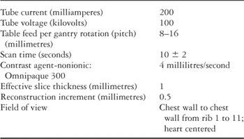
Table 1.
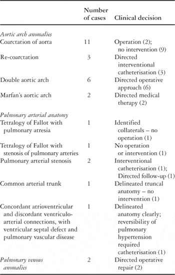
Table 2.

A double aortic arch completely encircles and compresses the trachea and oesophagus. Note that the left arch is smaller than the right.

Critical coarctation of the descending aorta, in association with hypoplasia of the transverse arch.

Totally anomalous pulmonary venous connection to the coronary sinus. The arrow shows the junction of the pulmonary venous confluence with the coronary sinus.
 You have
Access
- 9
- Cited by