INTRODUCTION
The first step in wisdom is to know the things themselves (Linnaeus).
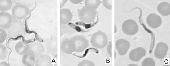
Trypanosomes like many other microorganisms have few morphological features detectable at the level of light microscopy that can be used to distinguish different species. For example, only a few small differences in cell size and shape, and the attributes of the flagellum and kinetoplast distinguish the 3 species of trypanosome shown in Fig. 1, although these are very different parasites in terms of host range, pathogenicity and transmission cycle. Moreover, Fig. 1A could depict the dividing forms found in the mammalian bloodstream of any of the trypanosome species or subspecies within subgenus Trypanozoon, comprising the 2 human pathogens Trypanosoma brucei gambiense and T. b. rhodesiense, and the livestock pathogens T. b. brucei, T. evansi and T. equiperdum. The difficulty of morphological identification is compounded by the disappearance of the few useful distinguishing features during trypanosome development in the vector. Thus, right from the start, it has been clear that more than just morphological examination is needed for identification of these parasites.
Trypanosomes in Giemsa-stained thin films of infected mouse blood. (A) Trypanosoma brucei brucei, (B) T. congolense and (C) T. vivax.

DNA-based identification methods tap into the intrinsic genetic identity of the organism, providing an unchanged genetic signature whatever the developmental stage examined – particularly useful for parasites where individual life cycle stages may show little resemblance to each other. Moreover, DNA-based methods still work even if morphology has been poorly preserved, or if there is very little parasite material in the sample, or if parasite DNA is contaminated with host material. Through the twin advantages of specificity and sensitivity, DNA-based methods have a wider range of applicability and efficacy in epidemiological studies than morphological examination alone, and are now indispensable tools for the study of trypanosomiasis as for so many other parasitic diseases. In this review, my aim is to trace the development of DNA-based methods for identification of the African tsetse-transmitted trypanosomes, starting from papers published in Parasitology in the late 1980s that addressed the problem of identification of trypanosomes in the vector (Kukla et al. Reference Kukla, Majiwa, Young, Moloo and Ole-Moiyoi1987; Gibson et al. Reference Gibson, Dukes and Gashumba1988).
DNA PROBES FOR IDENTIFICATION OF TRYPANOSOMES IN TSETSE
African trypanosome species can be distinguished by morphological characteristics of the bloodstream form parasites (Fig. 1). However, these differences are lost among the developmental stages in the tsetse vector and therefore trypanosomes within the fly were traditionally identified by their sites of development (Lloyd and Johnson, Reference Lloyd and Johnson1924), e.g. midgut and salivary glands for Trypanosoma brucei, midgut and proboscis for T. congolense and T. simiae, and proboscis only for T. vivax. There are obvious problems with this system, in that it fails to identify infections that have not developed beyond the midgut (immature infections) and to distinguish species that develop in the same locations in the fly (e.g. T. congolense and T. simiae); in addition, mixed infections of more than 1 species are rarely detectable without recourse to further investigations, e.g. animal infection. Notwithstanding the limitations, much of our understanding of tsetse infection rates with different trypanosome species in the field was based on this flawed methodology until the advent of DNA-based methods for identification.
Species-specific DNA probes for African trypanosomes were developed in the 1980s, typically using repetitive sequences such as satellite DNAs with very high copy number, which offered the advantage of great sensitivity as well as specificity, since these non-coding DNAs vary extensively between species (Kukla et al. Reference Kukla, Majiwa, Young, Moloo and Ole-Moiyoi1987; Majiwa and Webster, Reference Majiwa and Webster1987; Gibson et al. Reference Gibson, Dukes and Gashumba1988). Samples were prepared as dot blots of crude tissue preparations, such as macerated tsetse midguts, spotted directly onto nitrocellulose filters and air dried; such filters could conveniently be stored dry at ambient temperature until return to the laboratory; processing consisted of in situ treatment with alkali to release and bind the DNA, followed by hybridization of replicate dot blots with each radioactively-labelled species-specific DNA probe and exposure to autoradiography film to reveal the results. Although dot blots were relatively easy to prepare and store in the field, processing and analysis involved lengthy laboratory work and enough replicate dot blots were needed to allow hybridization with each species-specific probe.
Despite these drawbacks, field application of this methodology immediately proved its value for identification of the trypanosomes present in the midguts of wild-caught tsetse. A high proportion of infections were shown to consist of more than 1 species (Majiwa and Otieno, Reference Majiwa and Otieno1990), a feature missed by studies based on the dissection method of Lloyd and Johnson (Reference Lloyd and Johnson1924). The ability to distinguish T. congolense from the pig pathogen, T. simiae, which shares the same developmental sites in the fly, showed the power of the approach to provide relevant epidemiological data on the relative risk of trypanosome transmission to different livestock species from infected tsetse populations (McNamara et al. Reference McNamara, Dukes, Snow and Gibson1989). Some tsetse infections remained unidentified by the limited range of species-specific DNA probes available, but the unforeseen bonus of this apparent failure of the technique was the discovery of new trypanosome species. In The Gambia, the finding of a number of unidentifiable infected midgut squashes from Glossina morsitans submorsitans led to the discovery of the new species T. godfreyi, a suid trypanosome sharing development in the midgut and proboscis with T. congolense and T. simiae, and thus belonging to subgenus Nannomonas (McNamara et al. Reference McNamara, Mohammed and Gibson1994). Across in East Africa, a new genotype related to T. congolense was discovered in G. pallidipes caught in Tsavo West National Park (Majiwa et al. Reference Majiwa, Maina, Waitumbi, Mihok and Zweygarth1993), now called T. simiae Tsavo (Gibson et al. Reference Gibson, Stevens, Mwendia, Makumi, Ngotho and Ndung'u2001). These 2 new members of subgenus Nannomonas joined the 3 other distinct genotypes of T. congolense already described among trypanosomes isolated from livestock: T. congolense savannah, forest and kilifi or Kenya Coast (Young and Godfrey, Reference Young and Godfrey1983; Gashumba, Reference Gashumba1986; Gashumba et al. Reference Gashumba, Baker and Godfrey1988; Knowles et al. Reference Knowles, Betschart, Kukla, Scott and Majiwa1988). The development of species-specific DNA probes for each of these genotypes allowed epidemiological tracking in studies of their tsetse or livestock hosts.
Despite the major gains in the understanding of trypanosomiasis epidemiology brought about by the deployment of species-specific DNA probes in field studies, the limited sensitivity of the technique (about 100 trypanosomes in a dot blot (Gibson et al. Reference Gibson, Dukes and Gashumba1988)) made it less useful for identification of the low numbers of trypanosomes found, for example, in tsetse proboscis or animal blood samples. Moreover, dot blots, although easy enough to prepare and store in field conditions, required extensive lab processing and use of radioactive chemicals, restricting analysis to a few reference labs. For these reasons, the dot blot technique was quickly abandoned once the simplicity and superior sensitivity of methods based on the polymerase chain reaction (PCR) were appreciated.
PCR-BASED IDENTIFICATION OF TRYPANOSOMES
Existing knowledge about species-specific DNA sequences and their use as DNA probes for trypanosomes formed a solid foundation for the development of PCR-based methods of identification (Moser et al. Reference Moser, Cook, Ochs, Bailey, McKane and Donelson1989; Masiga et al. Reference Masiga, Smyth, Hayes, Bromidge and Gibson1992; Majiwa et al. Reference Majiwa, Thatthi, Moloo, Nyeko, Otieno and Maloo1994) and application to field-collected material soon yielded results that greatly extended the previous observations made using DNA probes. The previous dot blot sample preparation method needed to be changed to facilitate PCR, while still providing easy methods for sample collection and storage in the field: amplifiable DNA needed to be extracted with minimal processing and, in view of the sensitivity of PCR, it was now important to avoid sample-to-sample contamination. These considerations have been addressed by collection of samples spotted directly onto filter paper or placed in lysis buffer in individual tubes. Such samples can sometimes be used directly as PCR template after rehydration and/or heat treatment, but other types of sample, e.g. blood or tsetse midgut, contain substances that inhibit the PCR polymerase and need further purification, for example, incubation with Chelex resin, phenol extraction and/or alcohol precipitation.
At least 8 different genotypes of livestock trypanosome are identifiable by PCR (T. brucei sspp.; T. congolense savannah, forest and kilifi; T. simiae; T. simiae tsavo; T. godfreyi; T. vivax) with a sensitivity of detection of less than 1 trypanosome present in the sample used for PCR (Masiga et al. Reference Masiga, Smyth, Hayes, Bromidge and Gibson1992). Mixed infections of 2, 3 and even 4 different trypanosomes were recorded and occurred at much higher frequencies than ever expected from previous studies using the dissection technique (Majiwa et al. Reference Majiwa, Thatthi, Moloo, Nyeko, Otieno and Maloo1994; Masiga et al. Reference Masiga, McNamara, Laveissiere, Truc and Gibson1996). For example, Lehane et al. (Reference Lehane, Msangi, Whitaker and Lehane2000) found that 36% of the 352 proboscis infections from G. pallidipes they identified by PCR were mixed infections, the majority of 2 different trypanosome genotypes. This raises important epidemiological questions as to how such mixed infections are acquired by the flies – from a single feed on a host carrying a mixed infection or from sequential feeds on different infected hosts. In support of the single feed hypothesis, susceptibility of tsetse to infection with T. brucei sspp. and T. congolense is known to be highest in flies just after emergence from the puparium and to drop dramatically after the fly's first bloodmeal (Wijers, Reference Wijers1958; Maudlin, Reference Maudlin1991), probably as a consequence of maturation of the teneral fly and increasing activity of the insect's innate immune system. On the other hand, infection rates with both T. vivax and T. congolense were found to increase with age (Woolhouse and Hargrove, Reference Woolhouse and Hargrove1998), indicating that flies continue to acquire infection after the first feed. The likelihood of a new trypanosome infection being able to take hold may also be influenced by the presence of an established infection, particularly if the first infection has heightened the level of activity of the immune system. While these considerations confound interpretation to some extent, the nature of the mixed infections observed may provide interesting insights into the feeding habits of a particular tsetse population. For example, Lehane et al. (Reference Lehane, Msangi, Whitaker and Lehane2000) found statistically significant co-occurrence of the suid trypanosomes, T. simiae, T. simiae Tsavo (aka T. congolense Tsavo) and T. godfreyi, in proboscides of infected G. pallidipes from Tanzania; a second group of flies showed an association between T. congolense savannah and Kenya Coast (aka kilifi), both trypanosomes typically found in ruminants. Taken together, these results indicate different feeding preferences among particular populations of G. pallidipes on the Tanzanian coast.
Even with 8 species-specific PCR tests, it was apparent that many infections remained unidentified (Solano et al. Reference Solano, Argiro, Reifenberg, Yao and Duvallet1995; Morlais et al. Reference Morlais, Grebaut, Bodo, Djoha, Cuny and Herder1998; Lehane et al. Reference Lehane, Msangi, Whitaker and Lehane2000; Malele et al. Reference Malele, Craske, Knight, Ferris, Njiru, Hamilton, Lehane, Lehane and Gibson2003). The identification of T. vivax was particularly problematic. This species completes its entire developmental cycle in the tsetse proboscis/upper oesophagus where relatively few trypanosomes are present (estimated maximum 103) compared to the large numbers found in midgut infections with subgenera Trypanozoon or Nannomonas (estimated maximum 106). It became evident that the initial DNA sequence used as a hybridization probe and for PCR primer design was only specific for the West African form of T. vivax (Dickin and Gibson, Reference Dickin and Gibson1989; Masiga et al. Reference Masiga, Smyth, Hayes, Bromidge and Gibson1992). Although this kind of T. vivax is widespread in tropical Africa and has also established itself in South America, other T. vivax genotypes are present, particularly in East Africa (Masake et al. Reference Masake, Majiwa, Moloo, Makau, Njuguna, Maina, Kabata, Olemoiyoi and Nantulya1997), and hence a generic PCR test for T. vivax was required. The DNA target now favoured for PCR identification of T. vivax is a gene encoding an antigen used for immunodiagnosis of T. vivax infection (Masake et al. Reference Masake, Nantulya, Pelle, Makau, Gathuo and Ole-Moiyoi1994; Masake et al. Reference Masake, Majiwa, Moloo, Makau, Njuguna, Maina, Kabata, Olemoiyoi and Nantulya1997; Morlais et al. Reference Morlais, Ravel, Grebaut, Dumas and Cuny2001); in a direct comparison with the West African T. vivax PCR test on a series of tsetse proboscis samples from Cameroon, the antigen gene T. vivax PCR test proved far superior, identifying 86% of 36 proboscis samples compared to 42% with the old PCR test (Morlais et al. Reference Morlais, Ravel, Grebaut, Dumas and Cuny2001). A further complication revealed by PCR identification on a wide scale was the realization that midgut infections of T. congolense could disappear after the trypanosomes colonized the proboscis (Lehane et al. Reference Lehane, Msangi, Whitaker and Lehane2000), and thus all proboscis infections were not necessarily T. vivax as assumed by the dissection method (Lloyd and Johnson, Reference Lloyd and Johnson1924).
The success of species-specific PCR tests prompted the development of tests to identify the human infective trypanosomes, T. b. gambiense and T. b. rhodesiense, to allow the incrimination of reservoir hosts and vectors of these pathogens. Group 1 T. b. gambiense causes the majority of human trypanosomiasis cases and is widely distributed across West and Central Africa. The genotype of this parasite has many conserved characteristics suitable for development of specific PCR tests (Bromidge et al. Reference Bromidge, Gibson, Hudson and Dukes1993; Mathieu-Daude et al. Reference Mathieu-Daude, Bicart-See, Bosseno, Breniere and Tibayrenc1994; Radwanska et al. Reference Radwanska, Claes, Magez, Magnus, Perez-Morga, Pays and Buscher2002b), but the most satisfactory test at present is that based on a conserved gene for a flagellar pocket glycoprotein, TgsGP (Radwanska et al. Reference Radwanska, Claes, Magez, Magnus, Perez-Morga, Pays and Buscher2002b). There is as yet no specific test for the less common Group 2 T. b. gambiense, of which only a handful of isolates are known from Ivory Coast (Mehlitz et al. Reference Mehlitz, Zillmann, Scott and Godfrey1982); the heterogeneity of this group has so far hindered discovery of molecular markers. For many years this was also true for T. b. rhodesiense, which is genetically heterogeneous between and within foci in East Africa. However, the discovery of the SRA (serum resistance associated) gene, the single gene that confers human infectivity on this subspecies (De Greef et al. Reference De Greef, Imberechts, Matthyssons, Van Meirvenne and Hamers1989; Xong et al. Reference Xong, Vanhamme, Chamekh, Chimfwembe, Van Den Abbeele, Pays, Van Meirvenne, Hamers, De Baetselier and Pays1998), was a gift for the development of a simple PCR test for identification of T. b. rhodesiense (Welburn et al. Reference Welburn, Picozzi, Fevre, Coleman, Odiit, Carrington and Maudlin2001; Gibson et al. Reference Gibson, Backhouse and Griffiths2002; Radwanska et al. Reference Radwanska, Chamekh, Vanhamme, Claes, Magez, Magnus, De Baetselier, Buscher and Pays2002a), and this single gene PCR test has proved invaluable for identifying this zoonotic subspecies in its animal reservoir hosts (Welburn et al. Reference Welburn, Picozzi, Fevre, Coleman, Odiit, Carrington and Maudlin2001; Njiru et al. Reference Njiru, Ndung'u, Matete, Ndungu and Gibson2004). The sensitivity and applicability of these specific gene targets for direct diagnosis of T. b. gambiense and T. b. rhodesiense in the field has been greatly improved by redesigning the assays for loop-mediated isothermal amplification (LAMP) rather than conventional PCR (Njiru et al. Reference Njiru, Mikosza, Armstrong, Enyaru, Ndung'u and Thompson2008a, Reference Njiru, Mikosza, Matovu, Enyaru, Ouma, Kibona, Thompson and Ndung'ub; Thekisoe et al. Reference Thekisoe, Kuboki, Nambota, Fujisaki, Sugimoto, Igarashi, Yasuda and Inoue2007).
GENERIC PCR IDENTIFICATION
The usefulness of species-specific PCR tests in epidemiological studies meant that this methodology was soon being applied to large sample collections. With 8 species-specific PCR tests, hundreds of individual PCR's were needed to process even a fairly modest sample collection. It therefore became desirable to develop a generic method of identification whereby only a single PCR test was required for each sample. Size variation among trypanosome species in the internal transcribed spacer (ITS-1) of the ribosomal RNA (rRNA) locus provided the necessary discriminatory power (Desquesnes et al. Reference Desquesnes, McLaughlin, Zoungrana and Davila2001) and this methodology has been widely applied to identification of both tsetse midgut and livestock blood samples (Cox et al. Reference Cox, Tilley, McOdimba, Fyfe, Eisler, Hide and Welburn2005; Njiru et al. Reference Njiru, Constantine, Guya, Crowther, Kiragu, Thompson and Davila2005; Adams et al. Reference Adams, Malele, Msangi and Gibson2006). In the ITS-1 generic PCR test, the PCR primers flank the ITS-1 spacer, which is variable in both length and sequence; the primers are chosen in the 18S and 5·8S rRNA genes such that they match all trypanosome species of interest (Fig. 2); the size of the single PCR fragment amplified from a trypanosome sample is then measured by gel electrophoresis against a DNA size marker and compared with known values from reference species. The high copy number (>100) of the rRNA locus ensures that the PCR has a good level of sensitivity. Problems arise when the amplified fragments from 2 different species are too close in size for resolution by gel electrophoresis, and in this case additional species-specific PCR tests need to be done.
Diagram of part of the trypanosome ribosomal RNA locus showing locations of 18S, 5·8S and 28S rRNA genes and ITS-1 PCR primers (arrowheads).

As with species-specific PCR tests, the ITS-1 PCR is able to detect mixed infections when 2 or more fragments are amplified, but, in addition, the generic PCR test gives a band rather than merely a negative result with unknown trypanosome species. For example, ITS-1 PCR detected an unknown trypanosome genotype among samples collected from tsetse in Tanzania by virtue of the unique size of the PCR fragment amplified (Adams et al. Reference Adams, Malele, Msangi and Gibson2006); this new trypanosome had previously been found when tsetse samples that remained unidentified by species-specific PCR tests were subject to DNA sequence analysis of the 18s rRNA gene (T. godfreyi-like; Malele et al. (Reference Malele, Craske, Knight, Ferris, Njiru, Hamilton, Lehane, Lehane and Gibson2003) ).
On the other hand, ITS-1 PCR will fail to discriminate species where amplified fragments cannot be resolved by size and this can have major consequences. For example, the putative new species of trypanosome recently found in Tanzania (Hamilton et al. Reference Hamilton, Adams, Malele and Gibson2008) was missed by ITS-1 PCR identification, because the amplified fragment was mistaken for that of T. simiae (Adams et al. Reference Adams, Hamilton, Malele and Gibson2008). Instead, this trypanosome was detected by the more discriminatory technique, fluorescent fragment length barcoding (FFLB) (Hamilton et al. Reference Hamilton, Adams, Malele and Gibson2008). FFLB relies on size polymorphisms in multiple small regions of the 18S and 28S rRNA genes detected by analysis on an automated sequencer, so that differences as small as a single base pair can be measured; gene fragments are amplified by PCR using fluorescently-labelled primers in conserved regions of the rRNA genes that flank regions of variable size, allowing detection of PCR products by fluorescence (Hamilton et al. Reference Hamilton, Adams, Malele and Gibson2008). Although this technique requires more specialized equipment and reagents than conventional PCR, gains are made not only in accuracy but also in the capacity for high throughput, with the possibility of analysing 96 samples simultaneously within a day or two.
The development of the FFLB technique was possible because of the vast resource of rRNA gene sequences already available on the database. Alignment of the 18S and 28S rRNA genes from the tsetse-transmitted trypanosomes quickly identified regions of similarity and variability, allowing the positioning of suitable PCR primers. The sizes of amplified fragments were predicted from the database sequences and subsequently confirmed in reference trypanosome isolates, providing each known trypanosome species with a recognizable barcode of fragment sizes. Any non-matching barcode found among field isolates would represent an unknown genotype that could be investigated further by sequence analysis.
Comparison of rRNA gene sequences has allowed the relatedness of different trypanosome species to be quantified, and the phylogenetic trees constructed from these data have revealed the relationships between different taxa. This kind of analysis has put trypanosome taxonomy on an evolutionary footing, giving a new dimension to our previous understanding which was based on purely qualitative traits. Whereas, in the early days, DNA probes could provide only a positive or negative identification, the new sequence-based techniques also give a measure of relatedness and allow both known and unknown trypanosome species to be placed accurately in the evolutionary tree. These tools have not only provided a sound basis for epidemiological studies, but have also given us a much greater appreciation of trypanosome diversity.
I would like to acknowledge my debt to my Ph.D. supervisor and mentor David Godfrey for setting me off on the road to trypanosome identification in the first place, and to the many other colleagues who have contributed to the results reviewed here.