Introduction
In conjunction with an ever-growing global population, resource scarcity has become a global challenge that deserves attention. Resource scarcity was ranked No. 4 among the top 10 global trends in a survey conducted by the World Economic Forum Network of Global Agenda Councils.1 Our research over the past decade has centered on the development of effective strategies for achieving affordable and sustainable use of precious metals (Table I). While these elements only exist at a level of parts per billion (ppb) in the earth’s crust, they play critical roles in the economically important automobile, chemical, pharmaceutical, petroleum, and energy industries.Reference Cotton2 Furthermore, due to the ever-increasing demands from both existing and emerging applications, their long-term sustainability has emerged as a major societal concern.
Partial list of precious metals widely used for catalytic and plasmonic applications, including their abundances in the earth’s crust, current prices, and total amounts mined in human history.

* Based upon data released in July 2018.
Platinum (Pt), for example, has a crustal abundance of 37 ppb.Reference Zereini and Wiseman3 It takes almost five months to produce 30 g of Pt from about 10 metric tons of ore. If all of the Pt that had been mined in human history were collected and formed into a cube, the side of the cube would only measure 7.2 m. It is therefore unsurprising that the price of Pt has reached $USD30 per gram and doubled (and even tripled or quadrupled at certain points) over the past two decades.4 As one of the most versatile catalytic materials, Pt is widely used in the chemical industry (e.g., for the synthesis of nitric acid), petroleum industry (e.g., for both hydrogenation and dehydrogenation reactions), and the automobile industry (e.g., in catalytic converters). Since the last century, a grand challenge has been launched to identify cost-effective and sustainable products based on Pt. Proton-exchange membrane fuel cells (PEMFCs),Reference Scofield, Liu and Wong5 a technology that has been around for many decades and is widely recognized as the cleanest (zero emission) source of energy, relies upon Pt. Currently, a relatively large amount of Pt (roughly, 80 g for the PEMFC in a mid-range sedan) is required to accelerate the oxygen reduction reaction (ORR) and thus achieve the required power density for transportation use. As a matter of fact, the cost and sustainability issues around Pt have been a longstanding barrier to the industrial-scale commercialization of PEMFCs.Reference Shao, Chang, Dodelet and Chenitz6
Gold (Au) is another compelling example. Elemental Au has a crustal abundance of only 3.1 ppb.Reference Prior7 As of 2018, a total of 190,000 metric tons of Au has been mined in human history.Reference Prior7 When this amount of Au is evenly distributed among the world population of 7 billion, each person will receive roughly 25 g, which is just enough for crafting an ordinary wedding band. As one of the least reactive metals, elemental Au does not oxidize under atmospheric conditions.Reference Cotton2,8 After exposure to air, not even a single atomic layer of gold oxide(s) can be detected.
When processed as a nanomaterial, Au displays a range of surprising properties different from its bulk form.Reference Harden and Toynbee9,10 In 1857, Faraday reported his groundbreaking work on the chemical synthesis of Au nanoparticles,Reference Faraday11 opening new opportunities for Au. The unique optical properties of Au nanomaterials, known as localized surface plasmon resonance (LSPR), have enabled many applications in chemistry and medicine, including sensing, drug delivery, image contrast enhancement, and cancer theranostics.Reference Yang, Yang, Pang, Madeline and Xia12 Furthermore, the discovery of catalytic activity for Au nanoparticles with diameters of 6 nm and below has generated new applications for this ancient metal.Reference Ishida and Haruta13
Clever strategies will be needed to commercialize the vast applications enabled by Au nanomaterials. A good argument can be made for the use of Au nanoparticles as a drug carrier for cancer treatment. As reported for Doxorubicin (Dox), a popular anticancer drug, the most commonly used treatment involves intravenous injection every 21−28 days at a dosage in the range of 40−60 mg/m2 in terms of skin area. For solid Au nanospheres of 12.7 nm in diameter, the loading efficacy of Dox was approximately 2.68 × 10⁻² g of Dox per gram of Au.Reference Kim, Jeong and Jon14 In this case, by multiplying the skin area with the dosage and then dividing by loading efficacy, 2.67 g of Au will be needed for each patient every 3−4 weeks, corresponding to 766 metric tons per year in terms of the worldwide demand from all cancer patients (estimated to be ca. 24 million) in 2032. Given that the majority of Au is currently used as currency reserves and jewelry, it is simply impossible to find this large amount of Au for the drug-delivery application. As such, if approved for clinical use, the massive demand of Au for drug delivery and related biomedical applications may cause major changes to existing market shares of Au.
During the past decade, a number of research groups have sought to address the sustainability issue of precious metals by developing a novel class of nanostructures known as nanocages, characterized by hollow interiors, ultrathin and porous walls, and well-controlled sizes and surface structures.Reference Zhao, Wang, Yang, Gilroy, Qin and Xia15 Depending on the elemental composition, such hollow nanostructures show remarkable optical and catalytic properties that are often superior to those of their solid counterparts, while the amount of materials is markedly reduced. When made of Au, for example, the nanocages show LSPR peaks precisely tunable in the visible and near-infrared (NIR) regions to enable deep tissue penetration,Reference Skrabalak, Chen, Sun, Lu, Au, Cobley and Xia16 while their hollow interiors can be loaded with drugs and other chemical/biological effectors. If constructed from Pt, the nanocages show greatly enhanced catalytic activity toward both oxygen reduction and hydrogen oxidation,Reference Zhang, Roling, Wang, Vara, Chi, Liu, Choi, Park, Herron, Xie, Mavrikakis and Xia17–Reference He, He, Wang, Lin, Yin, Hong, Wu and Li19 two reactions essential to the operation of PEMFCs. By keeping the wall thickness below 2 nm (or roughly 10 layers of atoms), it is feasible to increase the size of the hollow nanostructures to 20 nm and above without compromising the utilization efficiency of metal atoms. The increase in dimensions makes it practical to create side faces with well-controlled packing for the surface atoms, including the presence of planar defects such as twin planes and stacking faults, to optimize the catalytic activity and selectivity.
In this article, we offer a brief account of our recent accomplishments in mechanistic understanding and experimental control related to the chemical synthesis of such nanostructures. We also highlight how the methodology that we developed can be extended to other metals, including their alloys, for the fabrication of nanomaterials with diverse compositions and properties.
Maximizing the utilization efficiency of atoms
The utilization efficiency of atoms refers to the fraction of atoms positioned on the outmost surface relative to the total number of atoms in the whole material; therefore, it is directly proportional to the specific surface area of the material. Traditionally, in heterogeneous catalysis, the utilization efficiency of atoms is increased by reducing the size of the particles.Reference Xia and Yang20 For example, by reducing the edge length of a Pt nanocube from 20 to 3 nm, the utilization efficiency will be increased from 6 to 39%, while the specific surface area is increased from 14 to 93 m2/g. This explains why the current Pt/C catalysts used in PEMFCs are typically based upon Pt particles of only 3–5 nm in diameter. While reducing the size of catalytic particles is effective in increasing the utilization efficiency of atoms, the “quality” of the atoms on the surface may be compromised as the increased proportions of atoms at corners and edges will result in lower catalytic activity due to the strong binding of oxygen to those low-coordination atoms.Reference Xia and Yang20 Additionally, the reduction in contact area between the Pt nanoparticles and the carbon support tends to make the catalytic system less durable due to particle migration, sintering, and detachment.Reference Dai, Lu, Cao, Campbell and Xia21
An alternative approach to increasing the utilization efficiency of atoms is based upon the development of nanosheets with a thickness approaching a few layers of atoms.Reference Xia and Yang20 One can easily understand this concept by considering the structure of graphene.Reference Allen, Tung and Kaner22 All of the carbon atoms in a graphene sheet are positioned on the surface, giving a utilization efficiency as high as 100%. For a nanosheet consisting of two layers of metal atoms, the utilization efficiency of atoms can still be kept at 100%. It was possible to hammer Au and other precious metals into ultrathin sheets (e.g., roughly 500 atoms in thickness) even by ancient goldsmiths, but not into nanosheets of only a few atoms in thickness. In a series of recent developments, however, nanosheets as thin as two atomic layers have been successfully synthesized for precious metals such as Pt, Pd, Rh, and Ru, by choosing the right combination of precursor, capping ligand, and solvent.Reference Yin, Liu, Pan, Walsh and Yang23–Reference Kong, Xu, Zhang, Dai, Oliaee, Li, Zeng, Wu and Peng26 These results point toward a new strategy for increasing the utilization efficiency of precious metals.
Learning from paper origami
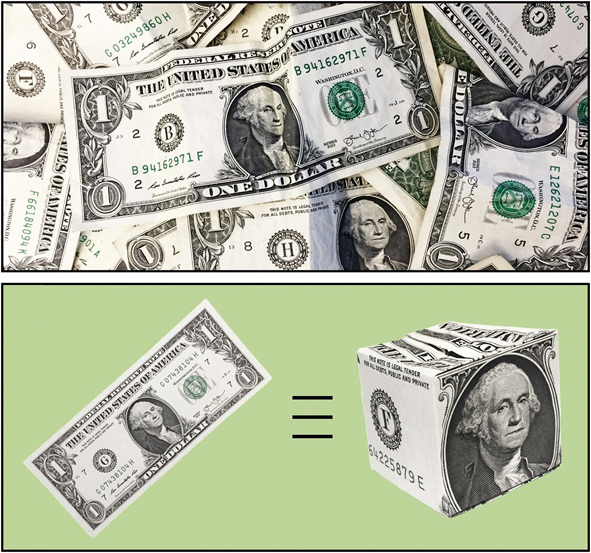
Most of the atoms in nanosheets may not be active toward catalytic reactions because they are inevitably blocked by long-chain ligands necessary for both passivation and stabilization. It is almost impossible to drop-cast nanosheets on a catalytic support in a manner that will allow full exposure of both sides of each nanosheet. Additionally, nanosheets easily crumble, fold, and stack during sample preparation, making their practical use a major challenge (Figure 1a).
Schematic illustration showing how the strategy of “paper origami” can be utilized to fold 2D sheets into 3D objects to solve issues such as easy stacking and poor dispersion on a supporting substrate.

These technical problems can be solved by “folding” a metal nanosheet into a three-dimensional, hollow structure, a trick that has its origin in paper origami (Figure 1b). In the folding process, the wall thickness of the resultant hollow structure can be kept essentially the same as the original sheet. The exceedingly high utilization efficiency of atoms should be largely retained while the folded structures can be easily dispersed on a catalytic support, just like conventional catalytic particles. While the concept of “folding” is intriguing, it is almost impossible in reality to implement it. An exception is graphene, which has been demonstrated to roll up to generate carbon nanotubes and related structures.Reference Kosynkin, Higginbotham, Sinitskii, Lomeda, Dimiev, Price and Tour27
Alternatively, the same hollow structures can be readily fabricated through template-directed synthesis. The atoms of another metal of interest can be conformally deposited on the surface of a nanocrystal template in a layer-by-layer fashion to obtain core–shell nanocrystals. Removal of the template then leaves behind a hollow nanocrystal whose shape and physical dimensions are essentially identical to those of the original template.Reference Zhao, Wang, Yang, Gilroy, Qin and Xia15–Reference Zhang, Roling, Wang, Vara, Chi, Liu, Choi, Park, Herron, Xie, Mavrikakis and Xia17 Such hollow nanostructures are also known as nanocages due to the generation of small holes in the walls during the removal of the core by chemical etching. Conceptually, a nanocage can be considered as a “folded” nanosheet, but with small holes in the walls. If the walls are thinned to ca.1 nm, the utilization efficiency of atoms can reach 40%, and the surface structure can be engineered to yield optimal catalytic activity and selectivity.
Synthesis of nanocages via galvanic replacement
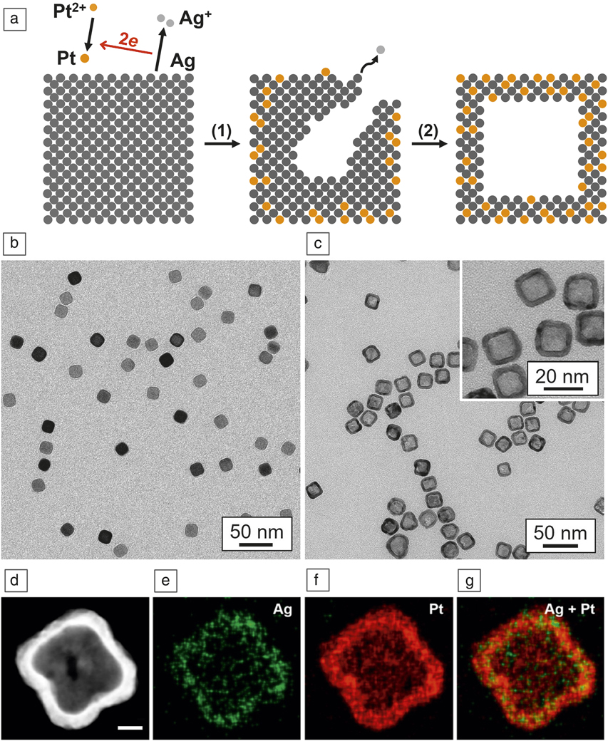
As an electrochemical process, galvanic replacement involves the oxidation of one metal, known as a sacrificial template, by the ions of another metal with a higher reduction potential (Figure 2a). Upon contact, the atoms in the template are spontaneously oxidized and dissolved into the solution phase, while the ions of the second metal are reduced to the elemental state and conformally deposited onto the template. In a single step, one can generate hollow nanostructures with porous walls within a few minutes. Since our first report in 2002 for a system involving Ag nanocubes and HAuCl4,Reference Sun and Xia28 this simple and versatile method has been applied to many different elements for the fabrication of a wide variety of nanostructures characterized by hollow interiors and porous walls.Reference Xia, Wang, Ruditskiy and Xia29 The only requirement is to choose the right electrochemical pairs to attain a favorable difference in reduction potential.
(a) Schematic illustration showing the morphological and structural changes at different stages of the galvanic replacement reaction between a Ag nanocube and PtCl42− in an aqueous solution. Step 1 shows the deposition of Pt atoms on the Ag template at the expense of oxidation of Ag atoms, generating a hollow interior; step 2 reveals the formation of a Pt-Ag alloy nanocage with a cubic shape. Transmission electron microscope (TEM) images of (b) the Ag nanocubes that served as a template for the formation of (c) Pt-Ag nanocages through the galvanic replacement reaction with PtCl42− precursor. The inset shows the nanocages at a higher magnification. (d) High-angle annular dark field-scanning TEM image of a single Pt-Ag nanocage. Energy dispersion x-ray mapping of (e) Ag, (f) Pt, and (g) Ag + Pt. (d–g) Scale bar = 5 nm. (b–g) Reprinted with permission from Reference Reference Yang, Roling, Vara, Elnabawy, Zhao, Hood, Bao, Mavrikakis and Xia32. © 2016 American Chemical Society.

For the synthesis of Au-based nanocages, one can simply titrate an aqueous solution of HAuCl4 into a suspension of Ag nanocrystals in water at a controlled temperature. Depending on the exact morphology of the Ag nanocrystals, holes can be created either on the side faces or at the corner sites. For example, when Ag nanocubes with slightly truncated corners are employed, the resultant nanocages tend to have small openings at the corner sites due to the site selectivity associated with the Ag dissolution and Au deposition processes.Reference Chen, McLellan, Siekkinen, Xiong, Li and Xia30 Since the redox potentials of both PtCl42–/Pt and PdCl42–/Pd (0.76 V and 0.59 V, respectively, versus the standard hydrogen electrode [SHE]) are more positive than that of the AgCl/Ag pair (0.22 V versus SHE), Pt- and Pd-based nanocages can also be prepared using a similar galvanic replacement reaction with Ag nanocrystals.Reference Chen, Wiley, McLellan, Xiong, Li and Xia31 Because the number of Au, Pt, or Pd atoms deposited is stoichiometrically related to the number of Ag atoms oxidized from the template, one cannot separately adjust the composition and wall thickness of the resultant nanocages. In addition, if the galvanic replacement reaction involves the imbalanced removal and deposition of atoms (in the case of Ag template, three Ag atoms are removed for every Au atom deposited when HAuCl4 is employed as the precursor), the inevitable generation of vacancies makes it difficult for the deposited atoms to faithfully replicate the surface structure on the template. As a result, the nanocages are often characterized by a nonuniform wall thickness and a rough surface.
Figure 2b–g shows transmission electron microscope (TEM) images of the template based upon Ag nanocubes and the nanocages obtained through the galvanic replacement route, as well as the energy dispersion x-ray mapping of an individual nanocage.Reference Yang, Roling, Vara, Elnabawy, Zhao, Hood, Bao, Mavrikakis and Xia32 The nanocages are made of an alloy due to interdiffusion between Pt and Ag, which tends to be accelerated in the presence of vacancies caused by the 1:2 stoichiometric ratio between the number of the deposited Pt atoms and number of the dissolved Ag atoms. The nanocages did not show any sign of collapsing when the wall thickness was reduced to a few nanometers. It was found that the density of pores created in the walls of a nanocage increased with the amount of the PtCl42– precursor added. Interestingly, the surface roughness of the resultant nanocages is dependent on the reaction temperature, a phenomenon that can be ascribed to the coalescence of atomic vacancies during the galvanic replacement and dealloying processes.Reference Xia, Wang, Ruditskiy and Xia29 Altogether, by introducing an aqueous PtCl42– solution dropwise into a reaction mixture containing Ag nanocubes at 40°C, we were able to obtain Pt-Ag alloy nanocages with a smooth surface and in high yield. The nanocages displayed a single-crystal structure essentially identical to that of the initial Ag nanocubes, enclosed by {100} facets.
Synthesis of nanocages via template-directed deposition and then etching
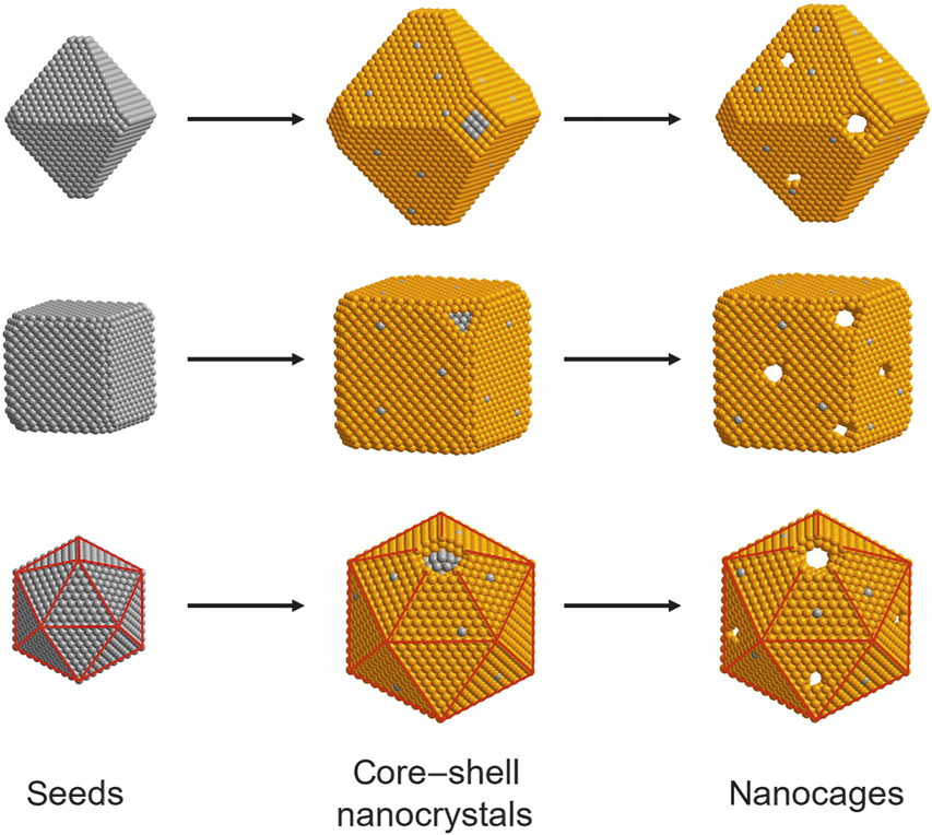
We have also developed a different approach to the production of metal nanocages by combining template-directed deposition with selective etching. While this approach is applicable to many different combinations of metals, we have mainly focused on Pt and Pd. As illustrated in Figure 3, a typical synthesis starts from the conformal deposition of Pt atoms on Pd nanocrystals with well-controlled surface structures in a layer-by-layer fashion, followed by selective removal of the Pd cores through chemical etching.Reference Zhao, Wang, Yang, Gilroy, Qin and Xia15–Reference Zhang, Roling, Wang, Vara, Chi, Liu, Choi, Park, Herron, Xie, Mavrikakis and Xia17 Nanocages with well-defined surface structures and porous walls of controlled thickness down to a few atomic layers (or below 1 nm) can be readily obtained.
Schematic illustrations showing the fabrication of nanocages enclosed by {111} facets (top row), {100} facets (middle row), and {111} facets plus twin boundaries (bottom row), respectively, through template-directed, conformal deposition, followed by selective etching of the template. The twin boundaries on the icosahedral nanocrystals are marked by red lines.

With regard to the template, we have mainly focused on Pd nanocrystals because of the ability to control their synthesis in terms of diverse morphologies, tunable sizes, and creation of different internal defect structures.Reference Jin, Liu, Zhang, Xie, Liu and Xia33–Reference Huang, Wang, Ruditskiy, Peng, Zhao, Zhang, Liu, Ye and Xia35 Additionally, the relatively small (0.77%) lattice mismatch between Pt and Pd enables epitaxial deposition of Pt atoms on a Pd template for the formation of Pd@PtnL core–shell nanocrystals (“nL” denotes the number of atomic layers in the shell). Afterward, different etchants such as FeCl3 and HNO3 can be employed to selectively remove the Pd cores, producing Pt-based nanocages characterized by hollow interiors, ultrathin and porous walls, and well-defined facet structures.17–19 One of the major challenges for such a synthesis is to overcome the relatively high Pt–Pt bond energy (307 kJ•mol−1), which tends to favor island growth rather than layer-by-layer deposition for the Pt atoms.Reference Xia, Xie, Liu, Peng, Lu, Wang, Kim and Xia36 Another challenge is to generate a Pt shell with an appropriate thickness while retaining a sufficiently large number of Pd channels for the selective removal of the Pd core through chemical etching. Both challenges were successfully addressed by elevating the reaction temperature to simultaneously accelerate the surface diffusion of Pt adatoms and facilitate the interdiffusion between Pd and Pt.Reference Zhang, Roling, Wang, Vara, Chi, Liu, Choi, Park, Herron, Xie, Mavrikakis and Xia17
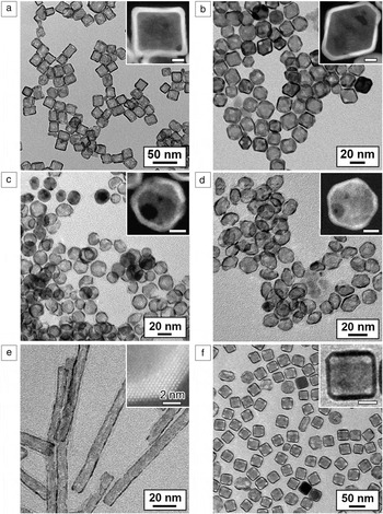
Figure 4a–d shows TEM images of the Pt-based nanocages derived from Pd nanocrystals with cubic, octahedral, icosahedral, and decahedral shapes, respectively.Reference Zhang, Roling, Wang, Vara, Chi, Liu, Choi, Park, Herron, Xie, Mavrikakis and Xia17–Reference He, He, Wang, Lin, Yin, Hong, Wu and Li19,Reference Wang, Vara, Luo, Huang, Ruditskiy, Park, Bao, Liu, Howe, Chi, Xie and Xia37 These nanocages were rich in Pt (>80 wt%). Significantly, by employing different Pd polyhedral nanocrystals as the templates, we were able to precisely control the atomic structures on the surface, including the presence of {100} and {111} facets, as well as twin boundaries. The synthetic method was also extended to other types of Pd templates such as nanorods and nanoplates for the generation of Pt-based nanotubes, prism-like nanocages, and nanorings.Reference Huang, Ruditskiy, Choi, Zhang, Liu, Ye and Xia38,Reference Wang, Luo, Huang, Chi, Howe, Xie and Xia39 Figure 4e shows a TEM image of a typical sample of Pt-based nanotubes, whose side faces and ends were covered by {100} and {111} facets, respectively, in addition to the penta-twinned structure.
Transmission electron microscope (TEM) images of Pt-based nanocages with (a) cubic, (b) octahedral, (c) icosahedral, and (d) decahedral shapes, which were fabricated by templating with cubic, octahedral, icosahedral, and decahedral nanocrystals of Pd, respectively. The insets show high-angle annular dark-field scanning TEM images of individual nanocages. Scale bars = 4 nm. (e) TEM image of Pt-based nanotubes derived from penta-twinned nanorods of Pd. The inset shows an atomic-resolution scanning TEM image of the corner region of a nanotube. (f) TEM image of Au-based nanocages derived from Ag@Au6L core–shell nanocubes, where the subscript 6L denotes six atomic layers in the shell. The inset shows a TEM image of an individual nanocage at a higher magnification. Scale bar = 15 nm. (a, b) Adapted with permission from Reference Reference Zhang, Roling, Wang, Vara, Chi, Liu, Choi, Park, Herron, Xie, Mavrikakis and Xia17. © 2015 AAAS. (c) Adapted with permission from Reference Reference Wang, Figueroa-Cosme, Yang, Luo, Liu, Xie and Xia18. © 2016 American Chemical Society. (d) Adapted with permission from Reference Reference Wang, Vara, Luo, Huang, Ruditskiy, Park, Bao, Liu, Howe, Chi, Xie and Xia37. © 2015 American Chemical Society. (e) Adapted with permission from Reference Reference Wang, Luo, Huang, Chi, Howe, Xie and Xia39. © 2017 American Chemical Society. (f) Adapted with permission from Reference Reference Sun, Kim, Gilroy, Liu, König and Qin40. © 2016 American Chemical Society.

In addition to Pd nanocrystals, templates made of less expensive metals such as Ag or Cu have also been explored in an effort to reduce the cost.Reference Sun, Kim, Gilroy, Liu, König and Qin40–Reference Aslam, Chavez and Linic42 However, these metals generally have lower reduction potentials than those of Pt2+/Pt or Au3+/Au, and thus the template is susceptible to destruction by the galvanic replacement reaction with PtCl42− or AuCl4− precursor. To address this issue, the Qin group at the Georgia Institute of Technology introduced a strong reductant, such as ascorbic acid, together with the use of an alkaline environment, to promote the reduction of the Pt or Au precursor and thereby suppress the galvanic replacement reaction.Reference Sun, Kim, Gilroy, Liu, König and Qin40 When the resultant Ag@PtnL or Ag@AunL core–shell nanocrystals were subjected to chemical etching with aqueous H2O2, the Ag cores could be selectively removed to generate Pt- or Au-based nanocages. In both cases, nanocages of approximately 40 nm in edge length could be obtained even when the wall thickness was reduced to 2 nm. Figure 4f shows a TEM image of a typical sample of the Au-based nanocages that were derived from Ag@Au6L core–shell nanocrystals.
Platinum-based nanocages for fuel-cell applications
As a novel class of catalytic materials, Pt-based nanocages showed substantially enhanced activity and durability toward ORR, a reaction key to the operation of PEMFCs. Our initial studies mainly focused on the cubic and octahedral nanocages fabricated from templates based upon Pd cubic and octahedral nanocrystals with edge lengths of 18 and 19 nm, respectively.Reference Zhang, Roling, Wang, Vara, Chi, Liu, Choi, Park, Herron, Xie, Mavrikakis and Xia17 Our results confirmed that the octahedral nanocages enclosed by {111} facets were more active than the cubic counterparts encased by {100} facets. The specific and mass activities of the octahedral nanocages were 1.98 mA•cm−2 and 0.75 A•mgPt−1, respectively, whereas those of the cubic nanocages were about 0.81 mA•cm−2 and 0.38 A•mgPt−1, respectively. For icosahedral nanocages, the surface is covered by 20 {111} facets and 30 twin boundaries, and their specific and mass activities went up to 3.50 mA•cm−2 and 1.98 A•mgPt−1, respectively. These values were almost 10× greater than those of the current state-of-the-art Pt/C catalyst.Reference Wang, Figueroa-Cosme, Yang, Luo, Liu, Xie and Xia18 The nanocage-based ORR catalysts also showed excellent stability when subjected to accelerated durability test. After 5000 cycles in the potential range of 0.6–1.1 V versus SHE, for example, the mass activity of the cubic and octahedral nanocages only decreased by 18% and 22%, respectively. When the number of cycles was increased to 10,000, 32% and 36% drops from the initial values were observed for the cubic and octahedral nanocages, respectively. A more substantial drop of nearly 40% was observed for the icosahedral nanocages after 5000 cycles. Nevertheless, the mass activity of the Pt-based icosahedral nanocages was still approximately four times (0.77 A•mgPt−1 versus 0.19 A•mgPt−1) as high as that of the pristine Pt/C catalyst.
The catalytic activity of the Pt-Ag alloy nanocages for ORR prepared through galvanic replacement showed variations as a function of time due to the change in elemental composition.Reference Yang, Roling, Vara, Elnabawy, Zhao, Hood, Bao, Mavrikakis and Xia32 When the initial Pt19Ag81 nanocages were subjected to electrochemical cycling in the potential range of 0.6–1.1 V versus SHE, the composition changed to Pt46Ag54, Pt56Ag44, Pt65Ag35, and Pt79Ag21 after 5000, 10,000, 20,000, and 30,000 cycles, respectively, due to the dissolution of Ag during electrochemical cycling. After 30,000 cycles, the Pt-Ag nanocages still showed mass activity twice as great as that of the commercial Pt/C catalyst. Based on density functional theory (DFT) calculations, the (100) surface with a composition of Pt50Ag50 gave the greatest specific activity because of the formation of the most stable O2 transition state, in agreement with the experimental result that showed peak activity for the Pt-Ag nanocages with elemental composition close to Pt50Ag50.
Gold-based nanocages for biomedical applications
Applying Au nanomaterials to cancer theranostics has been of great interest.Reference Faraday11 One can functionalize the Au surface with a variety of ligands through the established Au−S chemistry to present a well-defined and precisely engineered interface to interact with the biological system of interest.Reference Love, Estroff, Kriebel, Nuzzo and Whitesides43 One can systematically examine the effects of a given parameter on various biological processes, including cellular uptake, tissue penetration, blood circulation, biodistribution, tumor targeting, and body clearance. Other applications are based upon the unique optical properties of Au nanomaterials, including their strong absorption and scattering enabled by LSPR. The absorption cross-section of a Au nanostructure is typically 5−6 orders of magnitude greater than that of a dye molecule, making it a superb contrast agent for optical imaging modalities. More significantly, the LSPR peak of a Au nanostructure can be readily tuned into the NIR region (750–1200 nm) to meet the requirement for in vivo applications while its size, shape, and surface properties can be optimized to enhance the targeting efficacy. The high efficiency of photothermal heating intrinsic to Au nanostructures can also be leveraged to eradicate cancer cells and even ablate malignant tissue.Reference Chen, Glaus, Laforest, Zhang, Yang, Gidding, Welch and Xia44 Furthermore, Au nanostructures can serve as a carrier for the delivery of therapeutics, whose release at the diseased site can be controlled using NIR irradiation with good spatial and temporal resolutions.Reference Yavuz, Cheng, Chen, Cobley, Zhang, Rycenga, Xie, Kim, Song, Schwartz, Wang and Xia45–Reference Sun, Wang, Wang, Xu, Zhao, Vangveravong, Mach and Xia49
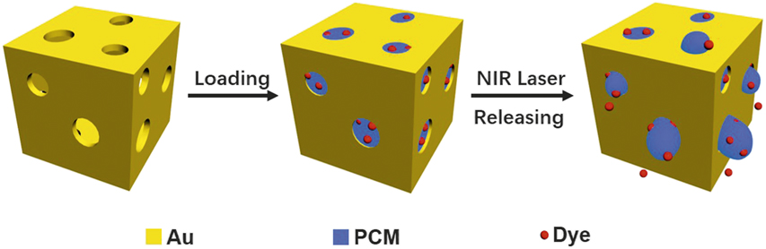
In one study, Au nanocages were exploited as a novel vector for drug delivery by leveraging their hollow interiors, porous walls, and strong absorption of light in the NIR region. Initially, we attached a thermoresponsive polymer brush to the outer surface of Au nanocages to control the release of an encapsulated drug using NIR irradiation.Reference Yavuz, Cheng, Chen, Cobley, Zhang, Rycenga, Xie, Kim, Song, Schwartz, Wang and Xia45 We then advanced the release system by switching to phase-change materials (PCMs) based upon fatty acids or alcohols (Figure 5).Reference Shen, Zhu, Huo, Yang, Xue and Xia46–Reference Sun, Wang, Wang, Xu, Zhao, Vangveravong, Mach and Xia49 As the PCM changes its physical state from solid to liquid at its melting point, the theranostic agent premixed with the PCM is released from the cavities. As long as the theranostic agent is miscible with the PCM, it can be readily loaded into the cavities of Au nanocages together with the PCM via diffusion. We could control the release profile by varying the irradiance of the NIR laser and the duration of exposure.Reference Shen, Zhu, Huo, Yang, Xue and Xia46 In an in vitro study, we further demonstrated that the surface of Au nanocages could be functionalized with a ligand to target the receptors overexpressed on breast cancer stem cells (CSCs), after their cavities had been filled with a mixture of Dox and PCM.Reference Sun, Wang, Wang, Xu, Zhao, Vangveravong, Mach and Xia49 The Au nanocages could target and effectively destruct CSCs through a combination of targeted photothermal heating and chemotherapy.
Schematic illustration showing how phase-change material, Au nanocage, and photothermal heating can be integrated for controlled release. Adapted with permission from Reference Reference Shen, Zhu, Huo, Yang, Xue and Xia46. © 2017 Wiley. Note: NIR, near infrared; PCM, phase-change material.

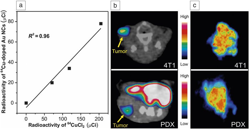
To track and quantify Au nanocages in vivo, we recently developed a robust method for their radio labeling with Cu-64 atoms and imaging by positron emission tomography (PET).Reference Yang, Huo, Gilroy, Sun, Sultan, Luehmann, Detering, Li, Qin, Liu and Xia50,Reference Yang, Liu, Fu and Qin51 The optical properties of the Cu-doped Au nanocages resembled those of the conventional nanocages, displaying strong and tunable absorption in the NIR region. When radioactive 64CuCl2 was partially used as a source for Cu deposition, we obtained Au nanocages with well-controlled radioactivity for imaging by PET (Figure 6a). After PEGylation (surface functionalization with poly(ethylene glycol) [PEG]), the radioactive Au nanocages were found to accumulate effectively in the tumor through the enhanced permeation and retention effect (see Figure 6b–c). We also sectioned the tumor at 24 h post injection into 40-μm-thick slices for autoradiographic analysis and found that the nanocages were uniformly distributed throughout the tumor regardless of the animal model.
(a) Linear correlation between the radioactivity of 64Cu-doped Au nanocages (NCs) and the amount of 64CuCl2 co-titrated with HAuCl4 into the reaction system. (b) Transverse positron emission tomography images of 40-nm PEGylated (surface functionalization with poly(ethylene glycol) [PEG]) 64Cu-Au nanocages in (upper) 4T1 (orthotopic, with the 4T1 tumor cells implanted in the breast fat pad) and (lower) patient-derived xenograft (PDX, where the cancer cells were derived from the tumor tissue of a human patient and implanted in flank) mouse models at 24 h post tail vein injection (30 μCi). (c) Auto-radiographs showing the intratumoral distributions of the Au nanocages. The color bars represent the signal intensity of accumulated 64Cu-doped Au nanocages. Adapted with permission from Reference Reference Yang, Huo, Gilroy, Sun, Sultan, Luehmann, Detering, Li, Qin, Liu and Xia50. © 2017 Wiley. Note: NIR, near infrared.

Ruthenium-based nanocages for potential use in ammonia synthesis
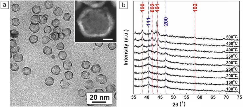
Ruthenium has found widespread use in an array of catalytic applications, including ammonia synthesis. However, extending the template-directed synthesis to Ru is challenging because it has a hexagonal close-packed (hcp) lattice rather than the face-centered-cubic (fcc) structure taken by other precious metals such as Pd, Pt, Ag, Au, Rh, and Ir. Nevertheless, our group recently demonstrated the synthesis of Ru-based cubic, octahedral, and icosahedral nanocages with walls as thin as five atomic layers (Figure 7a).Reference Zhao, Figueroa-Cosme, Elnabawy, Vara, Yang, Roling, Chi, Mavrikakis and Xia52–Reference Zhao, Lang, Vara, Elnabawy, Gilroy, Hood, Zhou, Figueroa-Cosme, Chi and Xia54 More interestingly, during deposition, the Ru atoms were packed into an fcc structure, rather than hcp, as a result of the influence of the Pd template. When the Pd core was removed through chemical etching, the resultant Ru nanocage was able to maintain the fcc structure. These transformed icosahedral nanocages enclosed by multiple twin boundaries were able to retain their icosahedral shape and the fcc structure at temperatures as high as 300°C.Reference Zhao, Lang, Vara, Elnabawy, Gilroy, Hood, Zhou, Figueroa-Cosme, Chi and Xia54 DFT calculations suggested that the fcc-structured Ru nanocages, when doped with some Pd impurities, were catalytically more active than the hcp-structured Ru nanoparticles toward nitrogen reduction for ammonia synthesis.
(a) Transmission electron microscope (TEM) image of the Ru-based icosahedral nanocages prepared by templating with Pd icosahedral nanocrystals. The inset shows a high-resolution TEM image of a nanocage. Scale bar = 5 nm. (b) X-ray diffraction patterns recorded from the Ru-based icosahedral nanocages upon in situ heating to different temperatures, indicating that the face-centered-cubic (fcc) structure could be retained up to 300°C. The characteristic peaks of the fcc and hexagonal close-packed structures were marked by blue and red dashed lines, respectively. Reprinted with permission from Reference Reference Zhao, Lang, Vara, Elnabawy, Gilroy, Hood, Zhou, Figueroa-Cosme, Chi and Xia54. © 2018 American Chemical Society.

Hollow versus core–shell nanocrystals
While we have mainly focused on nanocages in this article, the core–shell nanocrystals used to generate nanocages may also provide the same function in terms of catalysis.Reference Xie, Choi, Lu, Roling, Herron, Zhang, Park, Wang, Kim, Xie, Mavrikakis and Xia55–Reference Tan, Luo, Nan, Zou, Chen, Shu, Liu, Li, Song, Liao and Adzic58 Of course, blocking the inner surface of the shell will reduce the utilization efficiency of atoms and thus compromise the mass activity. However, the presence of the core, which serves as a support, allows one to reduce the shell thickness to one atomic layer, giving a utilization efficiency of 100%, without concerns about possible deformation of the shell.
In two recent studies, we demonstrated that the Pt shells in the Pd@Pt2.7L core–shell icosahedra could be well retained even after heating to 300°C, while pores started to appear on the surface of Pt-based icosahedral nanocages at 200°C.Reference Wang, Choi, Roling, Luo, Ma, Zhang, Chi, Liu, Xie, Herron, Mavirkakis and Xia57,Reference Vara, Wang, Howe, Chi and Xia59 As another benefit of core–shell nanocrystals, the inclusion of a second metal with an electronegativity different from the metal in the shell can modulate the electronic structure of the surface and thereby enhance its catalytic activity. Additionally, the strain field caused by the lattice mismatch between the core and the shell can also give rise to an enhancement in catalytic activity.
Despite these advantages, there are a number of issues in applying core–shell nanocrystals to catalytic applications at elevated temperatures. First, the shell composition might undergo constant changes during the operation of a catalytic device due to diffusion at elevated temperatures. The in situ electron microscopy study suggested that the Pd@Pt4L core–shell octahedra gradually evolve into Pd-Pt alloy octahedra under thermal stress, accompanied by compositional changes from Pt-rich to Pd-Pt alloys for the shell and the surface.Reference Vara, Wang, Howe, Chi and Xia59,Reference Vara, Roling, Wang, Elnabawy, Hood, Chi, Mavrikakis and Xia60 Second, the material used for the core has to be sufficiently abundant in order to achieve cost-effective and sustainable use of a precious metal. At the current stage of development, Pd is still the best choice of template material for the production of core–shell nanocrystals, but possible replacement candidates for Pd include Ag, Cu, Fe, Co, and Ni.
Conclusion
The past decade has witnessed significant progress in the use of precious metals in a broad range of applications by switching from poorly defined nanoparticles to nanocrystals with well-controlled compositions, sizes, shapes, and structures. Inspired by the concept of paper origami, we have synthesized nanocages with substantially increased utilization efficiency for the constituent atoms and tunable optical absorption in the visible and NIR regions. Depending on the elemental composition, the nanocages have shown promise for applications as diverse as energy conversion, ammonia synthesis, and cancer theranostics.
Despite impressive progress, we still need to address a number of issues. For example, since the nanocage structure is far from equilibrium, it is inevitably susceptible to changes during the operation of a catalytic device. This can potentially be addressed by employing an inexpensive material (metal or inorganic compound) as the template to produce core–shell nanocrystals, which can be directly used as catalysts with enhanced thermal stability. Another issue lies in the difficulty in producing nanocages at an industrial scale while still maintaining a tight control over their size, shape, and uniformity. This can be addressed by switching the synthesis from batch reactors to continuous-flow systems, where the throughput can be improved more than 200×.Reference Niu, Ruditskiy, Vara and Xia61–Reference Wang, Niu, Zhou, Wang, Park, Bao, Chi, Cai and Xia66 We hope this article offers new insights, from the perspective of materials science, for achieving cost-effective and sustainable use of scarce elements without compromising their functionality and performance.
Acknowledgments
This work was supported in part by research grants from the National Science Foundation (DMR-1505400 and CHE-1505441), the National Institutes of Health (R01 CA138527), and startup funds from the Georgia Institute of Technology. We are grateful to our collaborators for their invaluable contributions to this research.

Younan Xia has been the Brock Family Chair and Georgia Research Alliance (GRA) Eminent Scholar in Nanomedicine at the Georgia Institute of Technology since 2012. He holds joint appointments in the Department of Biomedical Engineering, School of Chemistry and Biochemistry, and School of Chemical and Biomolecular Engineering. He received his BS degree from the University of Science and Technology of China in 1987, MS degree from the University of Pennsylvania in 1993, and PhD degree in physical chemistry from Harvard University in 1996. He started as an assistant professor of chemistry at the University of Washington (Seattle) in 1997, and was promoted to associate professor and full professor in 2002 and 2004, respectively. His current research interests include nanomaterials, biomaterials, nanomedicine, regenerative medicine, optical imaging, electrospinning, and colloid science. Xia can be reached by email at younan.xia@bme.gatech.edu.

Ming Zhao joined the Georgia Institute of Technology in 2015 as a graduate student and is pursuing his PhD degree in chemistry in the School of Chemistry and Biochemistry. He received his BS degree in materials science and engineering and MS degree in materials physics and chemistry in 2012 and 2015, respectively, both from Nanjing University, China. His MS thesis work was focused on the durability testing of proton-exchange membrane fuel cells. His research interests include the synthesis of nanomaterials for catalysis- and energy-related applications. Zhao can be reached by email at mzhao70@gatech.edu.

Xue Wang is a postdoctoral fellow at the University of Toronto, Canada. She earned her BS degree in chemistry at Liaoning University in 2009, and received her PhD degree in physical chemistry from Xiamen University in 2015. She spent two years in the Xia group as a visiting graduate student. She was as an associate professor at Lanzhou Institute of Chemical Physics, Chinese Academy of Sciences, for two years. Her current research interests include the design and synthesis of nanomaterials for applications in electrocatalysis. Wang can be reached by email at xxue.wang@utoronto.ca.

Da Huo joined the Georgia Institute of Technology as a postdoctoral fellow in 2015. He received his BS degree in 2010 and PhD degree in 2015, both in materials science and engineering from Nanjing University, China. His current research interests include the development of novel nanomaterials for cancer theranostics. Huo can be reached by email at da.huo@bme.gatech.edu.