

1. Introduction
The marine vertebrate fauna of the Triassic Guanling Formation has become well known recently, occurring mainly in boundary area between Yunnan and Guizhou provinces in southwestern China (Jiang et al. Reference Jiang, Schmitz, Hao and Sun2006; Reference Jiang, Rieppel, Motani, Hao, Sun, Schmitz and Sun2008, Reference Jiang, Motani, Hao, Rieppel, Sun, Tintori, Sun and Schmitz2009; Motani et al. Reference Motani, Jiang, Tintori, Sun, Hao, Boyd, Hinic-Frlog, Schmitz, Shin and Sun2008; Wang et al. Reference Wang, Chen, Chen, Wang, Bachmann, Sander and Hagdorn2009; Wu et al. Reference Wu, Cheng, Li, Zhao and Sato2011; Liu et al. Reference Liu, Rieppel, Jiang, Aitchison, Motani, Zhang, Zhou and Sun2011; Shang, Wu & Li, Reference Li, Rieppel, Wu, Zhao and Wang2011). Conodonts dated the fossil yielding strata (Upper Member) of the Guanling Formation to the Pelsonian substage of the Anisian, Middle Triassic (Sun et al. Reference Sun, Hao, Sun and Jiang2006; Zhang et al. Reference Zhang, Zhou, Lu, Lou, Liu, Sun, Huang and Zhao2009). This fauna has been recently enriched by the discovery of a new member, Sinosaurosphargis yunguiensis Li et al. Reference Li, Rieppel, Wu, Zhao and Wang2011, of the Saurosphargidae Li et al. Reference Li, Rieppel, Wu, Zhao and Wang2011 from Luoping County, Yunnan. Early in 2012, a new marine reptile, Largocephalosaurus polycarpon Cheng et al. Reference Cheng, Chen, Zeng and Cai2012a was briefly described and attributed to the Eosauropterygia. Our further examination of the specimen suggests that this reptile is most probably referable to the Saurosphargidae and represents a new morphotype of the family. This is supported by the discovery of a new species Largocephalosaurus qianensis (see Section 3) from the same horizon of southwestern Guizhou, about 100 km northeast of the type locality of L. polycarpon in Luoping County, Yunnan.
L. polycarpon was originally established primarily based on an incomplete skull because the preparation of the postcranial skeleton of the specimen, WIGM (Wuhan Institute of Geology and Mineral Resources) SPC V 1009, was then incomplete. It was considered to be an eosauropterygian and closely related to the Nothosauroidea–Pachypleurosauria clade within the Sauropterygia (Cheng et al. Reference Cheng, Chen, Zeng and Cai2012a ). Whilst this specimen was being studied, research groups at GMPKU (Geological Museum of Peking University) and IVPP (Institute of Vertebrate Paleontology and Paleoanthropology, Chinese Academy of Sciences) were working on three similar specimens collected from the Upper Member of the Guanling Formation in Panxian, Guizhou Province in 2008. The GMPKU specimen was briefly reported as Saurosphargis cf. S. volzi (Jiang et al. Reference Jiang, Rieppel, Motani, Hao and Tintori2011). These new specimens are better preserved than that of the type species, and clearly show the presence of dorsal osteoderms and a closed dorsal ‘rib-basket’ formed by the underlying ribs. Our further examination demonstrates that those features are also present in L. polycarpon as shown by the partly prepared postcranial skeleton. Therefore, it is evident that Largocephalosaurus is clearly not an eosauropterygian but instead may be closely related to the Saurosphargidae. In morphology, the three new specimens are different enough from L. polycarpon and represent a new species; the most striking of those differences are: the presence of three premaxillary teeth, an oval orbit, a large parietal foramen, two (rather than three) sacral vertebrae, a unique pattern of osteoderms, and a short retroarticular process. Here, we describe the osteological anatomy of the new species, revise the systematics of Largocephalosaurus, and further test the phylogenetic relationships of the Saurosphargidae with other marine reptilian groups.
2. Material and methods
All three specimens were preserved in blocks of limestone or muddy limestone. One is a nearly complete skeleton, housed at the IVPP. As for the other two specimens housed at the GMPKU, one is represented by a skull and a cervical vertebra and the other is an incomplete skeleton with the skull and most of the tail missing. All specimens were mechanically prepared using airscribes with tips of various sizes. The IVPP specimen was exposed in ventral view but its skull was prepared on both dorsal and ventral sides. The two GMPKU specimens were exposed only in dorsal view.
3. Systematic palaeontology
Order DIAPSIDA Osborn, Reference Osborn1903
Family saurosphargidae Li et al. Reference Li, Rieppel, Wu, Zhao and Wang2011
Type genus. Saurosphargis Huene, Reference Huene1936.
Referred genera. Sinosaurosphargis Li et al. Reference Li, Rieppel, Wu, Zhao and Wang2011; Largocephalosaurus Cheng et al. Reference Cheng, Chen, Zeng and Cai2012a (originally described as an eosauropterygian).
Diagnosis. Aquatic diapsids characterized by the following combination of apomorphies: (1) dorsal ribs forming a closed basket; (2) presence of dorsal osteoderms; (3) external naris retracted, much closer to orbit than rostral tip; (4) median elements of gastral ribs often with a two-pronged lateral process on one side; (5) lateral most elements of gastral ribs broadened and contacting each other; (6) supratemporal extensively contacting quadrate shaft; (7) posterior margin of skull roof deeply emarginated; (8) jugal–squamosal contact; (9) presence of ectopterygoid; (10) presence of interpterygoid vacuity and open braincase-palatal articulation; (11) leaf-shaped tooth crown with convex labial surface and concave lingual surface; (12) dorsal vertebrae with elongate transverse process and a very low neural spine; (13) tip of neural spines table-like, covered by osteoderm(s); (14) large interclavicle boomerang-like or atypical T-shaped, with a small and sharp posterior process; (15) humerus not expanded at both ends; (16) nine carpals; and (17) four tarsals.
Remarks. Saurosphargis, the type genus of the Saurosphargidae, from the Middle Triassic of Europe, is a poorly known taxon; it is only represented by a section of 12 incomplete dorsal vertebrae with ribs (see Nosotti & Rieppel, Reference Nosotti and Rieppel2003, fig. 11). The diagnosis of the family was not defined when the family was established and is here largely based on two Chinese genera: Sinosaurosphargis and Largocephalosaurus.
Largocephalosaurus Cheng et al. Reference Cheng, Chen, Zeng and Cai2012a
Type species. Largocephalosaurus polycarpon Cheng et al. Reference Cheng, Chen, Zeng and Cai2012a .
Revised diagnosis. A saurosphargid genus differing from the others of the family in the following combination of apomorphies: (1) closed dorsal ‘rib-basket’ elongate oval; (2) oval supratemporal fenestra present, much smaller than orbit; (3) premaxilla excluded from external naris; (4) three or four premaxillary teeth; (5) posterolateral process of frontal elongate; (6) transverse process of dorsal vertebrae and proximal portion of the dorsal ribs slender, not much wider than inter-process or inter-rib spaces, respectively; (7) presence of a median row of large osteoderms on the top of neural spines; (8) coracoid and pubis similarly round in outline, separately with an open coracoid foramen and an open obturator fenestra; and (9) dorsal osteoderms not forming a complete carapace.
Remarks. Most of the diagnostic characters for Largocephalosaurus are unknown in Saurosphargis owing to the fragmentary nature of the latter. However, characters (1) and (6) distinguish the two genera. In Saurosphargis the body should have been broader than in Largocephalosaurus in terms of a large turning angle at the shoulder region of the dorsal ribs; this condition is comparable to that of Sinosaurosphargis. Similarly, the transverse process of the dorsal vertebrae and the proximal (dorsal) portion of the dorsal ribs are very similar between Saurosphargis and Sinosaurosphargis in morphology; they are much broader than the inter-process or inter-rib spaces.
Largocephalosaurus qianensis sp. nov.
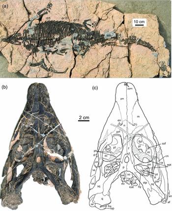
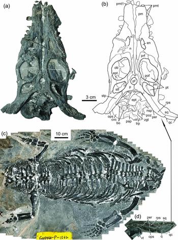
Figures 1, 2, 3, 4a–g, 5a–d, 6
Etymology. Specific name refers to the simplified name ‘Qian’ for Guizhou Province where the specimens were collected.
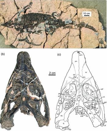
Type specimen of Largocephalosaurus qianensis sp. nov. (IVPP V 15638). (a) Skeleton in ventral view; (b) skull and mandible in dorsal view; (c) line drawing of (b). Abbreviations: af – articular fossa; an – angular; cn – coronoid; d – dentary; dr – dorsal rib; ec – ectopterygoid; en – external naris; eo – exoccipital; ept – epipterygoid; f – frontal; fpl – fossa on palatine; fra – foramen on retroarticular process; hy – hyoid; icat – atlantal intercentrum; j – jugal; m – maxilla; n – nasal; nat – neural arch of atlas; od – odontoid process; p – parietal; par – prearticular; pat – proatlas; pl – palatine; pm – premaxilla; po – postorbital; pof – postfrontal; ppm – palatal portion of premaxilla; pr – prootic; prf – prefrontal; pt – pterygoid; q – quadrate; rap – retroarticular process; sa – surangular; sim – possible skin impression; so, supraoccipital; sof – suborbital fenestra; sq – squamosal; st – stapes; stp – supratemporal.
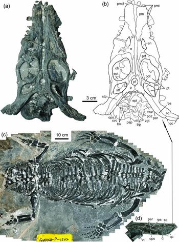
Referred specimens of Largocephalosaurus qianensis sp. nov. (a) Skull (GMPKU-P-1532-A) in dorsal view; (b) line drawing of (a); (c) skeleton (GMPKU-P-1532-B) in dorsal view; (d) right posterolateral portion of (a) in posterior and slightly dorsal view, interpretative lines have been added to highlight the structures of the relevant bones. Abbreviations as in Figure 1 plus: bo – basioccipital; cvc – centrum of an anterior cervical vertebra; cr – cervical rib; lg – lateral most gastral elements; nsp – neural spine; ob – orbital; ops – occipital portion of supratemporal; pap – paroccipital process; per – posterior edge of skull roof; pf – pineal foramen; pmt – premaxillary tooth; poz – postzygapophysis; qc – quadrate condyle; rps – roof portion of supratemporal; stf – supratemporal fenestra; trp – transverse process of vertebra; vt – vertebra; zgt – zygantrum.
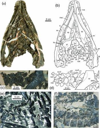
Skull and selected postcranial parts of Largocephalosaurus qianensis sp. nov. (a) to (d) and (f) type specimen (IVPP V 15638), (e) GMPKU-P-1532-B. (a) Skull in ventral view; (b) line drawing of (a); (c) most of cervical region in ventral view; (d) line drawing of (c); (e) cervical region in dorsal view, arrow indicating the displacement of cervical vertebra 8; (f) mid caudal vertebrae in right lateral and slightly ventral view, with many small, elongated granular osteoderms. Abbreviations as in Figures 1 and 2 plus: ar – articular; ax – axis; bs – basisphenoid; cav – caudal vertebra; cv – cervical vertebra; dv – dorsal vertebra; fec – facet for ectopterygoid; icat – intercentrum of atlas; icl – interclavicle; mec – Meckel's canal; nax – neural arch of axis; ncv – neural arch of cervical vertebra; ppicl – posterior process of interclavicle; prq – pterygoid ramus of quadrate; prz – prezygapophysis; qrp – quadrate ramus of pterygoid; sp – splenial; 1mt – first maxillary tooth; XII – cranial nerve 12.

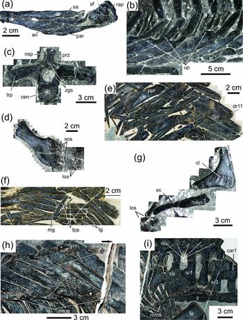
Various anatomical regions of Largocephalosaurus. (a) to (g) L. qianensis sp. nov.; (h) and (i) L. polycarpon (WIGM SPC V1009). (a) to (d) and (g) GMPKU-P-1532-B, (e) and (f) type specimen (IVPP V 15638). (a) Posterior portion of a right mandible in medial view, interpretative lines have been added to highlight the articular fossa; (b) lateral most portion of the right mid dorsal ‘rib-basket’ in dorsal view, showing the uncinate-like process of the dorsal ribs; (c) possibly the 8th cervical vertebra in anterior view, showing a single broad zygosphene; (d) left scapula in lateral view; (e) left mid dorsal ‘rib-basket’ in ventral view, showing a sharp rib angle (pointed by the arrow) between proximal and distal portions; (f) a disarticulated median gastral element with a two pronged lateral process on one side and the sheet-like structure formed by the lateral-most gastral elements; (g) right clavicle in anterolateral view and scapula in anterior view; (h) mid-portion of the left half of the trunk, showing two rows of osteoderms and dorsal crests of the trunk ribs in the type species; (i) left half of the sacral region, showing three sacral vertebrae and the connection of the last two dorsal ribs and the first sacral rib. Arrow in (e) and (h), indicates the turning angle and the head direction, respectively. Abbreviations as in Figures 1 to 3 plus: car – caudal rib; cdr – crest on dorsal surface of trunk ribs; cl – clavicle; cen – centrum of vertebra; dpr – distal portion of rib; dr – dorsal rib; fe – femur; il – ilium; ldr – the last dorsal rib; los – large osteoderms; lros – lateral row of osteoderms; mg – median gastral element; mros – medial row of osteoderms; os – osteoderm; ppr – proximal portion of rib; sc – scapula; sos – small osteoderms; sr – sacral rib; tpp – a two pronged lateral process of median gastral element on one side; up – uncinate process; zgs – zygosphene.

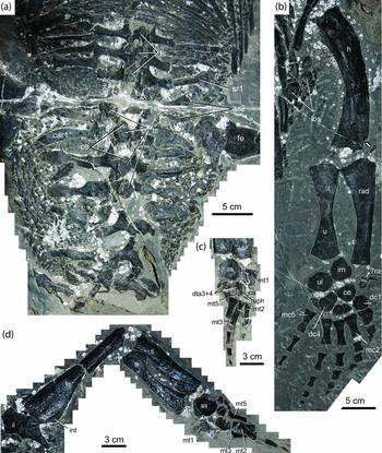
Girdles, limbs and caudal osteoderms of (a–d) Largocephalosaurus qianensis sp. nov. and (e) Sinosaurosphargis yunguiensis. (a), (b) and (d) IVPP V 15638 and (c) GMPKU-P-1532-B; arrows indicate dorsal lines or rows of small osteoderms in ventral view in (a) and (d); (a) anterior trunk in ventral view; (b) caudal osteoderms; (c) left manus in dorsal view; (d) sacral region in ventral view; (e) left side of the pectoral girdle of ZMNH M 8797 in dorsal view. Interpretative lines have been added to highlight the structures of relevant bones in (a), (d), (e). Abbreviations as in Figures 1 to 4 plus: as – astragalus; ca – calcaneum; ce – centrale; ch – chevron; co – coracoid; dc – distal carpal; dta – distal tarsal; fi – fibula; h – humerus; im – intermediate; int – internal trochanter; is – ischium; mc – metacarpal; mt – metatarsal; pu – pubis; ra – radiale; rad – radius; sv – sacral vertebra; ti – tibia; u – ulna; ul – ulnare.

Pelvic girdle and limbs of Largocephalosaurus qianensis sp. nov. in dorsal view (GMPKU-P-1532-B). (a) Sacral region, showing large and small osteoderms; (b) right forelimb; (c) left pes; (d) right hindlimb. Interpretative lines have been added to highlight the structures of relevant bones. Arrow in (b) indicates the ectepicondylar notch. Abbreviations as in Figures 4 and 5 plus: ts – dorsal table of neural spine; uph – ungual phalanx.

Holotype. IVPP V 15638, a nearly complete skeleton in ventral view, with posterior-most section of the tail missing.
Referred specimens. GMPKU-P-1532-A, a skull with a cervical vertebra in dorsal view. GMPKU-P-1532-B, an incomplete postcranial skeleton in dorsal view, with the posterior portion of the mandible but missing most of the tail.
Type locality and horizon. Xinmin District, Panxian County, southwest-most Guizhou Province, P. R. China; Member II of the Guanling Formation, Anisian, Middle Triassic.
Referred locality and horizon. As for the type.
Diagnosis. Differing from the type species in the following characters: (1) parietal paired; (2) pineal foramen large; (3) orbit oval in outline; (4) a distinct palatal fossa on the dorsal surface of palatine; (5) dorsal osteoderms elongated granular in outline and covering the lateral side of body, mixed with large and nearly round ones; (6) retroarticular process very short; (7) shoulder portion of dorsal ribs with no crest on dorsal surface but broadening as an uncinate-like process; (8) 33 presacral vertebrae (including 24 dorsals); and (9) two sacral vertebrae.
Remarks. The genotype species, L. polycarpon, has not been fully described because the preparation of the postcranial skeleton was not completed when it was erected. A number of characters listed in the diagnosis of the new species were recognized on the basis of the comparison with the partly prepared skeleton of the type species. A full description of L. polycarpon is in preparation.
4. Morphological description
The following description is based on IVPP V 15638 (the type specimen) unless the referred specimens (GMPKU-P-1532-A and GMPKU-P-1532-B) are identified.
4.a. Skull and mandible
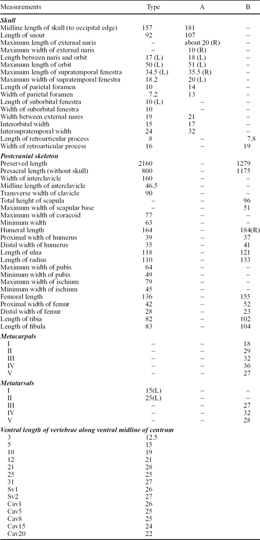
The skull is dorsoventrally compressed. The fronto-parietal skull table is well preserved in both IVPP V 15638 and GMPKU-P-1532-A. The region around the external nares is badly crushed in the former (Fig. 1c, d), but it is relatively better preserved in the latter (Fig. 2a, b). The rostrum, mainly formed by the premaxillae, is distinct and anteriorly rounded with parallel lateral margins as in Sinosaurosphargis. Posterior to the premaxilla–maxillary suture, the skull gradually widens, reaching its maximum width at the point between the posterior third of the orbits; the latter are of oval contour in lateral view, differing from the round outline in the type species (Cheng et al. Reference Cheng, Chen, Zeng and Cai2012a , fig. 2). The orbital margins form a thickened, granulated rim as in Sinosaurosphargis (Li et al. Reference Li, Rieppel, Wu, Zhao and Wang2011, fig. 2A). Our observations suggest that the smooth orbital margins of the type species are not original but a result of surficial erosion. The supratemporal fenestra remains open but is small, reaching a size of less than half that of the orbits, as in the type species. The pineal foramen is located anteriorly on the parietal skull table as in Sinosaurosphargis but much larger than in the latter, and even larger than that of the type species. The ventral cheek region may have been deeply embayed in life, suggesting a large infratemporal fenestra as in Sinosaurosphargis and Hanosaurus (Rieppel, Reference Rieppel1998a , fig. 2) although it is obscured by the dorsoventral compression of the skull. The posterior margin of the skull roof is strongly emarginated, showing a deep V-shaped outline. The braincase is obscured by the extensive overlap of the skull roof bones, which has been further exaggerated by the displacement of the relevant elements owing to the dorsoventral compression (for skull measurements see Table 1).
Selected measurements (in mm) of Largocephalosaurus qianensis sp. nov. from the type (IVPP V 15638), referred specimens GMPKU-P-1532-A (A) and GMPKU-P-1532-B (B)

Cav – caudal vertebra; R – right; L – left; Sv – sacral vertebra.
The paired premaxillae are relatively large as in many of the other aquatic sauropterygians (Rieppel, Reference Rieppel2000a ). Their dorsal surface around the rostral end is distinctly granulated, but pitted and grooved in other areas. The palatal surface of the rostrum is not exposed. The premaxilla, as shown in GMPKU-P-1532-A, is excluded by the anterior maxilla–nasal contact from the margin of the external naris (Fig. 2a, b), as in the type species; in contrast, no such a maxilla–nasal contact separates the bone from the naris in Sinosaurosphargis. The premaxilla meets the maxilla in the ventral margin of the upper jaw far posterior to the anterior end of the rostrum as in the latter, but the suture of the two bones is gently curved, extending posterodorsally as in the type species. The premaxilla bears only three teeth, unlike four teeth reported for the type species, five or six in Sinosaurosphargis, or five preserved in Eusaurosphargis, a similar aquatic reptile (Nosotti & Rieppel, Reference Nosotti and Rieppel2003); all three teeth are in situ on the left side but there are two plus an alveolus on the right side (Fig. 3a, b). The teeth are implanted in shallow sockets, in a sub-thecodont pattern. The tooth morphology is similar to that of Sinosaurosphargis (Li et al. Reference Li, Rieppel, Wu, Zhao and Wang2011, fig. 2E) and Eusaurosphargis (Nosotti & Rieppel, Reference Nosotti and Rieppel2003, fig. 3), with a basally expanded crown that is set off from a distinctly waisted peduncle and striations on the labial surface of the crown. The monocuspid tooth crown has a convex labial and a strongly concave lingual surface covered by striated enamel. In GMPKU-P-1532-A, the anterior-most tooth of the left premaxilla is exposed and the three teeth of the right premaxillary are also visible in dorsal view although none of them is complete (Fig. 2a, b). Our examination indicates that none of the preserved teeth is complete in the type species and the description of the tooth morphology for the species was not accurate. Striations and a waisted peduncle traceable in some teeth suggest that the type species most probably had similar tooth morphology.
The nasals of GMPKU-P-1532-A are complete and similar to those of the type species. They form a pair of rather large, plate-like elements that are slightly shorter but broader in the mid portion than the frontals (Fig. 2a, b). The nasal becomes narrower at both anterior and posterior ends. It forms the posterior and much of the dorsal margins of the external naris. Laterally, the strongly expanded mid-part meets the ascending (facial) process of the maxilla. Posterolaterally, the nasal-prefrontal suture is simple, convex towards the nasal. Posteriorly, the nasal–frontal suture is interdigitating. The dorsal surface of the nasals is distinctly grooved and ridged; the grooves and ridges are arranged in longitudinal rows on the posterior part of the bone.
The paired frontals contribute, compared with those of Sinosaurosphargis, a smaller part to the dorsal margin of the orbit. The broader contribution of the bone to the orbit in the type species may be not true because surface erosion has obscured the sutural patterns of the frontal with the neighbouring elements. Laterally, the frontal contacts the prefrontal anteriorly and the postfrontal posteriorly. Posteriorly, the frontal forms an elongate process that extends posteriorly and slightly laterally beyond the pineal foramen and approaches the supratemporal fenestra, as in the type species. This results in strongly concave posterior margins of the frontals and deeply interdigitating fronto-parietal sutures. Such a process is commonly present in European pachypleurosaurs such as Neusticosaurus and Serpianosaurus (Carroll & Gaskill, Reference Carroll and Gaskill1985; Sander, Reference Sander1989; Rieppel, Reference Rieppel1989) or the pachypleurosaur-like Chinese forms such as Wumengosaurus (Wu et al. Reference Wu, Cheng, Li, Zhao and Sato2011) and Diandongosaurus (Shang, Wu & Li, Reference Shang, Wu and Li2011) but not in Sinosaurosphargis. The dorsal surface of the frontals shows grooves and ridges arranged in a similar pattern to that seen in the nasals.
The parietals are paired (Figs 1b, c; 2a, b); unlike the type species in which they are fused posterior to the pineal foramen, or Sinosaurosphargis where they are entirely fused. The parietals form narrow anterior processes inserting into the frontals; each has a small anterolateral process to meet the postfrontal along the anteromedial margin of the supratemporal fenestra. The parietal broadens between the supratemporal fenestrae. Posterior to the end of the dorsal midline, the parietal forms a short posterolateral process along the deeply excavated occipital edge as in the type species which is different from the condition seen in Sinosaurosphargis (but see below). The dorsal surface of the parietal is weakly ridged and grooved.
The right maxilla of GMPKU-P-1532-A is nearly complete. It is large and forms a well-defined ascending process that extends dorsally posterior to the external naris and inserts a tip between the nasal and the prefrontal as in the type species (Fig. 2a, b). The maxilla sends a small anterodorsal process to meet the nasal and form the anterodorsal margin of the external naris posteriorly and meets the premaxilla anterodorsally. The lateral margin of the external naris is entirely formed by the maxilla. Along the margin of the upper jaw, the maxilla forms a slender, tooth-bearing posterior process that tapers to a tip at the level of the anterior three-fifths of the ventral margin of the orbit (Fig. 1b, c). The suture separating the ascending process of the maxilla from the prefrontal is simple. The tooth count of the maxilla cannot be established in either IVPP V 15638 (due to the occlusion of the mandible) or GMPKU-P-1532-A (where the dentition is not exposed). There are four and two teeth exposed in situ in the left and right maxillae of IVPP V 15638, respectively, and two and three teeth separately seen in the right and left maxillae of GMPKU-P-1532-A. The morphology of the maxillary teeth is the same as those of the premaxilla.
The complete prefrontal of GMPKU-P-1532-A is roughly hook-like in outline, with a short but broad anterolateral process forming the anterior margin of the orbit and a slender dorsal portion forming most of the dorsal edge of the orbit, contributing much more to the orbit than the frontal. As in other aquatic reptiles such as sauropterygians, no lacrimal foramen is present at the anterior margin of the orbit.
The nearly complete postfrontal and postorbital of GMPKU-P-1532-A are both triradiate in dorsal view (Fig. 2a, b). The former forms the granulated posterodorsal rim of the orbit anteriorly and the anteromedial margin of the supratemporal fenestra posteriorly. The dorsal surface of the postfrontal is heavily ornamented with ridges. The ornamentation of this bone is weak in IVPP V 15638 (Fig. 1b, c).
The postorbital is slightly larger than the postfrontal. Its descending ramus is shorter than the other two rami which form the major part of the bar between the orbit and the supratemporal fenestra and the supratemporal arcade, respectively. The postorbital–jugal contact is clear; the former articulates with the anterodorsal side of the latter. The postorbital–postfrontal suture is sharply curved while the postorbital–squamosal suture is strongly interdigitating, with a pointed posterior end of the postorbital inserting into the squamosal. The dorsal surface of the postorbital is weakly rugose. The relationships of the postorbital with the postfrontal and squamosal were obscured in the type species. The entrance of the squamosal to the orbit as described for the type species is questionable. Our examination suggests that the morphology of the postorbital in the type species is similar to that of the new species, with a large posterior (squamosal) process nearly forming the entire lateral edge of the supratemporal fenestra.
The jugal is well articulated with the neighbouring bones in GMPKU-P-1532-A; it is a simply curved, rod-like element, differing from the ‘boomerang’ shape of the bone in Sinosaurosphargis. It forms the ventral border of the orbit anteriorly and tapers to a tip posterodorsally to insert between the postorbital and the squamosal as in Sinosaurosphargis. Anteroventrally, the jugal sits on the posterior process of the maxilla. The lateral surface of the jugal is strongly convex along its length, with very light ornamentation. As with other skull bones, the morphology of the jugal is obscured owing to poor preservation; the sutures of the jugal with the maxilla and postorbital cannot be verified in the type species.
The squamosal is a large and irregular bone that was flattened during the process of fossilization. It is characterized by an elongate anterolateral process that meets the jugal to form the dorsal border of the ventrally opened infratemporal fenestra; in other words, the postorbital is excluded from the infratemporal fenestra as in Sinosaurosphargis. The anterolateral process was described as entering the orbit in the type species, which was not supported by our examination. The parietal process is the smallest, forming the lateral fourth of the posterior border of the supratemporal fenestra. The ventral, or descending, process is huge and extends anteroventrally along the posterior border of the infratemporal fenestra, approaching the lateral side of the quadrate condyle (Figs 1b, c; 2a, b). The lateral surface of the squamosal is rough, with fine ridges and grooves.
The supratemporal is present, although its suture with the parietal is not always evident enough in all specimens including that of the type species. A strong line of evidence for the presence of this bone comes from its relationships with neighbouring elements. In reptiles, the parietal generally never meets the quadrate while the supratemporal, when it is present, always sits on the dorsal head of the quadrate medial to the squamosal; this relationship can be well-exemplified in thalattosaurs (Nicholls, Reference Nicholls1999; Wu et al. Reference Wu, Cheng, Sato and Shan2009). In the two skulls of the new species, the bone posteromedial to the squamosal clearly overlaps the dorsal surface of the quadrate shaft so that it cannot be referred to a large posterolateral process of the parietal but an independent supratemporal (Figs 1b, c; 2a, b). For the same reason, we argue that the supratemporal is also present in the type species as well as in Sinosaurosphargis. The supratemporal appears as an elongate bone forming the lateral portion of the deep embayment-like posterior edge of the skull roof in dorsal view (Figs 1b, c; 2a, b). The supratemporal extensively covers the shaft of the quadrate but does not reach the quadrate condyle. Antero-medially, the bone enters the supratemporal fenestra. In posterior view, the supratemporal forms a strap-like occipital portion and extends distally along the posteromedial edge of the quadrate shaft (Fig. 2d).
The quadratojugal is most probably absent as in Sinosaurosphargis. It cannot be traced in either IVPP V 15638 or GMPKU-P-1532-A.
The quadrate is largely covered by the squamosal and the supratemporal and dorsoventrally flattened. Its shaft is exposed only ventral to the supratemporal and posteroventral to the squamosal. The external surface of the shaft is not ornamented. The quadrate condyle is flattened and not so pronounced, the lateral half of the condyle must have been bigger than the medial half in life because of the different thicknesses. The medial/ventral surface of the shaft is notably concave. The lamina-shaped pterygoid ramus of the quadrate is visible in ventral view, and overlaps the lateral surface of the quadrate ramus of the pterygoid (Fig. 3a, b).
The palate can be observed only through the orbits in the type species but is exposed in ventral view in the type specimen of the new species. The edentulous palate is exposed between the two mandibular rami. Sutures between palatal elements are obscured in many places because of cracks. The pterygoid is a large, plate-like bone and separated from its counterpart along the midline anterior to the basisphenoid as in Sinosaurosphargis. The palatine and vomer appear entirely covered by the displaced splenial and the symphysial portion of the mandible. The interpterygoid vacuity is definitely present, probably with an elongate triangular shape (anteriorly narrow and posteriorly broad). Within the vacuity, the ventral surface of the frontal is exposed anterior to the basioccipital–basisphenoid complex (Fig. 3a, b). The central part of the pterygoid is broad and articulates with the basipterygoid process medially and meets the ectopterygoid and the palatine laterally; the latter is visible through the left orbit in dorsal view (Fig. 1b, c). The suborbital fenestra which is a common opening in diapsid reptiles is retained but was not confirmed in Sinosaurosphargis. The pterygoid contributes a small portion to the border of the suborbital fenestra. The large quadrate ramus of the pterygoid forms a deep blade-like lamina that is extensively overlapped laterally by the pterygoid ramus of the quadrate and, together with the latter, forms a nearly vertically oriented sheet to serve as the lateral wall of the middle ear chamber.
The palatine is visible through the orbits in dorsal view (Fig. 1b, c). It is broad, sheet-like and bears a shallow fossa on the dorsal surface. Our examination indicates that this fossa does not appear to be present in the type species. The bone forms the anteromedial half border of the oval suborbital fenestra.
The ectopterygoid is a small, disk-like element and exposed on both sides of the skull. Its maxillary suture is covered by the occluded mandible but its pterygoid suture is clear and visible in the left orbit in dorsal view. The ectopterygoid does not meet the palatine on the palate but it is unknown whether the two bones meet along the lateral border of the suborbital fenestra owing to the occlusion of the mandible. Both the ventral and dorsal surfaces of the bone are flat and smooth. The ectopterygoid was described as an anteroposteriorly elongate bone that sutures the palatine medially in Sinosaurosphargis. According to a further examination, the previously identified ectopterygoid is actually a jaw bone from the mandible, possibly the elongate coronoid. The true ectopterygoid is a broad, non-symmetrical rectangular bone, and largely overlapped by the pterygoid medially (see IVPP V 17040).
The epipterygoid is exposed in IVPP V 15638 and GMPKU-P-1532-A (Figs 1b, c; 2a, b; 3a, b). It is characterized by a broadened ventral portion and a peduncle-like dorsal shaft. The ventral portion is basally thickened and sits on a prominence on the dorsal surface of the pterygoid. The narrow dorsal shaft becomes thinner dorsally and bends slightly caudally. It may have contacted the ventral surface of the parietal just anterolateral to the prootic in life.
The braincase is exposed between the quadrate rami of the pterygoids (Fig. 3a, b). It is rather short and the basioccipital–basisphenoid suture is only partly identifiable. The basisphenoid projects into a pair of short, strong and anterolaterally directed basipterygoid processes as in Sinosaurosphargis. The cultriform process appears very short if present. The basioccipital part forms a large, rounded occipital condyle and a fan-shaped anteroventral portion as shown in GMPKU-P-1532-A, although the latter was slightly damaged (Fig. 2a, b). A pair of foramina is present in the fan-like portion. Posterior to the occipital condyle, the two exoccipitals are exposed but disarticulated. The exoccipital and opisthotic are separate and preserved on both sides. The concave medial margin of the left exoccipital marks the contour of the foramen magnum on one side. The left exoccipital shows a small foramen that must have served for the exit of the hypoglossal nerve (Fig. 3a, b). The vagus (jugular) foramen, developmentally located between the exoccipital and opisthotic, is obscured due to the separation of the bones. The opisthotic is well exposed in GMPKU-P-1532-A (Fig. 2a, b), narrowing into a blunt tip distally as it forms the posterolaterally trending paroccipital process to contact the squamosal. The supraoccipital is exposed in dorsal view. It is more or less square in outline. A ridge, although very weak, is present along the midline. The external surface of the bone is slightly ornamented. The left prootic is partly exposed in the supratemporal fenestra (Fig. 1b, c). It is posteriorly concave, which may mark the anterior border of the fenestra ovalis. Other features cannot be observed owing to the dorsoventral compression of the skull.
A rod-shaped element just posterior and slightly lateral to the prootic and medial to the quadrate ramus of the pterygoid on both sides is most probably the stapes (Fig. 1b, c). The proximal end of the stapes is thickened but slightly damaged, which has obscured the formation of a foot-plate in life. In ventral view, the stick-shaped hyoid is preserved on both sides (Fig. 3a, b), and the distal part of the right one can be also observed through the supratemporal fenestra medial to the stapes in dorsal view. The hyoid is proximally somewhat compressed, weakly grooved on its ventral surface and tapers distally.
The mandible cannot be entirely observed owing to the occlusion with the upper jaw in IVPP V 15638 (Figs 1, 3a, b); it is not preserved in GMPKU-P-1532-A and is fragmentary in GMPKU-P-1532-B (Fig. 4a). Two dentaries are split from each other and their anterior tips do not reach the first premaxillary teeth. This is also the case in Sinosaurosphargis and the type species (Li et al. Reference Li, Rieppel, Wu, Zhao and Wang2011, fig. 2B; Cheng et al. Reference Cheng, Chen, Zeng and Cai2012a , fig. 2), suggesting that the mandible may have been shorter than the upper jaw, or that the rostrum may have overhung the mandible to a certain degree in the Saurosphargidae in life. Anteriorly, the articular facets suggest that the symphysis of the dentaries is relatively long and stronger than in European pachypleurosaurs such as Neusticosaurus (Sander, Reference Sander1989). Posteriorly, the dentary is deeply V-shaped to receive the surangular and angular. The disarticulation of the splenial exposed a deep Meckel's canal in the dentaries. The surface ornamentation of the dentaries is granular, with weakly-defined ridges. The dentary teeth are morphologically similar to those of the upper jaw. There is a series of 10 teeth exposed in the right dentary. The length of the portion with teeth missing suggests that there may have been five or more additional teeth. If so, there may have been at least 15 dentary teeth in total in life.
The disarticulated, sheet-like splenial extensively overlaps the palate. The anterior tip of the left splenial is complete and does not show an articular facet, suggesting that the bone did not meet its counterpart anteriorly and was not involved in the mandibular symphysis in life. The splenial is anteriorly narrow (dorsoventrally) and posteriorly broad; its posterior-most portion is missing. It is dorsally thin and ventro-medially thickened, and its labial surface is very concave to form the medial wall of the Meckel's canal.
The surangular is an elongate bone, more than the half length of the mandible. It is posteriorly thick and broad, extending posteriorly nearly to the end of the mandible where the bone terminated by a blunt end against the articular (Fig. 1b, c). Anteriorly, the surangular is narrow and tapers into a sharp process underlying the posterodorsal process of the dentary. Dorsally, the surangular shows a weak coronoid prominence, posterior to which the bone is concave. The external surface of the surangular (especially the posterior half) is smooth and lacking granules or ridges. The medial surface of the bone cannot be observed due to the displacement of the prearticular, and is poorly preserved in GMPKU-P-1532-B (Fig. 4a).
The nearly complete angular is even longer than the surangular, anteriorly reaching to the level of the fifth last dentary tooth of the dentition. As with the surangular, the angular has a broad posterior portion and narrow anterior portion. The former extends nearly to the end of the mandible and broadly covered the articular (Fig. 3a, b), and the latter terminates in a pointed end that inserts between the dentary and splenial. The angular–surangular suture is more or less straight. The external surface of the angular is lightly granulated, forming weak longitudinal ridges.
The prearticular is another long bone although it is shorter than the surangular or angular. It is also posteriorly broad and anteriorly narrow (Fig. 3a, b). The bone forms the posteromedial half of the ventral side of the mandible and joins the formation of the medial wall of the adductor chamber. The prearticular–angular suture is slightly curved. The broad posterior portion narrows into a triangular process to overlap the angular. The narrow anterior portion tapers into a sharp end to contact the splenial. The smooth external surface of the prearticular is posteriorly concave and anteriorly convex.
The coronoid is elongate and located at the dorsal prominence of the surangular and extends anteromedially to meet the splenial anteriorly and the prearticular ventrally (Fig. 1b, c). Its anterior-most end is covered by the upper jaw. The occlusal surface of the coronoid appears granulated, with weak ridges along the dorsal length.
The articular is a stout element and transversely broadened for the articular fossa as seen in GMPKU-P-1532-B (Fig. 4a). In ventrolateral view, the bone is not entirely covered by the prearticular and angular, sending a sharp anterior process between the two bones (Fig. 3 a, b). In ventromedial view, a ridge-like prominence extends antero-medially, resulting in a convex surface. Posteriorly, a short but broad retroarticular process is formed, which is thickened distally. In contrast, the retroarticular process of the type species is pronounced, with a sharply pointed end (Cheng et al. Reference Cheng, Chen, Zeng and Cai2012a , fig. 2). In dorsal view, the articulated quadrate covers most of the articular, but the retroarticular process is visible, with a slightly concave dorsal surface. A foramen just posterior to the edge of the articular fossa may represent the foramen aërum.
4.b. Vertebrae, ribs, gastralia and chevrons
Vertebrae. The vertebral column includes a section of 52 vertebrae from the atlas to the 52nd vertebra (in ventral view) and 23 posterior caudal vertebrae in two sections. The section of 52 vertebrae includes 9 cervical, 24 dorsal, 2 sacral, and 17 caudal vertebrae (Fig. 1a). The last cervical (the ninth) is distinguished from the first dorsal on the basis of the morphology of the rib attached; i.e. the rib is relatively short and slender, and tapers distally, while the first dorsal rib is clearly longer, more massive than the last cervical rib and distally thickened for the attachment of a cartilaginous segment presumably connected with the cartilaginous sternum ventromedially in life (Fig. 5a). In addition, the transverse process of the last cervical is also shorter and more slender than that of the first dorsal. The identification of the two sacral vertebrae was also based on the morphology of their ribs, or transverse processes, which are short but massive and bear a somewhat expanded distal end (Figs 5d, 6a). With the same criterion, three sacral vertebrae are recognized for the type species (Fig. 4i). The centrum of all vertebrae is deeply amphicoelous, as indicated by disarticulated vertebrae (Figs 2a, b; 3c, d; 4c).
The preserved part of the proatlas is hexagonal in outline, with a convex dorsal surface (Fig. 1b, c). The elements of the atlas are disarticulated. The nearly complete atlantal neural arches are still closely associated with the axial neural arch (Fig. 3c, d). The atlantal neural arches are medially very concave and laterally convex, showing an arc-like structure. Their base is thick and their spine portion is thin and antero-posteriorly expanded. It is difficult to know if the atlantal neural spine meets its counterpart of the other side. The atlantal intercentrum is wedge-shaped, anteriorly thick and posteriorly thin (Fig. 3a, b); it shows two facets for the articulation of the neural arch (anterolateral one) and the first cervical rib (posterolateral one).
The elements of the axis are largely articulated or closely associated (Fig. 3a–d). The stout odontoid process is partly wrapped by the atlantal intercentrum and an exoccipital anteriorly; the process bears a tongue-shaped projection anteriorly and a slightly convex posterior surface for the concave anterior surface of the axial centrum. The axial centrum is wider than long and shorter than that of cervical 3; it bears a facet for the neural arch. The axial neural arch is slightly displaced from the centrum, showing a part of the sutural facet with the centrum. The axial transverse process is invisible in IVPP V 15638 but exposed in GMPKU-P-1532-B, which is short and dorsoventrally broad (Fig. 3e).
A cervical vertebra attached to the occiput in GMPKU-P-1532-A is most probably a post-axial cervical on the basis of a short and strong transverse process for the rib and a stout neural spine (Fig. 2a, b). This vertebra is preserved in posterior view, showing a deeply concave surface of the centrum as in Sinosaurosphargis. The base of the neural arch just dorsal to the neural canal and medial to the postzygapophysis formed a broad and inclining surface that faces posteroventrally. The surface is nearly entirely occupied by a deep concavity (i.e. the zygantrum) which is not divided but well demarcated on both sides. The neural canal is not entirely exposed, and is nearly as wide as high in a disarticulated posterior cervical vertebra (Fig. 4c). The vertebra bears a broad but thin zygosphene which is anteriorly incomplete. The aforementioned vertebrae and others do not show an evident pachyostosis in the zygapophyses or transverse processes. A fine preparation of WIGM SPC V 1009 indicates that the zygosphene–zygantrum articulation is also present and the pachyostosis is absent too in the vertebrae of the type species.
The third to fifth cervical vertebrae are morphologically similar to each other in ventral view, with a short but broad centrum that is laterally constricted and lacks a ventral keel as in Sinosaurosphargis (Fig. 3c, d). As in the axis, the neural arch was not fused to the centrum in these cervicals and the sutural facet with the centrum is partly exposed owing to displacement. In dorsal view, the anterior cervical vertebrae of GMPKU-P-1532-B bear a relatively elongate transverse process when compared with those of Sinosaurosphargis (Fig. 3e). The transverse process is anteroposteriorly compressed, dorsoventrally broad and gradually becomes longer towards the dorsal vertebrae (Figs 2, 3e, 4c). The tip of the neural spine in those cervical vertebrae is somewhat broad, forming a table-like plate that becomes further broadened posteriorly. Cervical vertebrae 6 to 8 are mostly covered by the interclavicle in IVPP V 15638 but are exposed dorsally in GMPKU-P-1532-B (Fig. 3e). As in the anterior cervicals, the transverse process gets longer and the dorsal table of the neural spine becomes broader toward the dorsal series. These two structures of cervical 9 are similar to those of the first dorsal but the transverse process is just shorter and more slender than that of the latter. Both pre- and postzygapophyses in all cervicals except for the atlas are well-developed and positioned at nearly the same level; their articular facets appear more vertical than horizontal in orientation.
The 24 dorsal vertebrae are exposed in ventral view in IVPP V 15638 and in dorsal view in GMPKU-P-1532-B (Figs 1a, 2c). The centrum is slightly laterally compressed and, like the cervical series, lacks any ventral keel; it gets longer towards the mid dorsals and then shortens towards the sacrum. The neural spine is as low as that of the last two cervicals, the transverse process gets longer gradually until dorsal 9 and then changes little in length to dorsal 15. Thereafter, the transverse process shortens posteriorly, and those of the last dorsal are just slightly longer than that of the sacral vertebrae. Compared with those of Saurosphargis and Sinosaurosphargis, the transverse process of the dorsal vertebrae is more slender, not much wider than the space between the adjacent processes. In all dorsals, the top table of the neural spine is well-developed, reaching the greatest degree in the mid dorsals. Well-developed pre- and postzygapophyses are very similar to those of the posterior cervicals in position and orientation. It is evident that the neural arch is not fused to the centrum in those dorsals exposed in lateral view.
The two sacral vertebrae have shorter and more massive transverse processes and a broader top table of the neural spine when compared with the posterior dorsals. As shown in the preserved caudal vertebrae, the neural arches are still not fused to the centra (Fig. 3f), suggesting that IVPP V 15638 was not an old adult. In GMPKU-P-1532-B, the preserved anterior caudals (the first four) are similar to the sacrals and posterior dorsals in that the neural spine is narrow and its top table is still evident and covered by osteoderms (Fig. 6a); the top table narrows in the fifth caudal and disappears in the further posterior caudals in IVPP V 15638 (Fig. 3f). From the fifth backwards, the neural spine is relatively taller than in the sacral and posterior dorsal vertebrae, with a convex dorsal margin. The centrum is as broad as high in anterior caudals and becomes longer than high in posterior caudals, in which the neural spine becomes very low.
Ribs. A slender element mixed with skull bones on the right side in IVPP V 15638 was identified as an atlantal rib (Fig. 3a, b). It was displaced, with its thickened single articular head facing posteriorly and its slender shaft pointing anteriorly. The axial rib also appears to be single-headed, with a narrowed distal portion (Fig. 3c, d). The visible cervical rib 7 and further posterior ribs are all single-headed; as such, all cervical ribs are most probably single-headed and distally narrowed. Cervical ribs become longer towards the dorsal series. The first dorsal rib from vertebra 10 is much thicker and longer, with a blunt distal end for a cartilaginous segment to connect with the pectoral girdle, as mentioned earlier. As shown in GMPKU-P-1532-B (Fig. 2c), the dorsal ribs bear an uncinate-like process along the curved posterior margin of the shoulder region. In detail, this uncinate process is not as pronounced as in Saurosphargis but is much more laterally expanded (Figs 2c, 4b). The uncinate process of Eusaurosphargis is very different; it is very narrow, extremely pronounced and projecting from the convex margin of the shoulder region of dorsal ribs. No uncinate process is present in Sinosaurosphargis. Further preparation of WIGM SPC V 1009 revealed that the type species of Largocephalosaurus does not have such an uncinate process in dorsal ribs but a strong crest on the external (dorsal) surface of the shoulder region of the dorsal ribs (Fig. 4h). As in the other saurosphargids, the lateral portions of the dorsal ribs are broadened to abut each other, forming a ‘rib-basket’. The proximal portion of the dorsal ribs is relatively narrow when compared to that of Saurosphargis and Sinosaurosphargis, matching the slender transverse process of the dorsal vertebrae as in the type species. The turning angle at the shoulder of the dorsal ribs is evidently smaller than that in Saurosphargis and Sinosaurosphargis, suggesting a body shape that is a more elongated oval than that of the latter two (Figs 1a, 2c, 4b, e). The last three or four dorsal ribs are slender and short, with a pointed distal end not joining the formation of the ‘rib basket’. The two sacral ribs are shorter than the last dorsal but much more robust, with a slightly expanded and slightly thickened distal end (Figs 5d, 6a). In contrast, the last two dorsal ribs and the first sacral rib are connected together by processes in the type species (Fig. 4i), which is unique within the Triassic aquatic reptiles. All caudal ribs are not fused with the caudal vertebrae; the anterior caudal ribs are longer than the sacral ribs. All caudal ribs taper off distally and curve posteriorly, become shorter and shorter posteriorly and disappearing probably at the 10th or 11th caudal vertebra.
Chevrons. Displaced chevrons are stick-shaped in lateral view, with a slightly expanded proximal and a distal end (Fig. 1a). They are of the typical Y-shape in anterior or posterior view, with the forked proximal portion evidently shorter than the distal symphysis (Fig. 5b).
Gastralia. Gastral ribs are disarticulated and scattered in IVPP V 15638 and partly visible in dorsal view in GMPKU-P-1532-B (Figs 1a, 2c). As in Sinosaurosphargis, the lateral-most segments broaden to form a sheet-like structure and the angulated median segment does not form an evident anterior knob or process but often has a two-pronged lateral process on one side (Figs 4f, 5a). Such a lateral process of the median gastral segment was not described for Sinosaurosphargis (Li et al. Reference Li, Rieppel, Wu, Zhao and Wang2011). Our examination of the specimens found that a scattered median gastral segment of the paratype (IVPP V 16076) clearly bears a two pronged lateral process on one side, which was also shown in a photo of the specimen in Li et al. (Reference Li, Rieppel, Wu, Zhao and Wang2011, fig. 3B). Such a median gastral segment was also present in Saurosphargis and Eusaurosphargis (Nosotti & Rieppel, Reference Nosotti and Rieppel2003), Simosaurus and Nothosaurus (Rieppel, Reference Rieppel1998b ) and Corosaurus (Storrs, Reference Storrs1991, fig. 10H, I). It was not reported in Hanosaurus (Rieppel, Reference Rieppel1998a ) but a photo (Rieppel, Reference Rieppel1998a , fig. 3C) clearly shows that one or two median gastral segments had a two-pronged lateral process on one side. Our examination of the type specimen IVPP V 3231 further confirmed the presence of such a median segment in Hanosaurus.
4.c. Osteoderms
Osteoderms are present on the dorsal surface of the body but they do not form a carapace to completely cover the trunk, as was described for Sinosaurosphargis. They consist of many small, elongated granular elements that are arranged in lines or rows on the dorsal surface of the trunk and assembled together as a sheet to cover the lateral side of the body, especially in the regions close to the pelvis and along the side of the tail where they are mixed with some large elements (Figs 3f, 5b, d, 6a). In addition, there is a median row of large elements running along the dorsal tables of the neural spines although the osteoderms of this row are poorly preserved and appear not always to contact each other (Fig. 2c). The dorsal rows of small osteoderms seen in IVPP V 15638 (Fig. 5a) are widely separate from each other. These rows of small osteoderms were not preserved in GMPKU-P-1532-B, where they may have been cleaned out during preparation. The osteoderms of the median row are well-preserved in the neck region and sacral region in the type species, showing an elongate oval contour. The osteoderms meet each other at both ends and have a one to one relationship with the vertebrae; each osteoderm covers one half of the dorsal table of each of the two neighbouring neural spines (Fig. 4i). This indicates that the new species may have had a similar median row of osteoderms in life. There is no evidence of osteoderms on the ventral surface but something like ‘skin impression’ in IVPP V 15638, some of which was intentionally not removed during preparation (Figs 1a, 5a, d). The ‘impression’ has no evident structure but is in lightly greenish colour and restricted to the ventral surface of the body. We have no convincing evidence to support the suggestion that the coloured patches represent the impression of the true skin of the animal.
Osteoderms are also present on the dorsolateral surface of the body in the type species, as shown in the partly prepared trunk region (Fig. 4h). They differ from those of the new species in that they are much larger and regularly arranged, forming two longitudinal rows separately along the lateral side of the trunk region. The medial row runs along the aforementioned crests on the rib shoulders and the lateral row runs along the lateral-most side of the trunk (Fig. 4h). The osteoderms of the medial row are large and connected to one another, while those of the lateral row are small and massed together along the body side. The two rows of the osteoderms finally converge together when they reach the pelvis.
4.d. Pectoral girdle
Each element of the pectoral girdle is well represented in IVPP V 15638 or GMPKU-P-1532-B. The scapula is not significantly different from that of the type species, with a stout dorsal blade and a broadened base (Fig. 4d, g). The dorsal blade is slightly constricted and distally truncated, and the ventral base is laterally concave, with a weak prominence, the acromion, along the anterodorsal margin. The glenoid surface is nearly vertical to the articular facet for the coracoid and also nearly parallel to the acromion in orientation. Compared with that of other marine reptiles, the scapula is most similar to that of Corosaurus, an eosauropterygian in which the distal end of the dorsal blade is relatively more expanded (Storrs, Reference Storrs1991, fig. 12A, B).
The coracoid was displaced; it is simply a round plate with an open coracoid foramen. The foramen was probably bordered by the articulation of the scapula in life (Figs 1a, 5a). The nearly circular outline differs from that of any other Triassic marine reptiles including Eusaurosphargis in which the coracoid is roughly a rectangular or bilaterally constricted bone with an open coracoid foramen. No coracoid was exposed in Sinosaurosphargis. The semi-oval outline of a partly exposed bone on the ventral surface of the only specimen of Saurosphargis appears comparable to a coracoid in size and shape (Nosotti & Rieppel, Reference Nosotti and Rieppel2003, fig. 11). If so, it is more similar to the coracoid of Eusaurosphargis than the coracoid of the new species here. The roughly round coracoid of Helveticosaurus from the Middle Triassic of Europe is the most similar in outline although its open coracoid foramen does not reach so deeply into the bone (Rieppel, Reference Rieppel1989, fig. 6).
The clavicle differs little from that of the type species, showing an angulated outline. Its two arms enclose an angle of about 90° and are distally pinched off (Figs 4g, 5a). As shown in GMPKU-P-1532-B, the clavicle articulates with the dorsomedial side of the scapula (Figs 2c, 4g). In lateral view, the surface of the bone is rough, with fine ridges or striations. Its internal surface is not exposed. Compared with that of Sinosaurosphargis, the anterolateral corner of the clavicle is rounder. The clavicle was described as articulating with the dorsolateral side of the scapula in Sinosaurosphargis (Li et al. Reference Li, Rieppel, Wu, Zhao and Wang2011). Our further examination of a referred specimen (ZMNH M 8797) of this taxon did not support this view, but revealed the same pattern of articulation as in the new species (Fig. 5e).
The interclavicle is well exposed and shows an intermediate outline between a typical T-shape and boomerang-shape in ventral view (Figs 1a, 3c, d, 5a). Its anterior margin is very concave and its posterior ‘shaft’ is very short and sharply pointed. Its external surface is slightly convex, with fine striations laterally and its internal surface is not exposed. In detail, the interclavicle differs from that of Sinosaurosphargis. In the latter, the bone is relatively broad, more triangular in outline (see ZMNH M 8797); its anterior margin is slightly concave and its two posterolateral margins are nearly straight.
4.e. Forelimbs
The forelimbs are well-preserved in dorsal view in GMPKU-P-1532-B (Fig. 2c) and partly preserved in ventral view in IVPP V 15638 (Figs 1a, 5a). The humerus resembles that of the type species and is similar to that described for Sinosaurosphargis (Li et al. Reference Li, Rieppel, Wu, Zhao and Wang2011), with a shaft strongly curved posteriorly, convex anteriorly and slightly concave ventrally. Both proximal and distal portions of the humerus are slightly expanded but it appears slightly constricted near the proximal and distal extremities and has no epiphysis-like structure to form a prominent articular head proximally or condyle distally. No ectepi- and entepicondylar foramina are present but an ectepicondylar groove is evident and anteriorly notched (Fig. 6b). In contrast, the entepicondylar foramen is present in the type species (Cheng et al. Reference Cheng, Chen, Zeng and Cai2012a , fig. 3).
Both the radius and ulna are, again, very similar to those of the type species and also comparable to those of Sinosaurosphargis. They are laterally convex and medially concave (Figs 5a, 6b). The radius is slightly longer than the ulna (Table 1). The former is proximally expanded, with an articular surface asymmetrically convex, and distally narrow, with a flat surface for the carpals. The ulnar side of the bone is concave and its lateral side is straight. The ulna is similarly expanded at both the proximal and distal ends and its articular facets for the humerus and carpals are convex.
There are nine carpals preserved in GMPKU-P-1532-B (Figs 2c, 5c, 6b). The ulnare and intermedium distal to the ulna are similar in size, both with an atypical circular outline. The former bears a convex surface for the ulna and slightly concave surface for distal carpals; the latter also has a convex surface for the ulna and a curved surface facing the radius. The triangular carpal between the intermedium and distal carpals 2 and 3 was identified as a centrale, which is smaller than the aforementioned two carpals. The distal carpal series consists of five elements, with the fourth one being the largest and then the first one; the second is the smallest and the other two are similar in size. The fourth carpal is more or less rectangular, the first is nearly circular and the others are irregular in outline. There is a small bone, similar to distal carpals 2 or 5 in size, between the radius and distal carpal 1 in both forelimbs although it is closer to the distal carpal 1 and the central than to the radius in the left manus. We assume that this bone is most probably the radiale as in the type species and the latest to fully ossify among all carpals in the individual. In Sinosaurosphargis (Li et al. Reference Li, Rieppel, Wu, Zhao and Wang2011, fig. 1B), there is also a small bone between the radius and distal carpal 1 and it is similar to distal carpals 4 and 5 (the latter was not counted as one in Li et al. Reference Li, Rieppel, Wu, Zhao and Wang2011) in size. If it is considered as the radiale then the manus of Sinosaurosphargis had the same number of carpals as described here. The presence of eleven carpals is the most peculiar feature of the type species (Cheng et al. Reference Cheng, Chen, Zeng and Cai2012a , fig. 3). If the pisiform and distal carpal 5 elements described by Cheng et al. (Reference Cheng, Wu, Sato and Shan2012a ) are not counted and some of the others are renamed, the number and arrangement of the carpals are essentially identical in the type and new species. As arranged in GMPKU-P-1532-B, the type centrale 3 should be distal carpal 4; distal carpal 4 should be distal carpal 5; and centrale 2 is the only centrale as described here. Our further examination indicates that the pisiform and carpal 5 elements described by Cheng et al. (Reference Cheng, Wu, Sato and Shan2012a ) in the type species, are actually three elements. Do these three elements represent three displaced osteoderms or extra ossifications of the carpus? The surface morphology is similar to that of other carpals, favouring the latter explanation. Unfortunately, the manus of the other side was not preserved and more specimens are therefore needed to verify whether the carpus of the type species had extra ossifications.
The five metacarpals do not overlap each other at their base as in Sinosaurosphargis (Figs 5c, 6b), suggesting an expanding pattern of the manual fingers in saurosphargids. The fourth metacarpal is the longest, slightly longer than the third and then followed by the second and the fifth. The first metacarpal is the shortest, evidently shorter than the second, but the most robust. In thickness, the other four are similar. The aforementioned metacarpal features are also true in Sinosaurosphargis; the only visible difference is that the first metacarpal is relatively less massive in the latter. As for the phalangeal formula, the pattern of 2–3–4 for the first three fingers is convincing because they are terminated by an ungual phalanx in both forelimbs. The fourth finger has four phalanges preserved, of which the fourth phalanx is much shorter than the others as is the penultimate phalanx of the type species; with the addition of the ungual phalanx, the fourth finger should have had five phalanges in life. Among the preserved three phalanges of the fifth finger, the third one is still much longer than wide as in the type species; in the latter, the fourth phalanx of the finger is small and square in outline and followed by the terminal ungual phalanx. If it is also true for the new species, then the fifth finger may have had five phalanges and the phalangeal formula of the manus may have been of 2–3–4–5–5 as in the type species.
4.f. Pelvic girdle
Three elements of the pelvic girdle are well-exposed in IVPP V 15638 and GMPKU-P-1532-B. The right ilium of the latter is exposed mainly in dorsal and slightly medial view (Fig. 6a, d). It has a low dorsal blade with a convex dorsal edge and an evident process directed posteriorly and slightly dorsally, and being distally pinched into a pointed end. Medially, the bone surface is very convex but no articular facet can be traced owing to many small covering osteoderms. For the type species, further preparation clarified the morphology of the ilium which differs little from that of the new species; there is a crest projecting above the acetabulum, but this region is not exposed in the new species. The ilium of the Middle Triassic Sanchiaosaurus appears similar in outline (Rieppel, Reference Rieppel1999, fig. H).
The pubis is almost a round plate as in type species and its nearly circular outline is also similar to the coracoid although slightly smaller in size (Figs 1a, 5d). Its obturator foramen is open and its sutural margin with the ischium is thickened. Among the Triassic marine reptiles from China, the round pubis of Hanosaurus is most comparable, nearly identical in morphology (IVPP V3231; Rieppel, Reference Rieppel1998a , fig. 5).
The ischium is kidney-like but asymmetrical in outline, with a large proximal/lateral portion and a narrow distal/medial portion; in other words, the bone is anteromedially convex and posterolaterally concave (Fig. 5d). In morphology, the ischium is again most comparable to that of Hanosaurus if the evidently concave margin between the iliac and pubic facets illustrated in Rieppel (Reference Rieppel1998a ) is not considered. Our examination of the Hanosaurus specimen IVPP V 3231 reveals that there is actually no such concave margin but a tiny shallow notch in its place (also see Rieppel, Reference Rieppel1998a , fig. 1b). As with the pubis, the sutural margins are thickened and facets for the ilium and pubis are not evidently separated.
4.g. Hindlimbs
Elements of the hindlimbs are well-preserved except for those of the autopodium; they are exposed in ventral view in IVPP V 15638 but in dorsal view in GMPKU-P-1532-B (Figs 1a; 5d; 6c, d). The femur morphologically resembles that of the type species, having a straight shaft with a strongly expanded proximal head and a slightly broadened distal portion. Its anterolateral margin is straight but its posteromedial edge is concave. Anterolaterally (extensor aspect), the shaft surface is very convex, with an elongate prominence along the bone length. The internal trochanter is developed and proximally positioned as in Corosaurus but not Hanosaurus where it is located far from the proximal end. As shown in IVPP V 15638, an intertrochanteric fossa is very shallow but evident. Distally, no evident tibial condyle is formed and the end is not broadened, a condition which is different from the well-expanded distal end seen in Eusaurosphargis (Nosotti & Rieppel, Reference Nosotti and Rieppel2003, fig. 18). Posteriorly/medially, the shaft surface is somewhat concave.
The fibula and tibia are in articulation in GMPKU-P-1532-B and basically similar in morphology to those of the type species (Fig. 6d). The two ends of the fibula are expanded. The proximal surface is asymmetrically convex with a facet for the femur and a facet for the tibia, and the distal surface is slightly convex for the astragalus. It is very concave along the tibial side and nearly straight along the lateral side. The tibia is column-shaped, with no expansion at both ends. This is not the case in the type species where the proximal end is evidently broader than the distal end and the fibular margin is concave. Both the proximal and distal surfaces of the tibia are slightly concave.
There are four tarsals, consisting of the astragalus, calcaneum and two distal tarsals (Figs 5d, 6d). The astragalus is the largest element and disk-like in outline; its facets for the fibula and tibia are slightly concave, and its posterior surface is concave. The calcaneum is complete in IVPP V 15638, smaller than the astragalus but larger than the two distal tarsals; it is nearly oval and structurally simple. The two distal tarsals are identified as distal tarsals 3 and 4, as in the type species.
The five metatarsals are better preserved in GMPKU-P-1532-B than in IVPP V 15638 although they are not in articulation. Metatarsal 1 is the most massive and shortest, with a base being widest among the metatarsals (Fig. 5d). Metatarsal 5 is slenderest, shorter than metatarsals 2 to 4. Metatarsals 2 and 4 are similar in length but the former is more slender than the latter (Fig. 6c, d). Metatarsal 3 is the longest and its shaft is thicker than metatarsal 2 but thinner than metatarsal 4. The shaft of all metatarsals is bilaterally somewhat constricted. In contrast, metatarsal 4 is the longest and also the slenderest among metatarsals in the type species. Furthermore, metatarsal 5 is second in thickness and as long as metatarsal 3 in the type species. According to our observation of WIGM SPC V 1009, metatarsal 5 of the type species is also unique in that it bears a projection at the lateral margin, just proximal to the distal end.
Pedal phalanges are not in articulation and poorly preserved in both type and GMPKU-P-1532-B, and the pedal phalangeal formula cannot be restored. Among the preserved phalanges, an ungual phalanx is present in GMPKU-P-1532-B, which is laterally compressed and its ventral side is not strongly curved.
5. Phylogenetic relationships
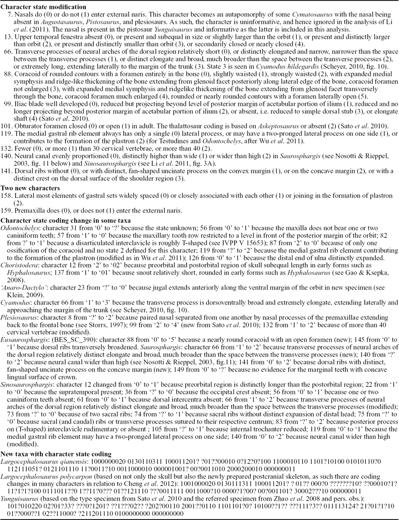
Largocephalosaurus was originally considered to be a sauropterygian, phylogenetically nested in a clade including typical pachypleurosaurs and nothosaurs (Cheng et al. Reference Cheng, Chen, Zeng and Cai2012a ). This result was based mainly on skull characters because the postcranial skeleton was not then available for study, and details of the well-preserved saurosphargid Sinosaurosphargis, had not yet been published for comparison. With the discovery of the new species, many anatomical features such as the close ‘rib-basket’, the lateral gastral elements forming a sheet-like structure, the deeply ‘V’-shaped posterior edge of the skull roof, the retention of the interpterygoid vacuity, the peculiar tooth morphology and well-retracted external naris strongly suggest that Largocephalosaurus is not a sauropterygian but may have had a close affinity to the Saurosphargidae. On the other hand, the phylogenetic relationship of the Saurosphargidae was hypothesized to be close to thalattosaurs (Li et al. Reference Li, Rieppel, Wu, Zhao and Wang2011), which differed from Nosotti & Rieppel (Reference Nosotti and Rieppel2003). For testing those competing hypotheses, we reanalysed the phylogenetic relationships based on a data matrix derived from that of Li et al. (Reference Li, Rieppel, Wu, Zhao and Wang2011). The derived data matrix included the addition of Largocephalosaurus and a Chinese pistosaur Yunguisaurus (Cheng et al. Reference Cheng, Sato, Wu and Li2006; Sato et al. Reference Sato, Cheng, Wu and Li2010), two new characters, the modification of some characters and character state coding changes in some taxa (see Table 2). In the current analysis, the new and type species of Largocephalosaurus are treated as two separate terminal taxa; for the type species, the character state coding is based on the skull as well as the newly prepared postcranial skeleton. The current data matrix consists of 39 terminal taxa (including the Ichthyopterygia) and 159 characters.
Changes to the data matrix of Li et al. (Reference Li, Rieppel, Wu, Zhao and Wang2011); character state modification, new characters, changes to coding, new characters and coding for new taxa, used to analyze the phylogenetic relationships of Largocephalosaurusi.

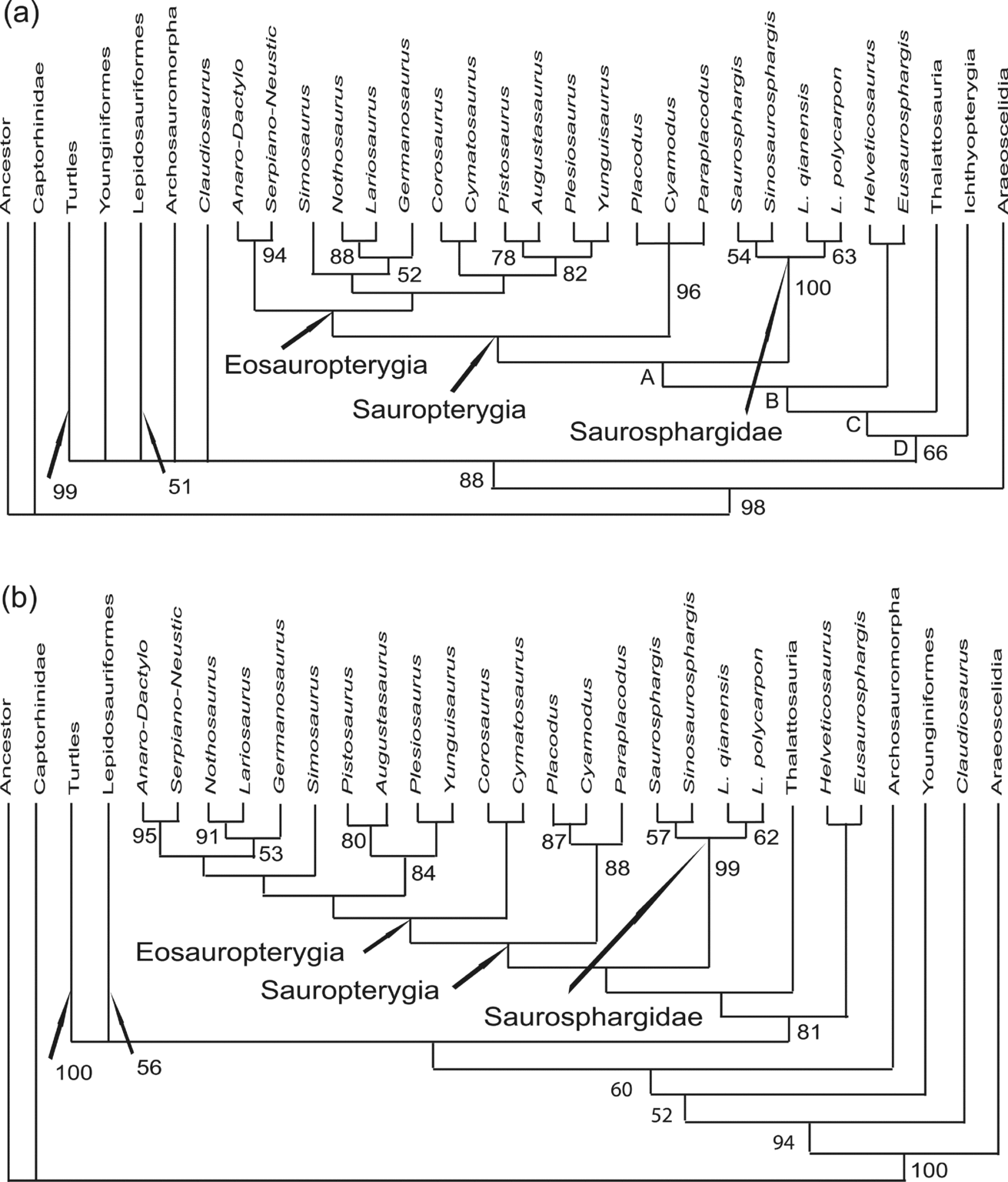
In our reanalysis, using PAUP 4.0b10 (Swofford, Reference Swofford2002), the resultant trees were rooted on an all-0-ancestor. An heuristic search with all characters equally weighted and all multistate characters unordered yielded 11 most parsimonious trees, each with a tree length of 570 steps, a consistency index of 0.3842, and a retention index of 0.6710. Interrelationships among the clades of the various marine reptiles included (i.e. Sauropterygia, Saurosphargidae, Helveticosaurus–Eusaurosphargis clade, Thalattosauria and Ichthyopterygia) are well established but not for other groups in the strict consensus of 11 most parsimonious trees (Fig. 7a). In contrast to Li et al. (Reference Li, Rieppel, Wu, Zhao and Wang2011), the Saurosphargidae is the sister-group of the Sauropterygia rather than the Thalattosauria although this relationship is not further supported by the bootstrap search owing to a low value for the monophyly of the latter, as found by Li et al. (Reference Li, Rieppel, Wu, Zhao and Wang2011). This result also differs from Nosotti & Rieppel (Reference Nosotti and Rieppel2003) in which thalattosaurs formed the sister-group of the Sauropterygia. For other marine reptilian groups, the Helveticosaurus–Eusaurosphargis clade, thalattosaurs and Ichthyopterygia form a set of successive sister-group relationships toward the Saurosphargidae–Sauropterygia clade, which also differs from Li et al. (Reference Li, Rieppel, Wu, Zhao and Wang2011) who found that interrelationships among all included marine reptilian groups entirely collapsed when the Ichthyopterygia was included in their analysis (see Li et al. Reference Li, Rieppel, Wu, Zhao and Wang2011, fig. S1). Bootstrap support is 63% for the monophyly of a clade including all aforementioned marine reptilian groups; 100% for the monophyletic Saurosphargidae; 61% for Saurosphargis–Sinosaurosphargis sister-group relationship; and 65% for the sister-group relationship of the two species of Largocephalosaurus within the family. Monophyly of the Sauropterygia was not supported in the bootstrap analysis, and their relationships with the Helveticosaurus–Eusaurosphargis clade, thalattosaurs and Ichthyopterygia were not resolved. As Li et al. (Reference Li, Rieppel, Wu, Zhao and Wang2011) suggested, these low support values may have been the result of the large amount of missing data for Helveticosaurus and Eusaurosphargis, the extreme specialization of the Ichthyopterygia and a large amount of convergence amongst the taxa included in the analysis. To test the interference of the Ichthyopterygia, we did the second analysis with the group excluded and an identical setting of the data matrix as in the first analysis. This analysis produced 16 most parsimonious trees, each with a tree length of 546 steps, a consistency index of 0.4011, and a retention index of 0.6838. As shown in the consensus tree of the 16 most parsimonious trees, the resolution of the second analysis is better than that of the first, with the interrelationships of more non-marine groups established (Fig. 7b). With the exclusion of the Ichthyopterygia, interrelationships among the marine groups also changed, especially for subgroups within the Sauropterygia. However, the results of the two analyses (with the inclusion or exclusion of the Ichthyopterygia) did not, as a whole, fundamentally differ in terms of the interrelationships produced by the bootstrap search.
Strict consensus trees of (a) 11 most parsimonious trees (MPTs) of the analysis with all taxa included; and (b) 16 MPTs of the analysis with the exclusion of Ichthyopterygia. Arabic numbers indicate the bootstrap support values; clades without numbers have a bootstrap support value lower than 50%. Abbreviations: Anaro – Anarosaurus; Dactylo – Dactylosaurus; Serpiano – Serpianosaurus; Neustic – Neusticosaurus. Synapomorphies, as optimized under accelerated transformation(ACCTRAN) assumptions in tree 1 of the 11 MPTs obtained in the first analysis (* indicates unequivocal character state): Sauropterygia, character states 13 (2)*, 18(0)*, 22(1), 29(0)*, 41(1), 43(0)*, 51(2)*, 54(1)*, 73(1), 78(1)*, 95(0)*, 98(2)*, 115(1), 135(1), 142(1), 147(1); Saurosphargidae, character states 12(1), 16(1), 25(1)*, 33(1)*, 38(0), 63(0)*, 68(1)*, 71(0), 72(1)*, 74(1)*, 85(1), 93(2)*, 104(0), 119(1)*, 126(1)*, 136(1)*, 141(2), 145(2)*, 149(1)*, 158(1)*; A, character states 17(2), 49(0), 51(1), 81(1)*, 83(2)*, 84(1), 90(1), 92(2)*; B, character states 37(0), 42(1), 66(1)*, 89(1)*, 98(1), 99(1), 101(1)*; C, character states 9(2), 29(1), 31(1)*, 38(1), 45(1)*, 48(1)*, 49(1), 62(1),64(1), 97(1), 139(1); D, character states 1(1)*, 9(0), 12(0), 13(3)*, 18(3)*, 58(1)*, 61(1)*, 75(0), 98(2)*, 102(1)*, 105(1)*, 109(1), 110(1), 112(0), 113(1), 114(1), 137(1)*.

6. Discussion and conclusions
Compared to Sinosaurosphargis, Largocephalosaurus is morphologically less modified from a typical diapsid reptile; it still retains an elongate body shape, a well-developed supratemporal fenestra, a suborbital fenestra (although reduced in size) and an incomplete osteoderm covering. As a group, the Saurosphargidae is also less specialized than the sister-group Sauropterygia in terms of the short neck (nine cervical vertebrae probably in all members) and the presence of the interpterygoid vacuity, the articulation between the braincase and palate, the pronounced transverse process of the pterygoid and the ectopterygoid.
The monophyly of the Saurosphargidae is well supported by 20 synapomorphies including 13 unequivocal character states (see Fig. 7). There are seven synapomorphies (with two unequivocal character states: the transverse processes of the dorsal vertebrae distinctly elongate and broad, much broader than spaces between the transverse processes [character 66] and the neural canal distinctly wider than high [character 140]) to support the close relationship of Saurosphargis with Sinosaurosphargis, while four synapomorphies (including three unequivocal character states: the parietal skull table weakly constricted [character 19], the internal trochanter well developed, [character 105] and the premaxilla excluded from the external naris [character 159]) support the monophyly of Largocephalosaurus itself (see Fig. 7). It is obvious that the fragmentary nature of Saurosphargis has obscured the consistency index of the other synapomorphies shared with Sinosaurosphargis and those for Largocephalosaurus. As for the close affinity of the Saurosphargidae to the Sauropterygia rather than thalattosaurs, eight synapomorphies are recognized, of which three are unequivocal, all with a consistency index of 0.5 or 1.0: the clavicle applied to the anteromedial surface of the scapula, [character 81(1)]; the posterior process of boomerang-like or atypical T-shaped interclavicle rudimentary [character 83(2)] and the humerus curved [character 92(1)]. The clavicle was described by Li et al. (Reference Li, Rieppel, Wu, Zhao and Wang2011) as articulating with the anterolateral surface of the scapula in Sinosaurosphargis. As argued earlier, based on our further examination of the specimen, the clavicle is also applied to the anteromedial surface of the scapula in this genus.
Although the bootstrap values are higher than 50% for the clades of the turtles (100%) and Lepidosauromorpha (57%), interrelationships between them as well as with the Archosauromorpha, Younginiformes, Claudiosaurus and the clade including all marine reptilian groups are unresolved in this study. Interestingly, the addition of Largocephalosaurus and Yunguisaurus did not change much in terms of the interrelationships within the clade of all related marine reptilian groups but significantly alters the phylogenetic positions of the turtles, Lepidosauromorpha, and Archosauromorpha. This, as suggested by Cheng et al. (Reference Cheng, Wu, Sato and Shan2012b ), may have been related to the unbalance of the current data matrix that mainly focuses on the marine reptiles. It is also interesting that the Ichthyopterygia did not play a significant role in establishing the interrelationships among the included aquatic groups, which is in sharp contrast to the findings of Li et al. (Reference Li, Rieppel, Wu, Zhao and Wang2011).
As indicated by the low bootstrap values, the interrelationships recognized here for all marine reptilian groups are not very stable and may alter with the discovery of new forms or better specimens of those fragmentary taxa. One of the most striking characteristic features of the Saurosphargidae is the dorsal osteoderm covering, which sometimes forms a closed carapace; among other marine reptilian groups, a similar situation is present in only the Placodontia, the basal group of the Sauropterygia. Recent studies of the placodontian phylogeny (such as Rieppel, Reference Rieppel2000b ) have indicated that the closed dorsal carapace was formed in the derived members of the Placodontia, which implies that this characteristic novelty may have independently evolved in the two groups.
Acknowledgements
We are deeply grateful to Mr Jinzhao Ding and Yinfang Chen (IVPP) and Ms Tian-fen Hu (GMPKU) for the preparation of the specimens and to Mr Wei Gao (IVPP) for taking the photos. X.-C.W particularly wishes to thank the IVPP for hospitality during his visit. T. Sato and an anonymous referee carefully reviewed the manuscript, offering critical comments and suggestions that led to its great improvement. This work was supported by research grants from the National Natural Science Foundation of PR China (NNSFC-41072014 and 40772015 to CL and X-CW; 40920124002 and 40672002 to D-YJ), the CAS/SAFEA International Partnership Program for Creative Research Teams of China (to CL and X-CW), the Program for New Century Excellent Talents in Universities (NCET-07–0015 to D-YJ), CMN (to X-CW), and FMNH (to OR).