Introduction
Atrial septal defects are a common form of CHD, accounting for about 10% of all congenital heart defects in children. Reference Hoffman and Kaplan1 Symptoms vary widely; some patients are asymptomatic, while others may experience shortness of breath, fatigue, exercise intolerance, or palpitations. The primary reason for closing the defect is significant left-to-right shunting that causes right heart overload, regardless of symptom presence. Over time, transcatheter closure has become the preferred method for suitable defects, offering better outcomes with fewer complications, lower costs, and shorter hospital stays compared to surgery.
Nonetheless, the decision to proceed with closure relies heavily on measuring the left-to-right shunt, typically expressed by the pulmonary to systemic blood flow ratio (Qp/Qs). Although cardiac catheterisation provides an accurate measurement, it is invasive, requires anaesthesia in children, and involves procedural risks. Non-invasive assessment using Doppler echocardiography is the standard alternative; however, calculating Qp/Qs can be technically challenging, time-consuming, and subject to observer variability. Consequently, there is a clear clinical need for simpler, reliable, and easily accessible surrogate markers—such as the inferior vena cava (IVC) collapsibility index—to aid decision-making and haemodynamic monitoring.
Therefore, this study aims to evaluate early changes in pulmonary and systemic venous flow parameters following transcatheter atrial septal defect closure and to explore the potential of the IVC collapsibility index as a non-invasive alternative to the Qp/Qs ratio.
Materials and methods
Echocardiographic recordings obtained 24 hours before and 24 hours after the procedure were retrospectively analysed in patients who underwent transcatheter atrial septal defect closure at our clinic between January 2023 and June 2024, and the impact of these parameters on acute-phase cardiac remodelling was evaluated.
A CONSORT-style flow diagram illustrating patient selection, enrolment, allocation, and analysis is presented in Figure 1( a ). Of the 35 patients assessed for eligibility, 31 underwent transcatheter atrial septal defect closure and were included in the final analysis. Criteria for transcatheter occlusion required a haemodynamically significant ostium secundum atrial septal defect, defined by a Qp/Qs ratio of 1.5:1 or greater, or evidence of right ventricular volume overload even in the absence of symptoms. For safety and effectiveness, all patients needed suitable cardiac anatomy for device closure, particularly adequate tissue rims to ensure secure device anchoring.
CONSORT—style flow diagram of patient selection, enrolment, allocation, and analysis. ( a ). Schematic illustration of the standardisation of inferior vena cava measurement ( b ).

Patients were haemodynamically evaluated both 24 hours before and after the procedure. All procedures were performed under general anaesthesia and guided by transthoracic echocardiography. Balloon sizing and transthoracic echocardiography were used to determine the size of the defect. The Amplatzer Septal Occluder, Lifetech CeraFlex Septal Occluder, and Occlutech Figulla Flex II Atrial Septal Occluder devices were used for closure.
Standard 2D transthoracic echocardiography examinations were performed using the Philips EPIQ 7C ultrasound system (Philips Medical Systems, Andover, USA) with an S5-1 transducer (1.0–5.0 MHz multi-frequency range) and an S8-3 transducer (3.0–8.0 MHz multi-frequency range).
To ensure consistency and reduce inter-observer variability, a single experienced paediatric cardiologist performed all offline echocardiographic measurements. Because the closure device was clearly visible in post-procedural imaging, blinding the operator to the study time point (pre- vs post-procedure) was not possible. However, to minimise potential bias, the operator was blinded to the invasive catheterisation data during echocardiographic analysis.
Patients in whom the device was observed in situ by transthoracic echocardiography performed 24 hours after the procedure were included in our study. Qp/Qs was calculated in the catheterisation laboratory using the Fick principle.
Pulse wave (PW) Doppler gradient/velocity/ velocity-time integral in the main pulmonary artery in the parasternal short axis and PW Doppler gradient/velocity/ velocity-time integral in the right upper pulmonary vein in the apical 4-chamber window were measured and determined as other variables. All echocardiographic measurements were taken while patients were breathing spontaneously in the supine position. In younger children who could not follow specific respiratory instructions (e.g., the sniff manoeuvre), maximum and minimum IVC diameters were recorded during the natural respiratory cycle under quiet conditions. Distraction techniques were employed to ensure patient cooperation and haemodynamic stability during image acquisition. To maintain standardisation, the IVC diameter was measured in the subcostal long-axis view. The measurement callipers were positioned approximately 1–2 cm caudal to the junction of the hepatic veins, strictly perpendicular to the vessel’s long axis, avoiding the ostium of the right atrium. M-mode imaging was used to capture the respiratory phasic changes, as shown in the schematic representation provided in Figure 1( b ). To assess cardiovascular status, the collapsibility index was calculated as the ratio of the difference between the maximum (expiration) and minimum (inspiration) IVC diameters to the maximum diameter. The IVC collapsibility index was calculated using the formula: (IVC diameter maximum—IVC diameter minimum)/IVC diameter maximum.
Demographic data, general and cardiac examination findings, and growth assessment data (body weight, height) were obtained from patient records. All procedures performed within the scope of the study were performed according to the relevant national ethical standards and the 2008 revised version of the 1975 Declaration of Helsinki. Institutional Ethics Committee approval was obtained, and written informed consent was obtained from all patients or their legal guardians regarding the use of the procedures and clinical records in the final publication.
Statistical analyses were performed using IBM SPSS Statistics for Windows, Version 24.0 (IBM Corp., Armonk, NY). The normality of data distribution was assessed using the Shapiro-Wilk test. Continuous variables are presented as mean ± standard deviation (SD) for normally distributed data and as median (minimum–maximum) for non-normally distributed data. Comparisons between pre- and post-procedural values were conducted using the paired samples t-test for parametric data and the Wilcoxon signed-rank test for non-parametric data. Comparisons between independent groups were performed using the independent samples t-test. The relationships between variables were evaluated using Spearman’s correlation and linear regression analyses. A p-value of <0.05 was considered statistically significant.
Results
Thirty-one patients were included in our study. Seventeen of our patients were male, and 14 were female. Male/Female ratio was 1.21:1, mean age was 90.19 ± 56.01 months, mean procedure weight was 26.20 ± 15.37 kg, median defect size by transthoracic echocardiography was 11.00 mm (7–19), median defect size by balloon sizing was 13.80 mm (7.30–25), mean atrial septal defect occluder device size was 14.48 ± 3.36 mm, median Qp/Qs ratio measured during angiography was 1.80 (1.50–2.60).
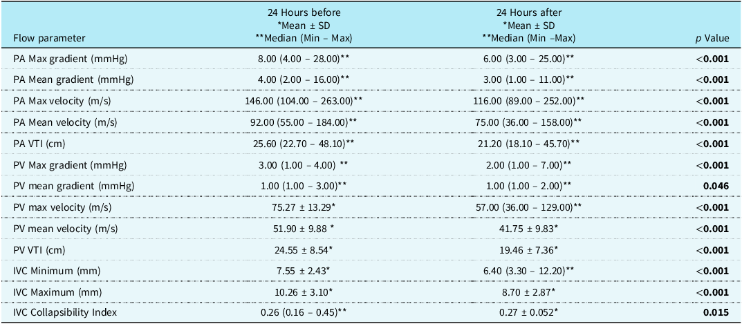
When the data obtained were evaluated, it was observed that pulmonary artery and pulmonary vein maximum-mean pressure gradient, pulmonary artery and pulmonary vein maximum-mean velocity, pulmonary artery and pulmonary vein velocity-time integral, IVC maximum and minimum parameters, and IVC collapsibility index changed statistically significantly after transcatheter closure compared to before (Table 1).
Flow parameters and inferior vena cava measurements assessed 24 hours before and after transcatheter atrial septal defect closure

PA = Pulmonary Artery; PV = Pulmonary Vein; IVC = Inferior Vena Cava; VTI = Velocity Time Integral.
A p-Value less than 0.05 was considered to indicate statistical significance.
* Parametrically distributed data are presented as mean ± standard deviation (SD).
** Non-parametrically distributed data are presented as median (min–max).
Parametric variables were analysed using the paired sample t-test, whereas non-parametric variables were evaluated using the Wilcoxon signed-rank test.
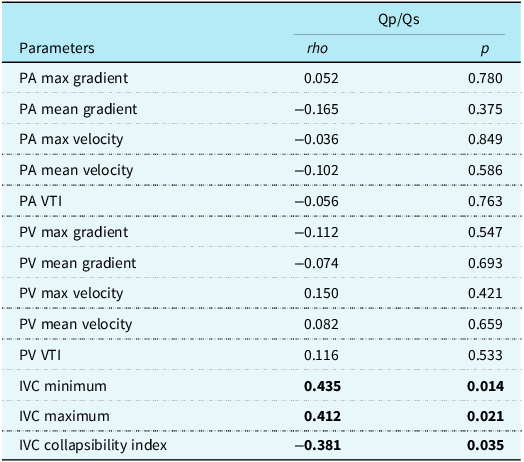
In the correlation analysis, there was a statistically significant moderate positive correlation between Qp/Qs values and IVC minimum values (rho = 0.435; p = 0.014). There was a statistically significant moderate positive correlation between Qp/Qs values and IVC maximum values (rho = 0.412; p = 0.021). There was a statistically significant weak negative correlation between Qp/Qs values and IVC collapsibility index values (rho = −0.381; p = 0.035). The other correlation coefficients in Table 2 are not statistically significant.
The correlation analysis between echocardiographic measurements obtained 24 hours before transcatheter atrial septal defect closure and the Qp/Qs ratio

PA = Pulmonary Artery; PV = Pulmonary Vein; IVC = Inferior Vena Cava; VTI = Velocity Time Integral.
Rho: Spearman’s correlation coefficient.
A p-value less than 0.05 was considered to indicate statistical significance.
Correlation analysis was performed using Spearman’s rank correlation test.
The results of the univariate linear regression analysis for estimating the Qp/Qs values based on IVC minimum, IVC maximum, and IVC collapsibility index were as follows:
Qp/Qs = 1.473 + 0.048* IVC Minimum
Qp/Qs = 1.498 + 0.033* IVC Maximum
Qp/Qs = 2.166 – 1.278* IVC Collapsibility Index
Graphical representations of the univariate linear regression analysis outcomes are presented in Figure 2.
Graphical representations for univariate linear regression analysis outcomes.

Subgroup analyses
In the subgroup analyses, a significant positive correlation was found between age and both the IVC minimum (p < 0.001) and maximum (p < 0.001) values using the Spearman correlation test. However, no statistically significant relationship was detected between age and the IVC collapsibility index. Additionally, independent samples t-tests assessing the effects of gender and device type revealed no significant associations with IVC minimum, maximum, or collapsibility index values. The Amplatzer Septal Occluder was excluded from device type analysis because it was used in only one patient.
Discussion
Atrial septal defects are among the most common types of CHD. In patients with an isolated secundum atrial septal defect and a significant left-to-right shunt, pulmonary blood flow increases, leading to enlargement of the right atrium and right ventricle, which causes volume overload and hyperkinetic circulation. Although most patients with atrial septal defect are asymptomatic, symptoms such as shortness of breath, exertional dyspnoea, and fatigue may occur due to this volume overload in patients with a significant left-right shunt. Previous studies have shown that transcatheter atrial septal defect closure significantly reduces flow parameters in the early period and contributes to the development of remodelling. In this study, we aimed to evaluate the pulmonary artery and pulmonary vein flow parameters, along with IVC diameter and collapsibility index, using transthoracic echocardiography before and after transcatheter atrial septal defect closure. These parameters were selected based on their potential to reflect volume overload and a hyperdynamic circulatory state. Furthermore, we aimed to identify a novel non-invasive marker by correlating these echocardiographic findings with the Qp/Qs ratio.
When examining the literature, it is noteworthy that few studies are based on flow parameters. Some studies use parameters such as myocardial perfusion index, ventricular or atrial volumes, left ventricular end-diastolic volume, and pulmonary artery diameter measurements, as well as studies that combine these parameters with basic flow parameters. Reference Sezer, Özyurt and Narin2 We chose to analyse the changes occurring after atrial septal defect closure using flow parameters only.
Considering the studies evaluating the effect of transcatheter closure on the remodelling of left structures, Salehian et al. Reference Salehian, Horlick and Schwerzmann3 evaluated the left ventricular myocardial perfusion index, left ventricular dimensions, ejection fraction, and left atrial volume in a group of adult patients and found that closure of atrial septal defects with a device led to a decrease in both left ventricular and left atrial volume. In the study by Pascotto et al. Reference Pascotto, Santoro and Cerrato4 which evaluated the effect of transcatheter atrial septal defect closure on cardiac remodelling in the early and mid-term, no significant decrease in left atrial volume was observed, but left ventricular diastolic diameter and the right ventricular/left ventricular diameter ratio decreased significantly.
In our study, pulmonary vein maximum and mean gradients, pulmonary vein maximum and mean velocities, and pulmonary vein velocity-time integral were analysed as flow parameters affecting the remodelling of left structures. When the literature is reviewed, it is noteworthy that no studies have evaluated all pulmonary vein flow parameters before and after transcatheter closure. In the study by Lam et al. Reference Lam, Fang, Yip, Li, Yang and Yu5 involving adult patients, the pulmonary vein velocity-time integral value yielded significant results both in evaluating the size of the shunt and in assessing remodelling after closure. In the study by Gobran et al. Reference Gobran, El-Saied, El-Shedoudy and Maria6 which included a paediatric age group, a significant decrease was observed in the mean peak systolic and diastolic wave velocities of the pulmonary vein after transcatheter atrial septal defect closure. When our data were evaluated, the pulmonary vein max gradient (p < 0.001), pulmonary vein mean gradient(p < 0.05), pulmonary vein max velocity (p < 0.001), pulmonary vein mean velocity (p < 0.001) and pulmonary vein velocity-time integral (p < 0.001) decreased significantly, consistent with the literature. The results of the present study, as well as those of other investigators, may be explained haemodynamically by the decrease in blood flow to the pulmonary artery after transcatheter atrial septal defect closure, and thus the decrease in the flow returning from the pulmonary veins to the left atrium.
When studies evaluating the effect of transcatheter closure on remodelling of right structures were evaluated, it was found that right ventricular function improved after closure in the study by Salehian et al., Reference Salehian, Horlick and Schwerzmann3 in which the myocardial perfusion index of the right ventricle was evaluated in a group of adult patients. In the study by Pascotto et al. Reference Pascotto, Santoro and Cerrato4 evaluating the effect of transcatheter atrial septal defect closure on cardiac remodelling in the early and mid-term, a significant decrease in right atrium volume was observed after the procedure.
In our study, we used pulmonary artery maximum and mean gradients, pulmonary artery maximum and mean velocities, pulmonary artery velocity-time integral, IVC minimum, and IVC maximum parameters to evaluate the right structures. In the study by Yadav et al. Reference Yadav, Gajurel and Poudel7 on adult patients in which flow parameters were studied, a significant decrease was observed in pulmonary artery Vmax, Vmean, and velocity-time integral values 48 hours after the transcatheter atrial septal defect closure compared to the pre-procedure period. In the study by Chen et al. Reference Chen, Sun, Cao, Zhang, Chen and Hu8 pulmonary artery Vmax, Vmean, and velocity-time integral values were found to be significantly decreased in echocardiography performed one week after atrial septal defect closure. The IVC collapsibility index is an important indicator of right atrial pressure. In a study conducted by Shafei et al. Reference Shafei, Kotby, Ganzoury, Habeeb and Elsayed9 when evaluated before and six months after transcatheter atrial septal defect closure, it was found that the IVC collapsibility index changed significantly. Our study builds upon these findings by demonstrating that haemodynamic adaptations—specifically, the recovery of venous compliance—begin as early as 24 hours after the procedure. This indicates that the IVC collapsibility index is a highly sensitive marker for immediate haemodynamic relief, preceding the gradual structural reverse remodelling observed in long-term follow-up studies. When the data obtained from all studies were evaluated, it was observed that the decrease in left-right shunt caused a decrease in pulmonary artery blood flow and led to significant changes in pulmonary artery flow parameters in the initial phase.
Based on our study, a significant decrease (p < 0.001) was found in the pulmonary artery maximum and mean gradients, pulmonary artery maximum and mean velocities, and pulmonary artery velocity-time integral parameters, and it was found to be consistent with the literature. The IVC maximum (p < 0.001) and IVC minimum (p < 0.001) values, as well as the IVC collapsibility index (p = 0.015), which was studied as a new parameter to evaluate the effect of right-sided structures on remodelling, decreased significantly. Because atrial septal defect is a left-to-right shunt defect, it increases pulmonary artery blood flow and leads to an increase in pulmonary artery diameter. This leads to a significant change in pulmonary artery flow parameters in the early period after atrial septal defect closure. In patients with atrial septal defect, increased pulmonary arterial flow leads to elevated pulmonary pressures, which in turn result in decreased right ventricular compliance and/or increased filling pressures. These changes expose the right atrium to pressure overload and dilation.
The physiological mechanism underlying the changes in the IVC collapsibility index following atrial septal defect closure is closely linked to the acute alteration in right atrial haemodynamics. The IVC acts as a compliant capacitance vessel, and its diameter and collapsibility are directly influenced by right atrial pressure and intravascular volume status. In patients with a significant atrial septal defect, chronic left-to-right shunting causes right ventricular volume overload and increased right atrial tension, which can blunt the respiratory phasic changes of the IVC. Transcatheter closure leads to an immediate cessation of the shunt, resulting in a significant and acute reduction in right ventricular preload. We postulate that this sudden relief of volume overload decreases the transmural pressure acting on the IVC walls. Consequently, the vessel becomes more compliant and responsive to the negative intrathoracic pressure generated during inspiration, leading to an increased collapsibility index. This improvement in the collapsibility index serves as an early surrogate marker of successful acute-phase cardiac reverse remodelling and the normalisation of right-sided filling pressures. Reference Sezer, Özyurt and Narin2,Reference Ciozda, Kedan, Kehl, Zimmer, Khandwalla and Kimchi10
Qp/Qs is the ratio of pulmonary to systemic blood flow, and it increases in conditions where pulmonary blood flow is elevated. Increased blood flow to the pulmonary arteries leads to a higher volume of blood returning to the right atrium, and the resulting increase in right atrial volume and pressure contributes to IVC dilation. The correlation analysis revealed a weak negative correlation between our Qp/Qs value and IVC collapsibility index, indicating that as Qp/Qs increases, the IVC collapsibility index decreases. This is expected from a haemodynamic perspective, as the blood flow through the IVC reduces collapsibility, which increases vascular stiffness.
When reviewing the literature, a study conducted in adult patients has shown that IVC dilatation and low IVC collapsibility index are generally associated with elevated central venous pressure, while normal venous dimensions and collapse indices support the conclusion of normal central venous pressure. Reference Ciozda, Kedan, Kehl, Zimmer, Khandwalla and Kimchi10 In a study by Egbe et al. Reference Egbe, Abozied and Miranda11 involving patients with tetralogy of Fallot, a dilated IVC was found to have the highest sensitivity (94%) in detecting severe right ventricular diastolic dysfunction. Although Egbe et al. focused on tetralogy of Fallot, our findings indicate that IVC mechanics are also a reliable reflection of right ventricular preload conditions in atrial septal defect physiology. In a study by Das et al. Reference Ali, Das and Naim12 which included patients in shock, a strong negative correlation was found between central venous pressure and the IVC collapsibility index, demonstrating the potential of the IVC collapsibility index as a non-invasive alternative to the invasive central venous pressure measurement. Our results align with those of Das et al., as we observed a similar negative correlation between shunt magnitude (volume load) and collapsibility. Regarding the reliability of these measurements in the paediatric population, Iwamoto et al. Reference Iwamoto, Tamai, Kohno, Masutani, Okada and Senzaki13 and Garcia et al. Reference Garcia, Meert, Safa and Aggarwal14 have provided evidence supporting the utility of IVC indices for estimating filling pressures in children. While Iwamoto et al. Reference Iwamoto, Tamai, Kohno, Masutani, Okada and Senzaki13 did not report a correlation in mechanically ventilated patients, Garcia et al. Reference Garcia, Meert, Safa and Aggarwal14 identified a significant inverse correlation between IVC collapsibility index and central venous pressure in spontaneously breathing patients. This distinction supports the validity of our measurement protocol, which was performed under spontaneous breathing conditions. Another study conducted in patients with chronic heart failure undergoing right heart catheterisation showed that the IVC diameter had the highest accuracy among the parameters used to identify patients with a pulmonary capillary wedge pressure ≥15 mmHg. Reference Goonewardena, Blair and Manuchehry15
In studies evaluating structural remodelling, it was observed that remodelling did not become significant in the early period and was prolonged until the middle period. In our study, all flow changes became significant in the early period. Crucially, the IVC collapsibility index offers clear advantages over traditional structural indices like right ventricular end-diastolic diameter or functional metrics like tricuspid annular plane systolic excursion (TAPSE). Structural changes usually take weeks to months to become evident due to the lag in myocardial remodelling. Similarly, while TAPSE measures longitudinal systolic function, it may not immediately detect volume unloading. In contrast, the IVC collapsibility index provides quick feedback on the normalisation of right-sided filling pressures. We believe that flow parameters are more useful than measures of structural remodelling in explaining early changes. However, some studies suggest that the IVC collapsibility index may be an alternative to current methods for assessing central venous pressure in ICU patients and pulmonary capillary wedge pressure via angiography in patients with chronic heart failure. We believe that flow parameters are more useful than structural remodelling measures in explaining changes in the early period. However, there are studies suggesting that the IVC collapsibility index may serve as an alternative to the methods currently used for assessing central venous pressure in intensive care patients and pulmonary capillary wedge pressure via angiography in patients with chronic heart failure.
Notably, to our knowledge, this study is among the first to comprehensively evaluate IVC parameters during acute atrial septal defect closure. The observed improvement in IVC dynamics suggests that these parameters may serve as sensitive, early indicators of remodelling in right-sided heart structures. The IVC collapsibility index may provide complementary, non-invasive information alongside established measures such as Qp/Qs when making decisions regarding transcatheter atrial septal defect closure and the timing of the procedure. With further validation in larger, prospective studies, the IVC collapsibility index may gain greater clinical relevance in guiding closure decisions and determining the optimal timing of the procedure. In this context, our findings may serve as preliminary evidence that helps inform and facilitate future prospective investigations in this area.
Limitations
In our study, the small sample size and the limited ability of younger children to perform standardised respiratory manoeuvres represent important limitations. The absence of strictly controlled respiratory efforts may have introduced variability in IVC measurements. In addition, the retrospective design restricted control over confounding factors such as respiratory effort and intravascular volume status at the time of image acquisition. Since the study population consisted solely of patients with haemodynamically significant shunts requiring intervention, a control group with insignificant shunts was not available, precluding Receiver Operating Characteristic analysis for defining a diagnostic cut-off value. Furthermore, the IVC collapsibility index was not compared with other established echocardiographic markers of right ventricular function. Prospective studies with standardised protocols are therefore needed to validate these findings.
Conclusion
In the acute phase after transcatheter atrial septal defect closure, significant reductions were observed in pulmonary artery, pulmonary vein, and IVC flow parameters. These changes reflect an immediate alteration in right atrial haemodynamics after defect closure. IVC measurements, including the collapsibility index, were sensitive to early haemodynamic unloading in the post-procedural period. When interpreted alongside established haemodynamic parameters such as Qp/Qs, the IVC collapsibility index may provide complementary, non-invasive information to support decision-making for transcatheter atrial septal defect closure and to determine the timing of the procedure.
Acknowledgements
The authors would like to thank Ferhan Elmalı for his contributions to the statistical analysis.
Financial support
This research received no specific grant from any funding agency, commercial or not-for-profit sectors.
Competing interests
None.
Ethical standard
The authors assert that all procedures contributing to this work follow the ethical standards of the relevant national guidelines on human experimentation and the Helsinki Declaration of 1975, as revised in 2008, and have been approved by the ethics committee of Health Sciences University Izmir Tepecik Education And Research Hospital (Approval Date: 02/09/2024 /Approval Number: 2024/ 08–23).