INTRODUCTION
Toxoplasma gondii is a widely distributed protozoan parasite capable of infecting humans as well as all warm-blooded animals. Toxoplasma gondii can cause toxoplasmosis, one of the most prevalent parasitic infections in humans. It has been estimated that approximately one-third of the world's population is infected with this parasite (Montoya and Remington, Reference Montoya and Remington1995; Petersen and Dubey Reference Petersen, Dubey, Joynson and Wreghitt2001; Robert-Gangneux and Dardé, Reference Robert-Gangneux and Dardé2012). Toxoplasmosis is generally asymptomatic in immunocompetent individuals; however, it may take a severe course in immunodeficient patients, as well as in immature foetuses and infants whose mothers suffered from primary infection during pregnancy (Luft and Remington, Reference Luft and Remington1992; Israelski and Remington, Reference Israelski and Remington1993; Wong and Remington, Reference Wong and Remington1994; Beazley and Egerman, Reference Beazley and Egerman1998).
Humans acquire the disease mainly via the oral route, through consumption of the raw or undercooked meat of infected animals or through the ingestion of sporulated oocysts that may be present in an environment contaminated with the infected feces of cats or other putative felid hosts (Dubey et al. Reference Dubey, Jenkins and Thayer1996; Cook et al. Reference Cook, Gilbert, Buffolano, Zufferey, Petersen, Jenum, Foulon, Semprini and Dun2000; Fayer et al. Reference Fayer, Dubey and Lindsay2004). Oocysts are excreted only by infected felids, the definitive hosts of T. gondii. Therefore, cats play a significant role in the epidemiology of toxoplasmosis (Dubey et al. Reference Dubey, Thayer, Speer and Shen1998). Oocysts, being resistant to unfavourable environmental conditions and chemical inactivation, are excellent survival reservoir forms of the parasite (Frenkel and Dubey, Reference Frenkel and Dubey1973; Frenkel et al. Reference Frenkel, Ruiz and Chinchilla1975; Dumètre and Dardé, Reference Dumètre and Dardé2003). Thus, basic environmental matrices may be an important source of human exposure (Benenson et al. Reference Benenson, Takafuji, Lemon, Greenup and Sulzer1982; Isaac-Renton et al. Reference Isaac-Renton, Bowie, King, Irwin, Ong, Fung, Shokeir and Dubey1998; Aramini et al. Reference Aramini, Stephen, Dubey, Engelstoft, Schwantje and Ribble1999). Several outbreaks of toxoplasmosis have been noted worldwide in which the direct source of infection in humans was soil or water contaminated with T. gondii oocysts (Benenson et al. Reference Benenson, Takafuji, Lemon, Greenup and Sulzer1982; Coutinho et al. Reference Coutinho, Lobo and Dutra1982; Bowie et al. Reference Bowie, King, Werker, Isaac-Renton, Bell, Eng and Marion1997; de Moura et al. Reference de Moura, Bahia-Oliveira, Wada, Jones, Tuboi, Carmo, Ramalho, Camargo, Trevisan, Graça, da Silva, Moura, Dubey and Garrett2006; Karanis et al. Reference Karanis, Kourenti and Smith2007; Baldursson and Karanis, Reference Baldursson and Karanis2011; Efstratiou et al. Reference Efstratiou, Ongerth and Karanis2017; Rosado-García et al. Reference Rosado-García, Guerrero, Karanis, Hinojos and Karanis2017). However, data accumulated to date concerning the prevalence of T. gondii in the environment is insufficient. Most studies on the occurrence of T. gondii in the environment have involved the detection of oocysts in water samples (Aramini et al. Reference Aramini, Stephen, Dubey, Engelstoft, Schwantje and Ribble1999; Dumètre and Dardé, Reference Dumètre and Dardé2003; Kourenti et al. Reference Kourenti, Heckeroth, Tenter and Karanis2003; Kourenti and Karanis, Reference Kourenti and Karanis2004, Reference Kourenti and Karanis2006; Villena et al. Reference Villena, Aubert, Gomis, Ferté, Inglard, Denis-Bisiaux, Dondon, Pisano, Ortis and Pinon2004; de Moura et al. Reference de Moura, Bahia-Oliveira, Wada, Jones, Tuboi, Carmo, Ramalho, Camargo, Trevisan, Graça, da Silva, Moura, Dubey and Garrett2006; Aubert and Villena, Reference Aubert and Villena2009; Gallas-Lindemann et al. Reference Gallas-Lindemann, Sotiriadou, Mahmoodi and Karanis2013; Karanis et al. Reference Karanis, Aldeyarbi, Mirhashemi and Khalil2013; Koloren and Demirel, Reference Koloren and Demirel2013; Koloren, Reference Koloren2014; Verant et al. Reference Verant, d'Ozouville, Parker, Shapiro, Van Wormer and Deem2014; Wells et al. Reference Wells, Shaw, Innocent, Guido, Hotchkiss, Parigi, Opsteegh, Green, Gillespie, Innes and Katzer2015), whereas only a few have described the parasite's presence in soil and plants (Afonso et al. Reference Afonso, Lemoine, Poulle, Ravat, Romand, Thulliez, Villena, Aubert, Rabilloud, Riche and Gilot- Fromont2008; Lass et al. Reference Lass, Pietkiewicz, Modzelewska, Dumètre, Szostakowska and Myjak2009, Reference Lass, Pietkiewicz, Szostakowska and Myjak2012; Karanis et al. Reference Karanis, Aldeyarbi, Mirhashemi and Khalil2013; Rosado-García et al. Reference Rosado-García, Guerrero, Karanis, Hinojos and Karanis2017). Moreover, it has been found that atopy and respiratory allergies are inversely related to gradients of exposure to oro-fecal or foodborne infections (including T. gondii) (Matricardi et al. Reference Matricardi, Rosmini, Riondino, Fortini, Ferrigno, Rapicetta and Bonini2000). The potential distribution of Toxoplasma oocysts in the air and their airborne transmission to the putative hosts is as yet unknown.
Therefore, the aim of the present study was to estimate the possible occurrence of T. gondii in environmental air samples collected from rural and urban areas of Poland, using sensitive molecular tools and microscopy, as well as to determine the genotype of detected parasites. The study was consequently aimed at determining whether T. gondii oocysts may be distributed within an air environment, thus constituting a potential risk of infection via the respiratory route or through nasal ingestion.
MATERIALS AND METHODS
Sampling system
Environmental air samples were collected with the use of disposable, pre-sterilized, individually packed membrane gelatine filters (Sartorius AG, Germany) with a pore size of 3 µm along with an MD8 Air Sampler mobile apparatus originally designed for the detection of airborne microorganisms (Sartorius AG, Germany). Air in a quantity of 1000 L was filtered from a height of about 30–50 cm above the surface of the ground for 30 min at an air flow rate of 30 L min−1. Following filtration, the filters were placed in disposable bags and transported to the laboratory.
Recovery experiments aimed at evaluating the methodology of airborne detection analysis
In initial experiments prior to evaluation of the detection of T. gondii oocysts in environmental samples, recovery tests were performed by using a suspension of T. gondii oocysts in sterile water. For these tests, oocysts of the PRU strain (genotype II) were used; these oocysts, approximately 2 years old, were obtained from the Laboratoire de Parasitologie-Mycologie, Université de Limoges, France. First, the oocysts were washed three times with sterile water and counted, using light and fluorescence microscopy as described below. Next, appropriate suspensions of oocysts in 1 mL of sterile water were prepared in order to obtain samples of 1 × 10⁴, 1 × 10³, 1 × 10² and 10 oocysts. Two series of four gelatine filters were then prepared and their surfaces experimentally contaminated with known numbers of T. gondii oocysts: 1 × 10⁴, 1 × 10³, 1 × 10² and 10. The surface of each filter was uniformly covered with 5 µL portions of the appropriate suspension of oocysts. Finally, the recovery procedure and specific detection of T. gondii was performed as described below. Material obtained from the first series of samples was investigated using microscopy and from the second series using molecular methods.
Sampling sites and study area
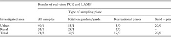
A total of 71 environmental air samples were collected between 2013 and 2015 at various sites located in the Pomerania (n = 60) and Warmia–Masuria (n = 11) provinces (northern and north-eastern Poland), including both urban (n = 40) and rural (n = 31) areas (Table 2, Fig. 2). In Warmia-Masuria province, all of the samples were collected from villages; in Pomerania province, from both urban and rural areas (40:20). Sampling was performed between May and September. Collected air samples originated from kitchen gardens and yards (n = 39), recreational areas (n = 12), and sandpits (where children were known to play) (n = 20) (Table 2).
Sampling sites were localized in places where cats (capable of excreting oocysts) were often observed and where humans might be exposed to the threat of infection. Usually at least a few cats were present on a permanent basis (information confirmed by local inhabitants) in yards and kitchen gardens in the investigated rural areas. Moreover, in urban localizations in Pomerania province investigated in this study, the presence of T. gondii had been previously confirmed in soil samples (Lass et al. Reference Lass, Pietkiewicz, Modzelewska, Dumètre, Szostakowska and Myjak2009). Additionally, studies performed on a domestic cat population from the urban area of Olsztyn, the capital of Warmia-Masuria province, showed that over 65% of animals were highly seropositive (Michalski et al. Reference Michalski, Platt-Samoraj and Mikulska-Skupień2010). This may indicate a permanent source of T. gondii infection in the cats’ environment. No similar data are available for Pomerania province. However, the data gathered from eastern, central and south-western Poland (Lublin, Lower Silesia, and Greater Poland provinces) indicate that the general percentage of seropositive cats (domestic and wild) ranges between 50 and 75% (Pawłowski, Reference Pawłowski2002; Sroka et al. Reference Sroka, Zwoliński and Dutkiewicz2007; Smielewska-Loś and Pacoń, Reference Smielewska-Loś and Pacoń2010; Sroka et al. Reference Sroka, Wójcik-Fatla, Szymańsk, Dutkiewicz, Zając and Zwoliński2010). All of these data prove the circulation of the parasite in the environment.
Purification and isolation of T. gondii oocysts from gelatine filters applied to the concentration of T. gondii oocysts from environmental air samples.
In order to concentrate trapped T. gondii oocysts and recover them from the surfaces of the gelatine filters, we employed the following procedure: first, the filter was cut to pieces with a clean scalpel and placed in a 50 mL Falcon tube. Then, 20 mL of warm water (80 °C) was added and mixed until the entire filter dissolved. Next, the suspension was centrifuged for 3 min at 1250 × g . The supernatant was removed, using an automatic pipette filler (Hirschmann Laborgeräte, Germany), and the pellet thus obtained suspended in 10 mL of warm water (80 °C) and centrifuged for 3 min at 1250 × g . The resulting pellet was placed in a 1·5-mL Eppendorf tube, suspended in sterile water, and stored at −20 °C for further analysis.
Microscopic detection of T. gondii oocysts
Three preparative methods were used to count T. gondii oocysts under the microscope in pellets obtained from the first series of the samples prepared in the recovery tests, including: (a) observation of fresh preparations using 40 × magnification; (b) staining of preparations according to the Ziehl-Neelsen staining method with the Henriksen–Pohlenz modification (Henriksen and Pohlenz, Reference Henriksen and Pohlenz1981), using 100 × magnification under an Opta-Tech MN 358 light microscope; (c) observation of fresh preparations under ultraviolet excitation at 400 × magnification using a ZEISS Imager D1 microscope equipped with appropriate fluorescence filters (exciter filter 330–385 nm) for detection of UV autofluorescence of oocysts (Lei et al. Reference Lei, Davey and Ellis2005).
Microscopic investigations of environmental air samples (final pellets obtained after oocyst recovery and concentration using gelatine filters) were performed using fresh preparations observed under light and fluorescence microscopes, as described above. Microscopy was applied for the first 22 collected air samples; the remaining samples were tested using molecular methods [real-time polymerase chain reaction (RT–PCR) and loop-mediated isothermal (LAMP)] only.
Molecular analysis
The same molecular methods (RT–PCR and LAMP), including DNA extraction and specific detection of T. gondii DNA, were applied for both recovery tests and investigation of environmental air samples.
DNA extraction
Prior to DNA extraction, the material obtained from air samples was prepared using three freeze–thaw cycles to disrupt the walls of the putative oocysts in the air samples and to improve the efficiency of DNA isolation. DNA extraction was performed using a Sherlock AX Kit (A&A Biotechnology, Gdynia, Poland) according to the manufacturer's instructions. All of the PCR templates were treated with an Anty-Inhibitor Kit (A&A Biotechnology, Gdynia, Poland), which removed polyphenolic PCR inhibitors using specific absorption particles, thereby removing factors capable of interfering with PCR. The PCR templates were stored at −20 °C.
Specific detection of Toxoplasma DNA
Detection using RT–PCR. For specific and sensitive T. gondii detection, RT–PCR was performed with the use of a pair of primers targeting a 129-bp fragment of the 35-fold repetitive B1 gene and the fluorescent-labelled TaqMan probe according to Arkush et al. (Reference Arkush, Miller, Leutenegger, Gardner, Packham, Heckeroth, Tenter, Barr and Conrad2003). Amplification was performed with an initial polymerase activation step (10 min at 95 °C), followed by 40 cycles of denaturation (15 s at 95 °C) and hybridisation/extension (1 min at 60 °C). The amplification reaction mixture consisted of 12·5 µL of Real-Time 2xHS-PCR Master Mix Probe (A&A Biotechnology, Gdynia, Poland), 400 nm of each primer (Metabion, Germany), 80 nm of TaqMan probe (Metabion, Germany) and 5 µL of template DNA in a 25-μL reaction volume. Amplifications were performed in an Mx3005P thermocycler (Stratagene, USA); PCR products were analysed using MxPro QPCR software. The cycle threshold (CT) value, which determines the cycle number at which the reporter's fluorescence exceeds the threshold value, was recorded. A sample was considered to be positive if the CT value was <40.
Detection using LAMP. The LAMP assay was performed using a Loopamp DNA Amplification Kit (Eiken Chemical, Tokyo, Japan) based on a set of primers specific to the T. gondii B1 gene (a 213-bp fragment of the AF179871 sequence) and thermal conditions as developed, evaluated and reported for the first time by Sotiriadou and Karanis (Reference Sotiriadou and Karanis2008). The reaction mixture consisted of 12·5 µL of 2 × reaction mix buffer, 40 pm of each of the primers FIP and BIP, 5 pm of each of the primers B3 and F3 (Sangon Biotech, Shanghai, China), 1 µL of Bst DNA polymerase and 2 µL of the extracted DNA in a 25-μL reaction volume. The reactions were performed in an Applied Biosystems 2720 Thermal Cycler (Thermo Fisher Scientific, USA). The amplified products were analysed using a WD-9413B gel imaging analysis system (Beijing Liuyi Biotechnology, China) following electrophoresis on 1·5% gel agarose (Biowest Regular Agarose G-10, Gene Company) stained with ExRed nucleic acid electrophoresis dye (Beijing Zoman Biotechnology, China).
All PCR experiments were performed with the inclusion of T. gondii positive controls (genomic DNA extracted from tachyzoites of the T. gondii RH strain, obtained from the National Institute of Hygiene, Poland) to ensure the correct functioning of the reaction and negative controls (water template) to prevent contamination of the PCR components. All of the negative samples were re-tested for the presence of PCR inhibitors by mixing 4 µL of the DNA template and 1 µL of T. gondii positive control.
Determining the sensitivity of the applied T. gondii molecular detection assays from suspensions of oocysts in water
DNA extraction and molecular detection using RT–PCR and LAMP were performed from three series of sterile water spiked with T. gondii oocysts in the following quantities: 1 × 10³, 1 × 10², 10. This step was performed to evaluate the detection limit of the applied molecular tools prior to application of the recovery procedure, and subsequently to estimate the approximate loss of oocysts caused by these procedures (as described below) and to define the detection limits of the applied detection methodology.
Quantitative RT–PCR for determining initial copy numbers of T. gondii DNA
In order to determine the initial copy numbers of the detected T. gondii DNA, each positive sample was tested with the real-time qualitative PCR (QPCR) method based on the standard curve. The standard template was generated by cloning the insert gene (a 129-bp fragment of the B1 gene amplified by means of conventional PCR using the set of primers described above) into a plasmid (A&A Biotechnology, Poland). To generate the standard curve, two series of eight dilutions of standard DNA in sterile water were prepared in a range of 5 × 10⁸ to 5 DNA copies per 1 µL. Following amplification of the standard dilution series, the standard curve was obtained by plotting the log of the initial template copy number against the CT value generated from each dilution. Amplification of the standard dilution series and of the DNA isolated from air samples was run on the same plate. Thus comparison of the CT values of the unknown samples with the standard curve enabled the quantification of the initial copy numbers.
Calculation of dispersive forms equivalent to T. gondii potentially present in the sample based on quantitative RT–PCR
Initial copy numbers of T. gondii DNA were then used to calculate dispersive forms equivalent to T. gondii potentially present in the examined samples. Thus, the number of T. gondii oocysts was estimated, taking into consideration the loss of oocysts during the recovery procedure. The oocyst charge of the sample was estimated using the formula:
 $${\rm OCS} = \left[ {\displaystyle{{N \times L} \over {35}} \times A \times B} \right] \times C$$
where: OCS = the oocyst charge of the sample; N = the initial copy number determined using real-time QPCR (per amount of DNA template taken for PCR); L = approximate loss of oocysts for gelatine filters; 35 = number of copies of the B1 gene per T. gondii cell; A = ¼ the number of T. gondii sporozoites per one sporocyst; B = ½ the number of T. gondii sporocysts per oocyst; C = total amount of extracted DNA (to estimate the total load of the environmental sample).
Sequencing of T. gondii DNA isolates
The final PCR product yielded by means of PCR of the positive samples was sequenced. Prior to sequencing, PCR products were cleaned with a Clean-Up Kit (A&A Biotechnology, Poland). The products of the sequencing reaction, performed using the primers described by Burg et al. (Reference Burg, Grover, Pouletty and Boothroyd1989), were cleaned with an ExTerminator Kit (A&A Biotechnology, Poland) and subjected to analysis using an ABI Prism 310 automated DNA sequencer (Applied Biosystems, USA). The results were analysed using ABI PRISM DNA Sequencing Analysis, version 3.7, for the Windows NT Platform (Applied Biosystems) and GeneStudioTM Professional (GeneStudio, Inc., USA).
Genotyping of detected T. gondii isolates
In order to identify T. gondii genotypes, the restriction analysis method was used, as described by Aspinall et al. (Reference Aspinall, Guy, Roberts, Joynson, Hyde and Sims2003). Nested PCR was performed to amplify two fragments of the T. gondii SAG2 gene (3′ and 5′ ends). The PCR reaction mixture consisted of 2·5 µL 10X GeneAmp PCR Buffer II (Applied Biosystems, USA), 2 mm MgCl2 (Applied Biosystems, USA), 0·2 mm of each dNTP (Fermentas, Lithuania), 0·4 µ m of each primer (Metabion, Germany), 1·25 U of AmpliTaq Gold Polymerase (Applied Biosystems, USA) and 2 µL of template DNA in a 25-μL reaction volume. Both amplification rounds were performed under the same conditions described by Aspinall et al. (Reference Aspinall, Guy, Roberts, Joynson, Hyde and Sims2003), with a change in the initial polymerase activation step (15 min at 95 °C). SAG2 genotypes were determined using restriction fragment length polymorphism analysis, using Sau3AI restriction enzyme (Promega, USA) to digest nested PCR products of the 5′ end and CfoI restriction enzyme (Promega, USA) to digest nested PCR products of the 3′ end (in separate reactions). All digestion was performed according to the manufacturer's instructions. The digested products were analysed using 2% agarose gel electrophoresis.
RESULTS
Limits to the detection of T. gondii oocysts using molecular assays
In the performed experiments, in which DNA was extracted from three series of sterile water spiked with T. gondii oocysts in the amounts 1 × 10³, 1 × 10² and 10, molecular detection was successful in the case of samples containing a minimum of 10 oocysts in 2 of 3 cases using RT–PCR and in 3 of 3 cases using LAMP, and in all tests at higher concentrations of oocysts using both methods.
Results of the recovery experiments
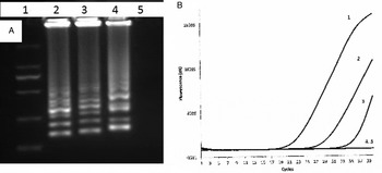
Two series of four gelatine filters experimentally contaminated with known numbers of T. gondii oocysts, i.e. 1 × 10⁴, 1 × 10³, 1 × 10² and 10, were investigated with the use of microscopic and molecular methods according to the protocols described above. These recovery tests showed that we were able, using microscopic methods, to detect T. gondii oocysts when the filter surface was contaminated with at least 1 × 10⁴ oocysts. At this level of contamination, the recovery rate was 1·7, 1·5 and 2% for an unstained preparation, stained preparation and a UV autofluorescence preparation, respectively. Recovery experiments with the use of RT–PCR and LAMP assays showed that T. gondii DNA was detectable when gelatine filter was contaminated with at least 1 × 10³ oocysts in two of three samples using RT–PCR and three of three samples using LAMP. T. gondii DNA was detected by both methods in all samples seeded at the level of 1 × 10⁴ oocysts (Fig. 1A, B).
Results of recovery tests using series of gelatine filters contaminated with T. gondii oocysts. (A) Result for LAMP: line 1 – molecular weight marker (2000 bp; Takara, Japan), line 2 – gelatine filter sample with 1 × 10⁴ oocysts, line 3 – gelatine filter sample with 1 × 10³ oocysts, line 4 – gelatine filter sample with 1 × 10² oocysts, line 5 – gelatine filter sample with 1 × 10 oocysts; (B) Result for RT–PCR: line 1 – gelatine filter sample with 104 oocysts, line 2 – gelatine filter sample with 1 × 10³ oocysts, line 3 – gelatine filter sample with 1 × 10² oocysts, line 4 – gelatine filter sample with 1 × 10 oocysts, line 5 – negative control. LAMP, loop-mediated isothermal; RT–PCR, real-time polymerase chain reaction.

Results of the investigations of environmental air samples
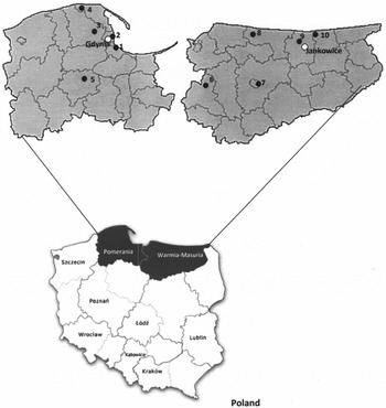
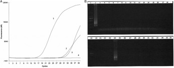
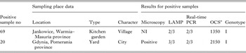
A total of 71 environmental air samples were collected and examined using RT–PCR and LAMP detection methods based on the T. gondii B1 gene. Toxoplasma gondii DNA was detected by both assays in two of the investigated samples (3%), one from an urban and one from a rural area. One positive sample (template no. 20) originated from a yard in Gdynia (a city in Pomerania province) and one (template no. 69) from a kitchen garden in Jankowice (a village in Warmia-Masuria province) (Table 1, Figs 2 and 3A, B). All air samples collected around sandpits and recreational areas were negative (Table 2).
Origin of positive air samples analysed in the Pomerania province and Warmia-Masuria province, Poland. White dots represent positive samples. Black dots and numbers represent localizations of sampling sites in the Pomerania province: 1 – area of Gdańsk and Sopot districts, 2 – area of Gdynia district, 3 – area of Wejherowo district, 4 – area of Puck district, 5 – area of Koscierzyna district; in the Warmia-Masuria province: 6 – area of Iława district, 7 – area of Olsztyn district, 8 – area of Bartoszyce district, 9 – area of Kętrzyn district, 10 – area of Węgorzewo district.

Results of the Toxoplasma gondii DNA detection in environmental air samples. (A) Results of RT–PCR: line 1 – positive control; line 2 – positive air sample collected from the yard in the Pomerania province (template No 20); line 3 – positive air sample collected from the kitchen garden in the Warmia-Masuria province (template No 69); line 4 – negative control; (B) Results of LAMP: line 1, 19 – molecular weight marker (2000 bp, Takara, Japan), line 2 – T. gondii positive control, line 4–23, 25–36 – negative samples, line 24 – positive sample (template No 20), line 4 – negative control. LAMP, loop-mediated isothermal; PCR, polymerase chain reaction.

Results of detection, quantification and genotyping of T. gondii in environmental air samples collected from the Pomerania province and the Warmia-Masuria province using molecular

LAMP, loop-mediated isothermal; PCR, polymerase chain reaction; NI, not investigated.
a Equivalent of number of oocysts, which could be present in whole air sample based on results of QPCR, assuming 100-fold loss of material during recovery procedures.
Detection of T. gondii DNA in the environmental air samples depending on type of sampling place

LAMP, loop-mediated isothermal; PCR, polymerase chain reaction.
The table shows number of positive samples per number of samples investigated in particular sampling sites.
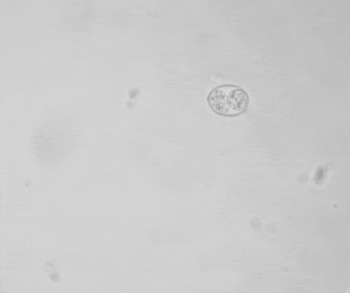
Microscopic examination confirmed the presence of T. gondii oocysts in one of the two positive samples (template no. 69) (Fig. 4).
Toxoplasma gondii oocyst visible in fresh preparation of the air sample No 20 collected from the yard in the Pomerania province.

PCR inhibition was not recorded in any of the tested negative samples.
Sequencing of positive samples and comparison with the T. gondii sequences available in the GenBank confirmed that the obtained PCR products represented T. gondii B1 gene fragments.
According to the results of our experiments, the approximate loss caused by the recovery procedure of oocysts from gelatine filters was at least a 100-fold. Therefore, the approximate level of contamination (equivalent of the total oocyst load) of positive air samples calculated based on the elaborated formula equalled 2150 or 1350 oocysts per one 1000-L air sample collected from the yard or kitchen garden, respectively (Table 1).
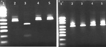
The two genotyped samples were positive for both 5′ and 3′ amplified fragments of the SAG2 gene and could be digested by Sau3AI and CfoI restriction endonucleases, respectively. According to the restriction patterns obtained following separation by electrophoresis, we classified the detected parasites as Toxoplasma SAG2 type I (Table 1, Fig. 5A, B).
Toxoplasma gondii SAG2 typing of the environmental air samples (A) Sau3AI digestions of 5′ SAG2 amplification products loaded onto a 2% gel agarose; (B) CfoI digestions of 3′ SAG2 amplification products loaded onto a 2% gel agarose: line 1 – molecular weight marker (pUC19 DNA/MspI (HpaII) Marker, 23, Fermentas, Lithuania), line 2 – genotype I positive control (RH strain), line 3 – genotype II positive control (DX strain), lines 4 and 5 – environmental air samples – SAG2 type I (no digestion).

DISCUSSION
The present paper provides useful results concerning T. gondii detection in environmental air samples, obtained through the application of two different sensitive molecular tools and microscopy for the detection of Toxoplasma oocysts and T. gondii DNA in this material. The paper presents an evaluation of the specific T. gondii detection system and analysis of air samples and sheds new light on the epidemiology and possible transmission of T. gondii via the respiratory route. To the best of our knowledge, this is the first study describing T. gondii detection in environmental air samples using molecular methods and microscopy. T. gondii DNA was found in 2 of 71 investigated samples; one of these positive samples was confirmed by microscopic investigation. The results of our findings provide evidence that air may play a role in toxoplasmosis epidemiology.
In this study, we present a procedure for collecting environmental air samples and for T. gondii oocyst recovery using a portable sampler and gelatine filters dissolvable in hot water. Some environmental air samples were investigated with both microscopic and molecular tools. However, for subsequent T. gondii detection, we decided to use molecular methods only, as they guaranteed high sensitivity and specificity in comparison with microscopic examinations. Moreover, we did not wish to divide and fragment environmental material, taking into consideration that oocysts may be dispersed in air and thus their concentration in samples was expected to be low. Recovery tests showed that a sample (gelatine filter) must be contaminated with at least 1 × 10³ oocysts to be detectable by molecular methods and with 1 × 10⁴ oocysts to be detectable by microscopic methods. Microscopy, while insufficient for sensitive detection, is capable of supporting molecular investigations. In general, T. gondii oocysts present in the air are most probably dispersed; recovery efficiency from air samples is limited, making this kind of study very difficult.
T. gondii positive air samples were found in one yard and one kitchen garden in urban and rural areas of Poland's Pomerania and Warmia-Masuria provinces (Tables 1 and 2), respectively. We noted that large numbers of stray cats were always seen near the sampling areas. These cats may excrete oocysts and contaminate their habitats. Our previous examinations, performed in Pomerania province, showed that sandpits, kitchen gardens, recreational areas and sand heaps were contaminated with Toxoplasma, which contributes to the spread of parasites in the environment (Lass et al. Reference Lass, Pietkiewicz, Modzelewska, Dumètre, Szostakowska and Myjak2009). It follows that T. gondii oocysts may float in wind and contaminate the air. This creates a potential risk that e.g. children playing in a sandpit or adults working in a kitchen garden will contract primary toxoplasmosis, not only through contact with contaminated soil but also through breathing contaminated air. Perhaps both factors are significant. This could account for reports of a considerable percentage of seropositive children (amounting to 8·2 and 23·2% in groups of between 1–5 and 6–10 years of age, respectively) in Poland (Szewczyk-Kramska, Reference Szewczyk-Kramska1999). Moreover, results of a survey performed in Europe on infected pregnant women showed that 17% of those questioned indicated contact with soil as a possible cause of infection (Cook et al. Reference Cook, Gilbert, Buffolano, Zufferey, Petersen, Jenum, Foulon, Semprini and Dun2000). We collected air samples relatively close to the ground. However, more samples, taken from different heights, should be tested in the future to estimate the degree of difference in the contamination of air layers at various levels.
Using quantitative detection via RT–PCR, an equivalent of the approximate oocyst charge (number of oocysts present) of samples originating in air was estimated. The results showed that contamination of the two positive samples did not exceed several thousand oocysts per 1000 L of air (taking the 100-fold oocyst loss into account). Thus, in one litre of investigated air, several oocysts are likely to be present. The average total lung capacity of an healthy adult human is several litres of air; the tidal volume (the volume of air inhaled or exhaled in a single breath during normal quiet breathing) is about 0·5 L; and the average human respiratory rate is 30–60 breaths per minute at birth (15–30 L of inhaled air per minute), decreasing to 12–20 breaths per minute in adults (6–10 L of inhaled air per minute) (DeBoer, Reference DeBoer2004; Lindh et al. Reference Lindh, Pooler, Tamparo and Dahl2009). In this study, samples were collected at a height of 30–50 cm above the ground, with a sampling rate of 30 L of air per minute. Thus, in positive samples several oocysts were gathered per minute of filtration. This suggests that a person present in a contaminated area (e.g. people working in gardens or children playing in sandpits) might inhale a comparable number of oocysts within 3–5 min. Therefore, long exposure to contaminated air may be a potential risk for humans, as well as animals; however, the infective dose (number of oocysts) responsible for the development of infection in humans is unknown, and more studies should be performed in this area to confirm these speculations. It must be also highlighted that quantitative RT–PCR makes it possible to count the number of DNA copies in the samples and to estimate the equivalent of T. gondii oocysts, sporocysts and sporozoites; however, their ratio in the sample has not been investigated. All the above-mentioned forms of this parasite may be present in the air.
The detection of T. gondii DNA in air samples is evidence for the presence of the parasite in the environment (in another matrix, apart from water and soil) and indicates a potential risk for humans and animals. Calculations performed based on quantitative RT–PCR assays enabled us to evaluate the probable intensity of contamination of air samples with T. gondii oocysts. However, detection of DNA itself does not enable estimation of the viability, biological activity, or virulence of isolates and dormant stages of parasites, or, consequently, their ability to infect humans and/or animals. The relative proportion of viable and non-infective Toxoplasma oocysts remains unknown.
T. gondii isolates are classified into three main clonal groups (I, II, and III), based on single- (SAG2 gene) or multilocus analysis (Howe et al. Reference Howe, Honore, Derouin and Sibley1997; Petersen and Dubey, Reference Petersen, Dubey, Joynson and Wreghitt2001). It is known that T. gondii strains representing SAG2 types I and II are regarded as pathogenic in humans. So far, little information has been gathered on the occurrence of Toxoplasma genotypes in Poland (Dubey et al. Reference Dubey, Jenkins and Thayer1996; Petersen and Dubey, Reference Petersen, Dubey, Joynson and Wreghitt2001; Nowakowska et al. Reference Nowakowska, Colón, Remington, Grigg, Gołąb, Wilczyński and Sibley2006; Sroka et al. Reference Sroka, Wójcik-Fatla and Dutkiewicz2006; Lass et al. Reference Lass, Pietkiewicz, Modzelewska, Dumètre, Szostakowska and Myjak2009, Reference Lass, Pietkiewicz, Szostakowska and Myjak2012). In this study, SAG2 type I was identified in both tested air samples. According to the literature, type II predominates in Europe, while type I is less frequent (Schwab and McDevitt, Reference Schwab and McDevitt2003; Ajzenberg et al. Reference Ajzenberg, Bañuls, Su, Dumètre, Demar, Carme and Dardé2004). In our previous studies concerning detection of T. gondii in soil and fresh fruits and vegetables, we found both genotypes (I and II), with a preponderance of type I (Lass et al. Reference Lass, Pietkiewicz, Modzelewska, Dumètre, Szostakowska and Myjak2009, Reference Lass, Pietkiewicz, Szostakowska and Myjak2012).
Further investigations are needed to pinpoint the circulation of Toxoplasma strains in environmental samples in Poland and their relevance to human and animal toxoplasmosis, as well as their actual implications for the contamination of the environment (water, food, air) and transmission via ororespiratory anatomical routes.
Concluding remarks
In this study, we present a procedure for the detection of T. gondii in environmental air samples and provide initial evidence that T. gondii may be present in the air; therefore inhalation of air contaminated with Toxoplasma oocysts may be one of the potential transmission routes for humans and animals. It remains to be investigated whether the high prevalence of toxoplasmosis infections around the world is associated with the respiratory route of transmission. Delineating the role of air distribution and of the respiratory tract in disease transmission may provide the evidence necessary to establish further guidelines for the prevention of toxoplasmosis.
While cats and other felids as final hosts are undoubtedly the principal means of acquiring infections and disease caused by Toxoplasma, this report presents ample evidence of respiratory transmission, which cannot be ignored and must be taken into consideration in future control studies. Our results could serve as a starting point for the improvement of T. gondii detection procedure in air samples as well as for further studies aimed at evaluating the actual airborne risk for humans and animals. The airborne transmission route of T. gondii to humans and animals via the dissemination of oocysts through environmental air and its epidemiological impact will likely now become a subject for debate. Future investigations and more extensive epidemiological data results will show whether this mode of transmission should be acknowledged as being of greater significance.
COMPLIANCE WITH ETHICAL STANDARDS
The manuscript does not contain the clinical studies or patient data.
The authors declare that they have no conflict of interest.
Informed consent was obtained from all individual participants included in the study.
FINANCIAL SUPPORT
We acknowledge the funding support from the Polish Ministry of Education and Science (Grant No. 2P05D00729) and One Thousand Talents Plan of the Chinese Government (Grant No. WQ2013630172).