Introduction
The official IMA List of Mineral Names (Pasero, Reference Pasero2025) reports four mineral species with the root-name chukhrovite, i.e., chukhrovite-(Ca), chukhrovite-(Ce), chukhrovite-(Nd), and chukhrovite-(Y). This root-name was first used by Ermilova et al. (Reference Ermilova, Moleva and Klevtsova1960) to describe a new mineral species from the Kara-Oba Mo-W deposit, Central Kazakhstan. This first finding was later renamed chukhrovite-(Y), following the dominance of Y among the Y+REE atoms (where REE = rare earth elements).
The name ‘chukhrovite-(Ce)’ was first introduced by Walenta (Reference Walenta1979) but, as noted by Fleischer et al. (Reference Fleischer, Cabri, Chao and Pabst1980), “Nothing whatever is said of the presence or absence of Y […]; previous analyses of chukhrovite are of chukhrovite-(Y), so the omission is a serious one”. Chukhrovite-(Ce) has been considered a valid mineral species since the special procedure of Nickel and Mandarino (Reference Nickel and Mandarino1987), assuming the type material to be that from Siberia described by Novikova (Reference Novikova1973). Actually, table 2 of this latter paper reports wt.% of Ce2O3 and Y2O3 as high as 3.83 and 2.92 wt.%, respectively, and then gives the normalised percentages as 27.1 and 20.7 wt.%, respectively. Considering the molar weights of Ce2O3 and Y2O3 (328.24 and 225.81 g/mol, respectively), these percentages correspond to 0.0826 mol Ce2O3 and 0.0917 mol Y2O3, respectively. Consequently, type material of chukhrovite-(Ce) has Y > Ce and thus corresponds to chukhrovite-(Y). This same discussion was previously reported by Pautov et al. (Reference Pautov, Bekenova, Karpenko and Agakhanov2005) who stressed that “refinement of crystal structure of rare-earth representatives of the chukhrovite group and re-study of chukhrovite-(Ce) are the most important tasks in the study of this group of minerals”.
Since then, no formal proposal for the redefinition of neotype material of chukhrovite-(Ce) has been proposed, notwithstanding some further occurrences have been reported (e.g. Niedermayr et al., Reference Niedermayr, Bauer, Bernhard, Blass, Bojar, Brandstätter, Gröbner, Hmmer, Koch, Kolitsch, Leikauf, Loranth, Poeverlein, Postl, Prasnik, Schachinger, Tomazic and Walter2008; Gentile et al., Reference Gentile, Vignola and Gatta2012). During the investigation of the mineral assemblages found in the abandoned Pb–Zn Tripi mine, located close to the hamlet of Alì, in the Peloritani Mountains, northeastern Sicily, Italy, chukhrovite-(Ce) was found again and this material was used for a full mineralogical characterisation of the species. These data were then used to propose to the Commission on New Minerals, Nomenclature and Classification of the International Mineralogical Association (CNMNC-IMA) the definition of the neotype material for this species (Proposal 24-C, Bosi et al., Reference Bosi, Hatert, Pasero and Mills2024), in accord with Dunn and Mandarino (Reference Dunn and Mandarino1987). Such proposal was accepted and a neotype specimen for chukhrovite-(Ce) was defined. Its type locality is the Tripi mine, Alì, Peloritani Mountains, Messina Province, Sicily, Italy. Neotype material is deposited in the mineralogical collection of the Museo di Storia Naturale of the University of Pisa under catalogue number 20075.
The aim of this paper is to give a full account of the crystal chemistry of chukhrovite-(Ce) from the Italian type locality, discussing its relationship with other members of the chukhrovite group.
Occurrence and physical properties
Chukhrovite-(Ce) was identified on some samples collected at the Tripi mine (38°00′50′′N, 15°24′27′′E), located some hundred metres SSW of the small hamlet of Alì. This mine exploited a stratabound fine-banded ore, hosted in phyllite, formed by sphalerite and galena, with minor pyrite and trace amounts of chalcopyrite, arsenopyrite, pyrrhotite and covellite in a gangue represented by quartz and fluorite (Saccà et al., Reference Saccà, Saccà, Nucera, De Fazio and D’Urso2007). Supergene minerals are smithsonite, gypsum, hydrozincite, ‘limonite’, malachite, kintoreite, austinite, conichalcite and zincolivenite (Saccà et al., Reference Saccà, Saccà, Nucera, De Fazio and D’Urso2007; Mauro et al., Reference Mauro, Biagioni and Zaccarini2022, Reference Mauro, Biagioni, Sejkora and Dolníček2023).
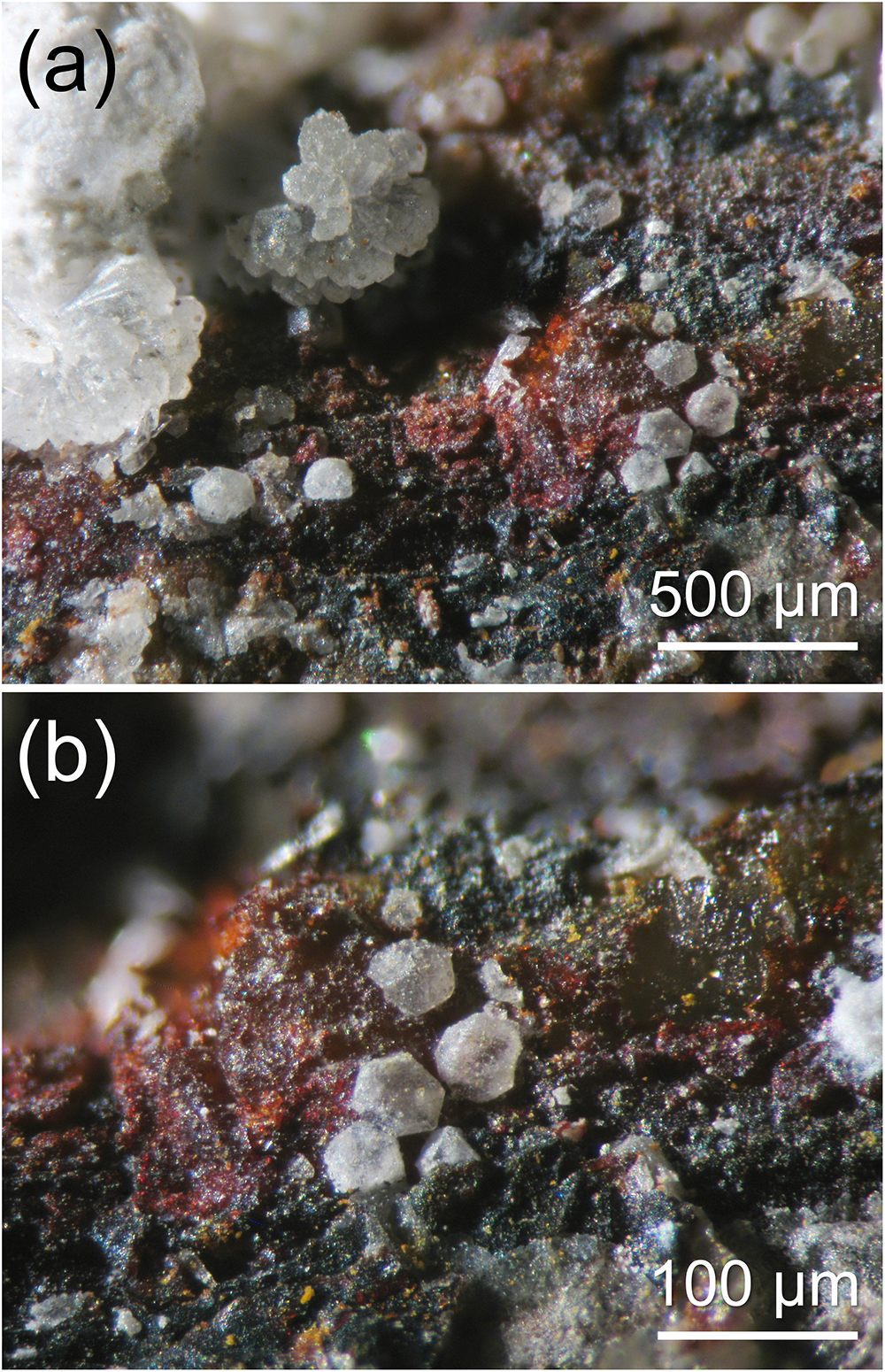
Chukhrovite-(Ce) occurs as cube–octahedral crystals, up to 0.05 mm in size, white in colour, with a white streak and a vitreous lustre, in thin fissures of quartz + fluorite veins, in association with creedite and fluorite (Fig. 1). Fluorescence under UV radiation was not observed. Mohs hardness is close to 3, in agreement with other chukhrovite-group minerals. Chukhrovite-(Ce) is brittle, with a poor-to-indistinct cleavage. Calculated density, based on the empirical formula and unit-cell parameters derived from single-crystal X-ray diffraction, is 2.460 g/cm3. The mean refractive index, obtained from Gladstone-Dale relationship (Mandarino, Reference Mandarino1979, Reference Mandarino1981), is 1.4799.
Chukhrovite-(Ce), whitish cube-octahedral crystals, up to 0.05 mm, with creedite and thin crusts of iron oxy-hydroxides. Tripi mine, Alì, Messina Province, Sicily, Italy. Holotype specimen, Museo di Storia of the University of Pisa. Catalogue number #20075. Photo D. Mauro.

Chemical data
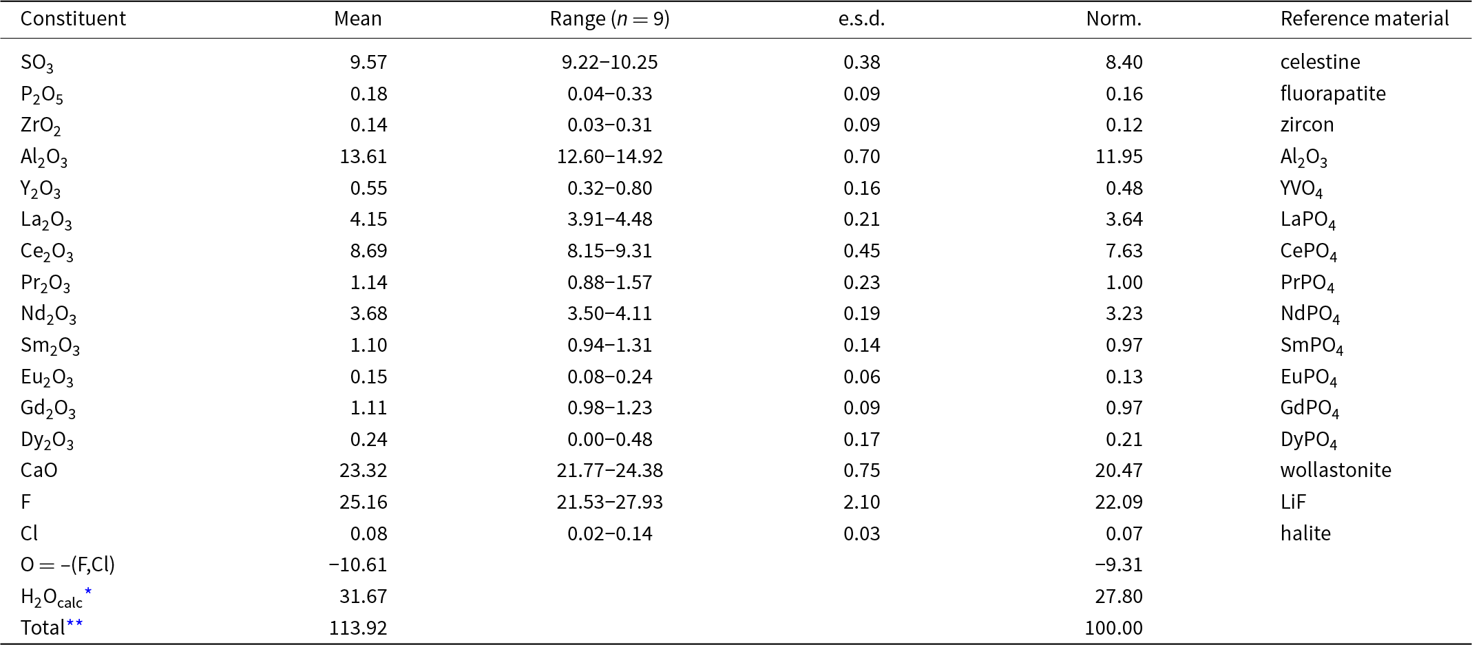
Quantitative chemical analyses of chukhrovite-(Ce) were carried out using a Cameca SX 100 electron microprobe (WDS mode, 15 kV, 5 nA and 10 μm beam diameter) on a polished crystal surface at the National Museum, Prague, Czech Republic. Results (average of 9 spot analyses) are given in Table 1. Other sought elements (As, Ba, Bi, Cu, Er, Fe, Hf, Ho, Lu, K, Mg, Mn, Na, Pb, Sc, Si, Sr, Tb, Ti, Tm, Th, U, V and Yb) were below detection limits. Matrix correction using the PAP algorithm (Pouchou and Pichoir, Reference Pouchou, Pichoir and Armstrong1985) as well as automatic corrections of overlaps for F-Ce, Pb-Y, Gd-La, Gd-Ce, Gd-Nd, Ba-Pr, La-Nd, Eu-Pr, Tm-Sm, Bi-Ce, P-Y and P-Ca were applied to the data.
Electron microprobe data for chukhrovite-(Ce) from the Tripi mine, Alì, Messina Province, Sicily, Italy

Notes: n = number of spot analyses;
* calculated in agreement with stoichiometry;
** high total due to partial dehydration; e.s.d. = estimated standard deviation.
Raman spectroscopy, as well as structure analysis (see below), indicated the absence of CO3 groups. Because insufficient pure material is available for a direct determination of H2O, it has been calculated based upon the known stoichiometry from structure analysis. Chukhrovite-(Ce) partly dehydrated in the electron microprobe chamber and it is also unstable under a focused electron beam.
The empirical formula of chukhrovite-(Ce) was calculated on the basis of 29 anions, assuming the presence of 13 (F+Cl+OH) atoms and 12 H2O groups per formula unit, and taking into account the results of the crystal structure study (see below). It is Ca3.17(Ce0.40La0.19Nd0.17Pr0.05Sm0.05Gd0.05Y0.04Eu0.01Dy0.01)Σ0.97Zr0.01Al2.04(S0.91P0.02)Σ0.93O4[F10.10Cl0.02(OH)2.88]Σ13.00·12H2O. The ideal, end-member formula, of chukhrovite-(Ce) is (Ca3Ce)[AlF6]2(SO4)F·12H2O, corresponding to (in wt.%) SO3 9.16, Al2O3 11.67, Ce2O3 18.79, CaO 19.26, F 28.27, H2O 24.75, O = F –11.90, sum 100.00.
Micro-Raman spectroscopy
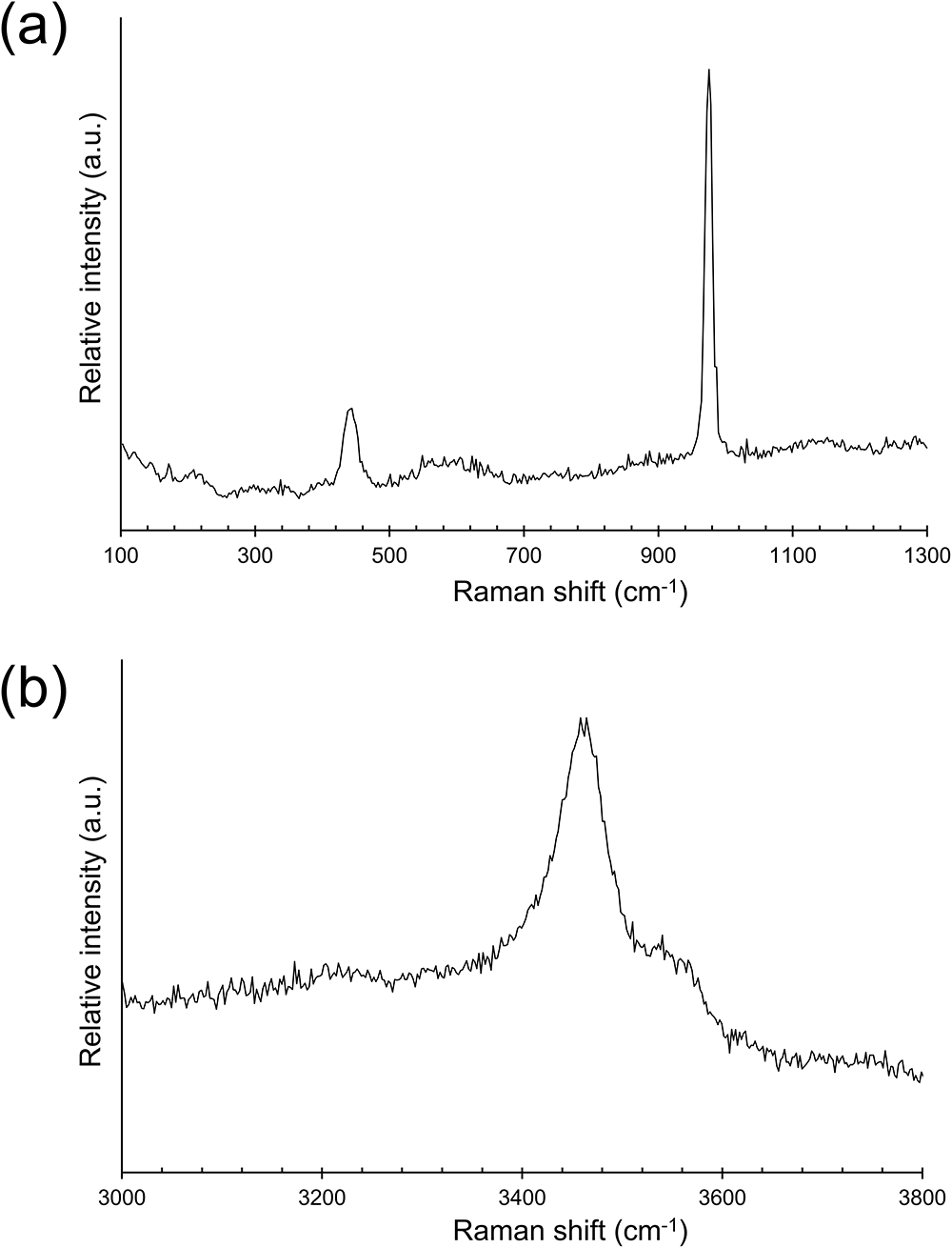
The Raman spectrum of chukhrovite-(Ce) was collected on an unpolished grain in the range 100–4000 cm–1 in nearly back-scattered geometry with a Horiba Jobin-Yvon XploRA Plus apparatus, equipped with a motorized x–y stage and an Olympus BX41 microscope with a 50× objective. The 532 nm line of a solid-state laser was used filtered at 25% (laser power 6.25 mW). The minimum lateral and depth resolution was set to a few μm. The system was calibrated using the 520.6 cm–1 Raman band of silicon before each experimental session. Spectra were collected through multiple acquisitions (3) with variable counting times of 30 and 60 s. Back-scattered radiation was analysed with a 1200 gr/mm grating monochromator. The possible thermal damage of the measured points was excluded by visual inspection of the excited surface after measurement, by observation of possible decay of spectral features in the start of the excitation and checking for thermal downshift of Raman lines. The Raman spectrum of chukhrovite-(Ce) is shown in Fig. 2.
Raman spectrum of chukhrovite-(Ce) in the range 100–1300 cm–1 (a) and 3000–3800 cm–1 (b).

The region between 100 and 1300 cm–1 is characterised by a strong band at 975 cm–1, related to stretching modes of SO4 groups. A weaker band occurs at 443 cm–1, and can be interpreted as due to the bending of SO4 groups. A weak and very broad band can be observed at ca. 580 cm–1, possibly still related to bending of SO4 groups. The position of these bands agrees with those reported by Vignola et al. (Reference Vignola, Hatert, Bersani, Diella, Gentile and Risplendente2012) in chukhrovite-(Ca). In the O–H stretching region, a broad band at 3460 cm–1, with a shoulder at 3540 cm–1, agrees with the occurrence of H2O groups suggested by the crystal structure refinement (see below).
X-ray crystallography
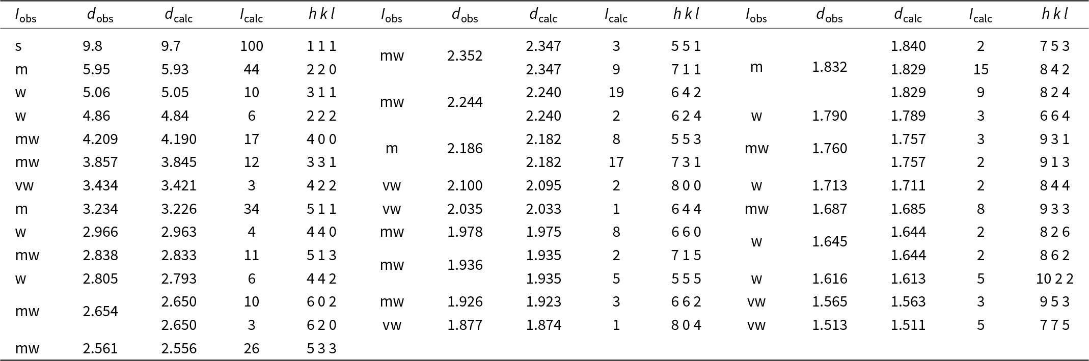
Powder X-ray diffraction data of chukhrovite-(Ce) were collected using a Bruker D8 Venture single-crystal diffractometer equipped with a Photon III CCD area detector (microfocus CuKα radiation) simulating a Gandolfi-like geometry (Centro per l’Integrazione della Strumentazione scientifica dell’Università di Pisa, CISUP, University of Pisa, Italy). Observed X-ray diffraction lines are reported in Table 2, along with the calculated pattern based on the structural model discussed below.
Powder X-ray diffraction data (d in Å) for chukhrovite-(Ce) from the Tripi mine

Notes: I calc and d calc were obtained using PowderCell 2.4 (Kraus and Nolze, Reference Kraus and Nolze1996) on the basis of the structural model of chukhrovite-(Ce) given in Table 4. Only calculated reflections with I calc > 2 (if not observed) are reported. I obs were visually estimated: s = strong; m = medium; mw = medium-weak; w = weak; and vw = very weak.
Single-crystal X-ray diffraction intensity data were collected using a Bruker D8 Venture diffractometer equipped with an air-cooled Photon III CCD detector, and microfocus MoKα radiation (CISUP, University of Pisa, Italy). The detector-to-crystal distance was 38 mm. Data were collected using ω and φ scan modes, in 0.5° slices, with an exposure time of 10 s per frame. A total of 788 frames was collected. The frames were integrated with the Bruker SAINT software package using a narrow-frame algorithm. Data were corrected for Lorentz-polarisation, absorption and background using the package of software Apex4 (Bruker AXS, Reference Bruker2022). Unit-cell parameters were refined on the basis of the XYZ centroid of 9157 reflections above 20σ(I) with 4.860° < 2θ < 60.87°.
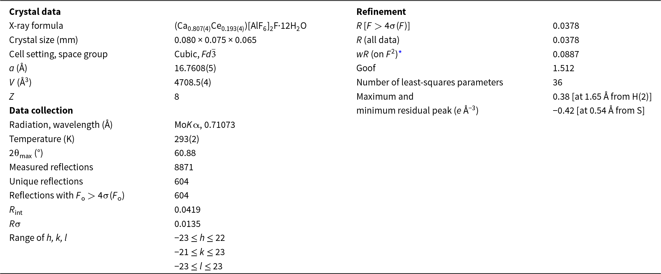
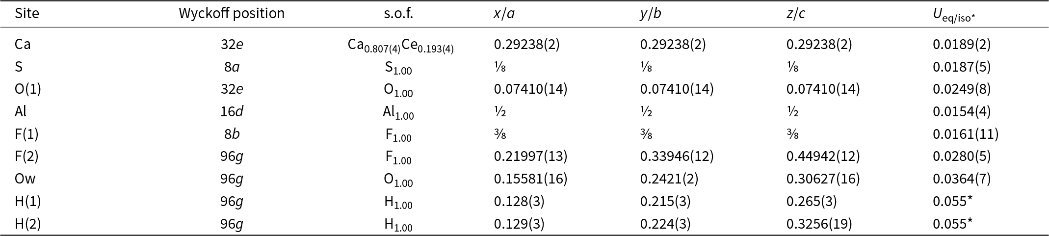
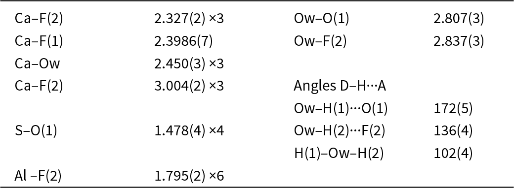
The statistical tests on the distribution of |E| values (|E 2 – 1| = 0.850) and systematic absences agree with the space group Fd  $\bar 3$. The crystal structure of chukhrovite-(Ce) was refined using Shelxl-2018 (Sheldrick, Reference Sheldrick2015) starting from the atomic coordinates of synthetic chukhrovite given by Mathew et al. (Reference Mathew, Takagi, Waerstad and Frazier1981). The following neutral scattering curves, taken from the International Tables for Crystallography (Wilson, Reference Wilson1992), were used: Ca vs. Ce at Ca(1); S at the S site; Al at the Al site; O at O(1) and Ow sites; F at F(1) and F(2) sites; and H at H(1) and H(2). The Ow–H distances were restrained and the isotropic displacement parameter of H atoms was constrained to be 1.5 times that of the bonded O atom. After several cycles of anisotropic refinement for all atoms but H, the R factor converged to 0.0378 for 604 unique reflections with F o > 4σ(F o) and 36 refined parameters. Details of the data collection and crystal structure refinement are given in Table 3. Atom coordinates and isotropic or equivalent isotropic displacement parameters are reported in Table 4, whereas Table 5 gives selected bond distances. Bond-valence calculation, shown in Table 6, was performed using the bond parameters of Brese and O’Keeffe (Reference Brese and O’Keeffe1991). The Crystallographic Information File (CIF) is available as Supplementary Material (see below).
Summary of crystal data and parameters describing data collection and refinement for chukhrovite-(Ce) from the Tripi mine

* Note: w = 1/[σ2(F o2) +55.2492P], where P = (F o2+2F c2)/3.
Sites, site occupancy factors (s.o.f.), fractional atom coordinates and isotropic (*) or equivalent isotropic displacement parameters (Å2) for chukhrovite-(Ce) from the Tripi mine

Selected bond distances (Å) and angles (°) for chukhrovite-(Ce) from the Tripi mine

Notes: D = donor; A = acceptor.
Weighted bond-valence sums (in valence units) for chukhrovite-(Ce) from the Tripi mine

Notes: left superscripts indicate the number of equivalent bonds involving anions. For the Ca site, the following site occupancy was used: Ca0.7875Ce0.1000La0.0500Nd0.0375Pr0.0125.
* Corrected for H-bonds.
Crystal structure of chukhrovite-(Ce)
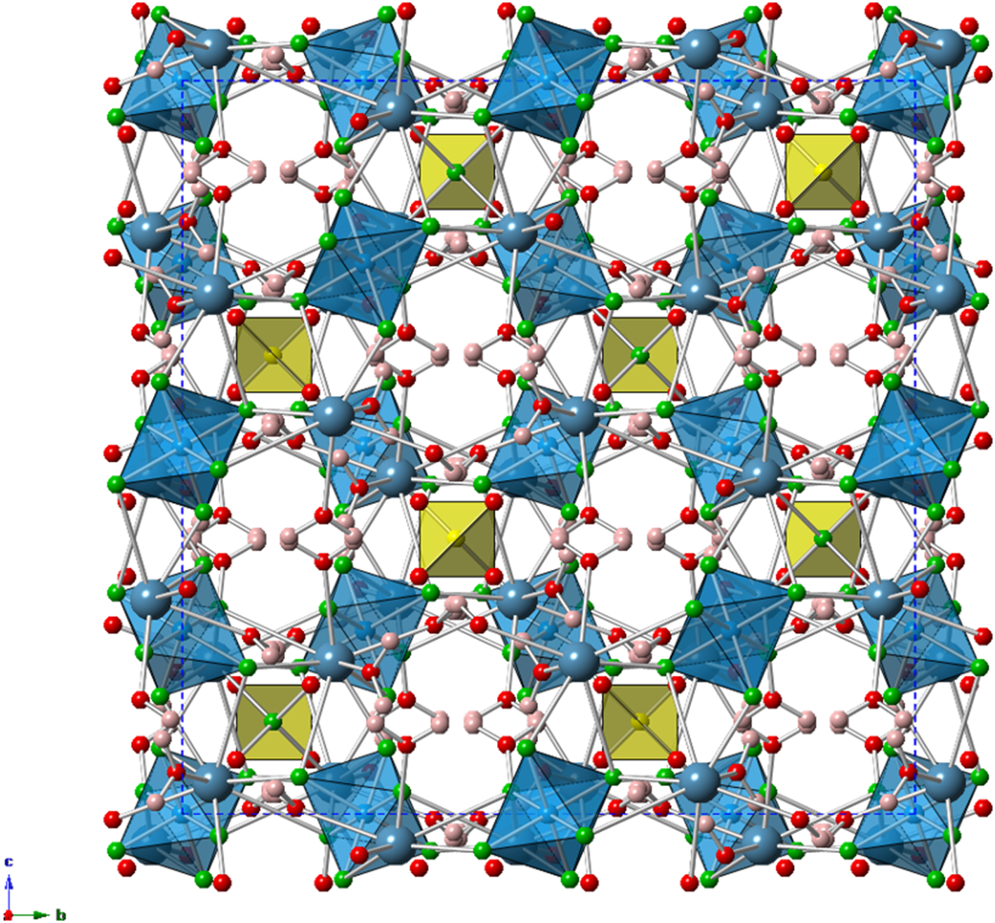
Three independent cation sites are present in chukhrovite-(Ce), namely the Al, Ca and S sites. The crystal structure is shown in Fig. 3.
Crystal structure of chukhrovite-(Ce) as seen down a. The unit cell is shown as dashed blue lines. Symbols: blue polyhedra = Al-centred octahedra; yellow = S-centred tetrahedra. Circles: green = F(1) and F(2) sites; red = O and Ow sites; pink = H(1) and H(2) sites. Drawn using CrystalMaker (Palmer, Reference Palmer2014).

Aluminium atoms are bonded to six F atoms hosted at F(2). Corners of AlF6 octahedra are shared with different Ca-centred polyhedra. Considering the long Ca–F(2) bond at ca. 3 Å, the linkage between Al- and Ca-centred polyhedra can be described as edge-shared. The bond-valence sum at the Al site is 3.06 valence units (vu).
The Ca site hosts Ca and minor REE. Among the latter, Ce is the most abundant. On the basis of electron microprobe data, the site population (Ca3.15Ce0.40La0.20Nd0.15REE*0.05□0.05) may be proposed, where REE* is the sum of remaining REE. The occurrence of a vacancy (□) is related to the substitution rule 3Ca2+ = 2Ce3+ + □. The mean atomic number should be 27.3 electrons (Z = 56 was used for REE*, as average of the atomic numbers of Pr, Sm, Gd and Y, neglecting minor Eu and Dy). This value can be compared with that obtained through the crystal structure refinement, i.e. 27.33 electrons. Both these values are slightly lower than that calculated on the basis of the empirical formula, i.e. 30.0 electrons. Atoms hosted at the Ca site show a seven-fold coordination, being bonded to three F atoms F(2), three H2O groups at Ow, and one F atom at F(1). In this way, a monocapped trigonal prism occurs, with one Ow being the capping ligand. Three other longer distances at ca. 3 Å may increase the coordination number to ten. Using the given site population, the bond-valence sum at the Ca site is 2.20 vu, in accord with a theoretical value of 2.18 vu.
Sulfur atoms show the typical tetrahedral coordination, with a <S–O> distance of 1.478 Å, agreeing with the value given by Hawthorne et al. (Reference Hawthorne, Krivovichev and Burns2000), i.e. 1.473 Å. The bond-valence sum is 5.92 vu, in agreement with the presence of S6+.
Four independent anion sites occur in chukhrovite-(Ce), i.e. O(1), F(1), F(2) and Ow. Oxygen atoms at O(1) are underbonded, and indeed they are acceptor of three H bonds from H2O groups hosted at Ow, with an O(1)···Ow distance of 2.807(3) Å, corresponding to a bond strength, calculated in agreement with Ferraris and Ivaldi (Reference Ferraris and Ivaldi1988), of 0.18 vu. The F(1) site is tetrahedrally coordinated by four Ca sites, and its bond-valence sum is 1.00 vu, in accord with the occurrence of F– anions. On the contrary, the anion at F(2) is three-fold coordinated and it is an acceptor of H bonds from Ow, with Ow···F(2) of 2.837(3) Å, with a bond strength of ca. 0.15 vu (Brown and Altermatt, Reference Brown and Altermatt1985). It is probable that minor OH groups suggested by electron microprobe analysis may be hosted at F(2), that could have a site occupancy close to [F0.76(OH)0.24]. However, in the calculation of bond-valence sums, only the F bond parameter was considered. Finally, the H2O group are bonded to the Ca site only and, as described, act as donors of H bonds to atoms hosted at O(1) and F(2).
The crystal-chemical formula of chukhrovite-(Ce) can be written as (Ca3Ce)[AlF6]2(SO4)F·12H2O (Z = 8).
Relationship with other species
Chukhrovite-(Ce) belongs to the chukhrovite group (Table 7), a series of fluoride–sulfates with cubic symmetry. In particular, chukhrovite-(Ce) is isotypic with chukhrovite-(Y) (Bokiy and Gorogotskaya, Reference Bokiy and Gorogotskaya1965) and with synthetic chukhrovite of Mathew et al. (Reference Mathew, Takagi, Waerstad and Frazier1981). The crystal structure of chukhrovite-(Nd) (Pautov et al., Reference Pautov, Bekenova, Karpenko and Agakhanov2005) and meniaylovite (Vergasova et al., Reference Vergasova, Semyonova, Epifanova, Filatov and Chubarov2004) are currently unrefined, but they are probably isotypic with chukhrovite-(Ce) and chukhrovite-(Y), with meniaylovite showing the substitution Al3+ + REE3+ = Si4+ + Ca2+. On the contrary, chukhrovite-(Ce) shows homeotypic relations with chukhrovite-(Ca) reported by Vignola et al. (Reference Vignola, Hatert, Bersani, Diella, Gentile and Risplendente2012), the latter having an additional partially occupied Ca site.
Minerals belonging to the chukhrovite group

Remarks on the origin of chukhrovite-(Ce)
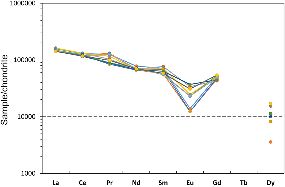
The association of chukhrovite-(Ce) with creedite and iron oxy-hydroxides suggests an origin from acidic solutions related to weathering of sulfides. The common lack of heavy metals (Cu, Pb, Zn) and As in both sulfates suggests that the parent solutions were sourced either from an ore portion rich in pyrite and poor in other sulfides, or, more likely, from outside of the ore body (e.g. from pyrite-bearing wall rocks). The latter possibility is also suggested from REE data of chukhrovite-(Ce). The chondrite-normalised REE patterns (Fig. 4) show a systematic decrease from La to Sm with a minimum variability, characterised by a narrow range of chondrite-normalised La/Sm ratios (2.1–2.7). Such a feature is surprising because chukhrovite-(Ce) also contains a high amount of F, which is one of the strongest REE-complexing ligands (Wood, Reference Wood1990). Poorly fractionated REEs are thus incompatible with a significant influence of F in the REE-bearing fluids, but can be compatible with weaker complexation caused by sulfate anions (Migdisov and Williams-Jones, Reference Migdisov and Williams-Jones2014; Migdisov et al., Reference Migdisov, Williams-Jones, Brugger and Caporuscio2016). Fluoride anions probably represented the key depositional agent allowing precipitation of a REE-bearing mineral (Williams-Jones et al., Reference Williams-Jones, Migdisov and Samson2012; Migdisov and Williams-Jones, Reference Migdisov and Williams-Jones2014; Migdisov et al., Reference Migdisov, Williams-Jones, Brugger and Caporuscio2016). In such scenario, chukhrovite-(Ce) crystallised from (REE,SO4)-bearing fluids after their interaction with the ore body of the Tripi mine containing fluorite as a gangue. The lack of Ce anomaly (Fig. 4) and the presence of a negative Eu anomaly (Eu/Eu* = 0.22–0.69; Fig. 4) preclude highly oxidising conditions during migration of REE-bearing solutions and precipitation of chukhrovite-(Ce) (Sverjensky, Reference Sverjensky1984; Bau, Reference Bau1991; Bau and Möller, Reference Bau and Möller1992). The limited variability of the Eu anomaly can reflect either minor fluctuations (or a continuous change) of redox conditions of fluids or inherited heterogeneity of source rocks from which REEs were leached.
Chondrite-normalised REE distribution patterns of individual spot anaylses for chukhrovite-(Ce) from the Tripi mine. The normalisation values are from Anders and Grevesse (Reference Anders and Grevesse1989).

Conclusions
Since the paper by Embrey and Hey (Reference Embrey and Hey1970), the role of type specimens in Mineralogy has become a central issue in the definition of new mineral species. Owing to the relative recentness of the practice of identifying type material, some species discovered before the 1970s still lack formally defined type specimens. However chukhrovite-(Ce) did not simply lack a formally defined type material, instead the selected one was actually Ce-rich chukhrovite-(Y). Consequently, the new finding at the Tripi mine, Sicily, Italy, allowed both the full characterisation of the Ce-member of the chukhrovite group and the definition of its neotype material, filling a gap in the systematics of this mineral group.
The Tripi mine is a scarcely known mineralogical locality in the Peloritani Mountains, northeastern Sicily. Recent investigations are revealing a mineralogy more complex than previously thought, with the identification of several species previously not reported from Sicily (e.g. Mauro et al., Reference Mauro, Biagioni and Zaccarini2022, Reference Mauro, Biagioni, Sejkora and Dolníček2023). Chukhrovite-(Ce) is the latest addition to these species, and it is the 45th mineral having its type locality in Sicily, and the first one discovered in the rocks of the Calabro–Peloritan Arc. Its finding shows that further studies on the mineral assemblages of the small ore deposits of this sector of southern Italy could allow interesting scientific discoveries.
Supplementary material
The supplementary material for this article can be found at https://doi.org/10.1180/mgm.2024.91.
Acknowledgements
The Centro per l’Integrazione della Strumentazione scientifica dell’Università di Pisa is thanked for the access to the single-crystal X-ray diffraction laboratory. This research has been supported by the Ministry of Culture of the Czech Republic (long-term project DKRVO 2024-2028/1.II.b; National Museum, 00023272) for JS and ZD. The authors wish to thank Fernando Cámara, Peter Leverett and an anonymous reviewer for their critical revision.
Competing interests
The authors declare none.